Review
. 2020 Apr 10;11(4):413.
doi: 10.3390/genes11040413.

Comprehensive History of CSP Genes: Evolution, Phylogenetic Distribution and Functions

Affiliations
Review

Comprehensive History of CSP Genes: Evolution, Phylogenetic Distribution and Functions

Guoxia Liu et al. Genes (Basel). .

Abstract

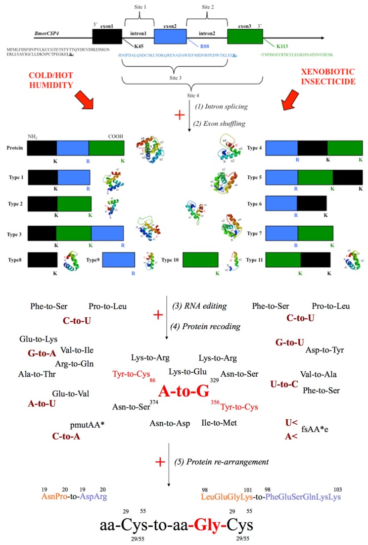
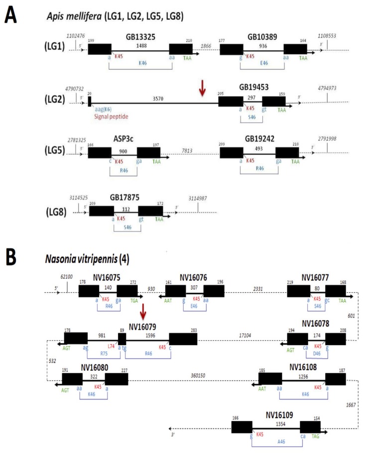
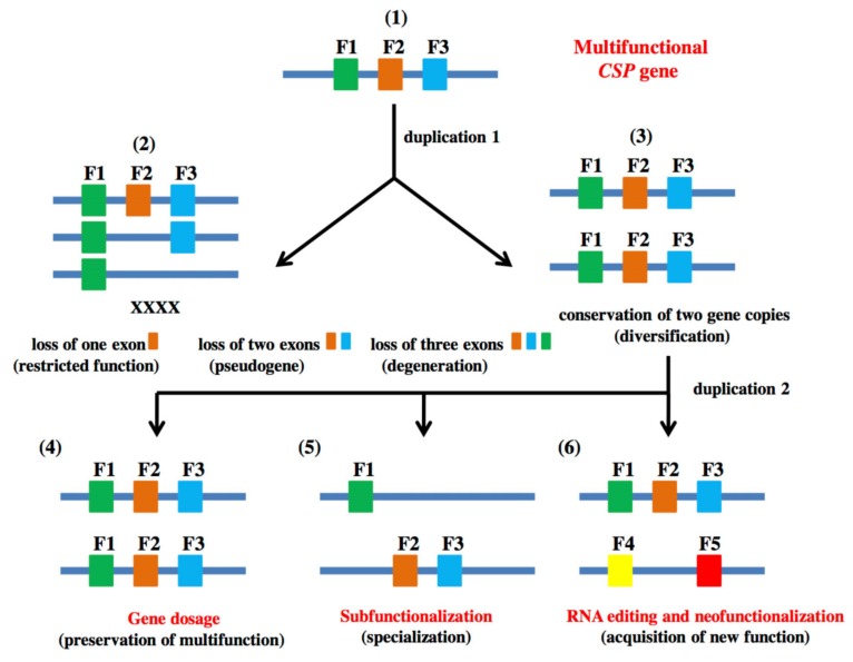
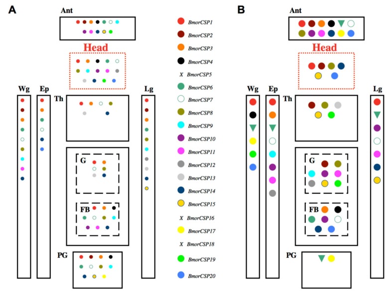
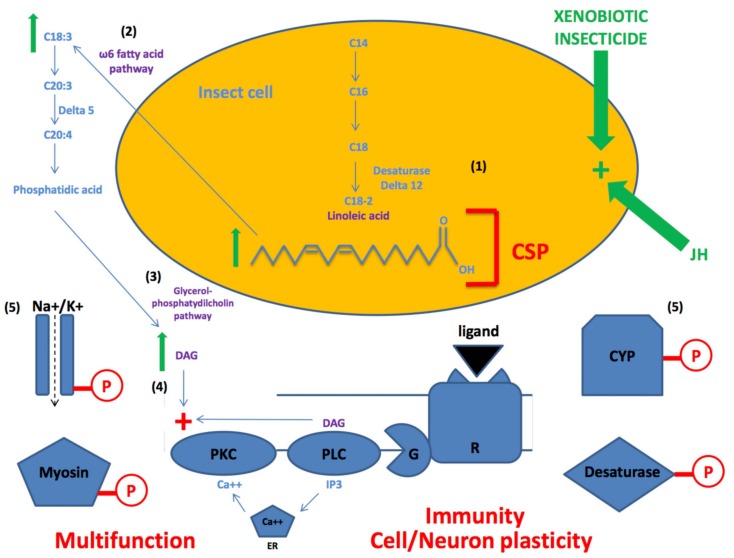
In this review we present the developmental, histological, evolutionary and functional properties of insect chemosensory proteins (CSPs) in insect species. CSPs are small globular proteins folded like a prism and notoriously known for their complex and arguably obscure function(s), particularly in pheromone olfaction. Here, we focus on direct functional consequences on protein function depending on duplication, expression and RNA editing. The result of our analysis is important for understanding the significance of RNA-editing on functionality of CSP genes, particularly in the brain tissue.

Keywords: RNA mutation; adaptive process; lipid transport; neuroplasticity; tandem duplication; xenobiotic resistance.


Conflict of interest statement

The authors declare no conflicts of interest.

Figures

Figure 1
Figure 1
Gene splicing and RNA editing mechanism for diversification of chemosensory proteins (CSPs) under environmental change. An insect CSP gene structure such as lepidopteran BmorCSP4 contains 2 introns and 3 exons [13,14]. (1) Genomic DNA (bold black line) is transcribed into premature mRNA yielding four possible sites for intron splicing. (2) The native protein sequence and eleven types of protein sequence variants can be produced by intron splicing, excision of non-coding regions (intron boundaries: K45, R88 and K113) and shuffling of coding regions (exon1 in black, exon2 in blue and exon 3 in green box). The folded shape of BmorCSP4 and a number of 11 new protein foldings (11 variants) can be generated from gene splicing. (3) The primary transcripts that are a faithful copy of the gene and variant mRNAs are all subject to further typo RNA editing, resulting in an increased number of genetic variants and protein subtypes [13,16,17,18,19,20]. Each mRNA is subject to mutations (A-to-G, A-to-U, C-to-A, C-to-U, G-to-A, G-to-U and/or U-to-C) depending on external conditions (cold/hot temperature, humidity and/or exposure to xenobiotic insecticides). (4) The substitutions A-to-G at positions 86 and 356 build proteins harboring tyrosine (Tyr) to Cysteine (Cys) mutations in two different regions of BmorCSP4. Base deletion mutations (A< and U<) result in an early stop codon (fsAA*e), thereby yielding shortened proteins. C-to-A mutation changes the position of the stop codon (pmutAA*) and enhances the number of truncated protein isoforms/edited variants [13]. (5) The protein is recomposed not only after the translation process, i.e., when mRNA is translated to produce a protein, but also after protein synthesis. Once the protein is synthesized, the Asparagine-Proline (Asn-Pro) motif switches to another amino acid motif, Aspartate-Arginine (Asp-Arg). The Leucine-Glutamate-Glycine-Lysine (LeuGluGlyLys) motif changes to Phenylalanine-Glutamate-Serine-Glutamate-Lysine-Lysine (PheGluSerGluLysLys) in the C-terminal tail. A Glycine residue (Gly) is inserted next to Cysteine at position 29, 55 or both [13,14,16,17,19,20]. Protein structures are generated by BmorCSP4 templates in SWISS-MODEL using the X-ray crystal structure of MbraCSPA6 (1kx9.1.A) as a reference model [7,18].
Figure 2
Figure 2
Genomic organization of Apis and Nasonia CSPs. (A) Genomic organization of A. mellifera CSPs on four different chromosomes (LG1, LG2, LG5 and LG8) (B) Genomic organization of N. vitripennis CSPs on chromosome 4. Exons are shown as black boxes, introns as bold black plain lines and intergenic intron regions as dotted lines. The numbers above the box and the plain line give the exon and intron size, respectively. The numbers in italics above the line give the distances between genes. Exon/intron sizes and intergenic distances are given in base pairs. The amino acid residue in red indicates the intron insertion site (K45, L74 or signal peptide). In blue is shown the triplet codon for the amino acid interrupted by intron insertion. Stop codons are indicated in green (Apis: TAA; Nasonia: TAA or TGA, A>G switch in stop codon). Horizontal arrow in black indicates the orientation of the gene: 5’–3’ (right) or 3’–5’ (left). The red vertical arrow points out the different position of the double-intron CSP gene in A. mellifera and N. vitripennis, respectively.
Figure 3
Figure 3
Schema of relationships from bacteria to insects and amino acid phylogenetic analysis of Apis and Nasonia CSPs. (A) Timeline of the evolutionary history of life from bacteria and prokaryote cells to multiple species of insects. (B) Gene phylogeny and orthology groups of bacterial/insect CSPs with focus on gene duplication profiling in honeybees and jewel wasps. Bacteria: Acinetobacter (WP_071212566, WP_071222707); Kitasatospora (WP_04307137, WP_07383810176) [29,30]. Insects: ants (EFN), beetles (AAJJ), flies (Dmel), moths (Bmor) and whiteflies (Btab) [12,13,14,15,37,38,44,45,46]. Crustacean: A. franciscana (AfraCSP; ABY62736, ABY62738); D. pulex (DpulCSP1, DpulCSP2; ABH88167, ABH88166). Phylogenetic trees are generated from a total of ninety protein sequences (IQ-TREE, UFBoot; 1000 replicates). Blue and green color circles represent Apis mellifera (Amel) and Nasonia vitripennis (NV) protein sequences, respectively. The gene structures are shown on the right for Amel (in blue) and NV (in green) CSPs. Branches are shown supported by >50% bootstrap value. Six major orthology groups are found corresponding to specific Amel and NV CSP sequences: group I (AmelGB19242, NV16079); group II (AmelASP3c); group III (AmelGB13325, NV16108); group IV (AmelGB17875, NV16075, NV16076, NV16077, NV16078); group V (AmelGB10389, AmelGB10453, NV16109); group VI (NV16080). Blue and green arrows indicate gene duplication profiling in Amel and NV, respectively. Supplementary Methods: Figure 3 The multiple sequence alignment was performed using Muscle global alignment (www.ebi.ac.uk/Tools/msa/muscle). Phylogenetic trees were constructed using IQ-TREE (http://iqtree.cibiv.univie.ac.at). The following parameters were used for phylogenetic tree construction, ultrafast bootstrap (UFBoot, using the –bb option of 1000 replicates), and a standard substitution model (-m TEST) was given for tree inference. The generated trees from IQ-TREE tool were visualized using Figtree (http://tree.bio.ed.ac.uk/software/figtree) and the branch-support values were recorded from the output treefile. The re-rooting was performed on WP_071212566 and WP_071222707 node. The trees were modified as cladogram and increasing order nodes were applied under trees section for better visualization.
Figure 4
Figure 4
Evolution of CSPs for neofunctionalization. (1) At some point far back in time (Bya), the original ancestor CSP gene retains three functions (three exons: F1, F2 and F3). The outcome of the first early duplication (duplication 1) is a pair of tandem paralogous genes strictly identical to the original gene. (2) Duplication 1 can lead to loss of one exon (restricted function), loss of two exons (unexpressed pseudogene: BmorCSP5, BmorCSP16, BmorCSP18 and AAJJ0269A1B [14,38]), or loss of the three exons (degeneration/loss of a complete gene copy). (3) Duplication 1 can also lead to the conservation of the two gene copies, increasing the original functions while providing a template for CSP diversification. Further successive genome duplications (duplication 2) allow expansion of the gene family. (4) Additional copies of the CSP gene are preserved and keep performing the same functions (F1–F3) as the ancestral gene, thus amplifying the original activity of “CSP”. (5) The different functions of CSP are divided over specific duplicated copies, leading to CSPs with more specialized functions (subfunctionalization). (6) Other duplicates are subjected to RNA editing events and multiple specific mutations that lead the CSP family to acquire new genes and functions (F4, F5).
Figure 5
Figure 5
Tissue expression profiling of CSPs. B. mori CSP gene expression profiling under normal conditions (A) and following exposure to abamectin insecticide (B). Data are from Xuan et al. [13,14]. Specific gene expression is shown by color code. X indicates no expression for truncated genes (BmorCSP5, BmorCSP16 and BmorCSP18). Up regulation in the expression levels of CSP genes is indicated by a larger circle. Down regulation in the expression levels of CSP gene (BmorCSP6) is indicated by a triangle oriented down. Ant: Antennae, Ep: Epidermis, FB: Fat Body, G: Gut, Lg: Legs, PG: Pheromone Gland, Th: Thorax, Wg: Wings.
Figure 6
Figure 6
Conjectural model of the role of CSP in activation of linoleic acid/omega6 fatty acid-diacylglycerol pathway upon xenobiotic insecticide or juvenile hormone exposure. (1) Some insect cells have the ability to synthesize linoleic acid (C18:2) de novo using fourteen-eighteen carbons-fatty acids (C14–C18) and specific desaturases. (2) C18:2 is fuel molecule for omega6 fatty acid pathways. Molecules such as arachidonyl-CoA ((Z5,Z8,Z11,Z14)-Icosatetraenoyl-CoA or C20:4) are products of a ∆5 desaturase reaction from eicosatrienoyl-CoA (C20:3) as a direct substrate. (3) Synthesis of these fatty acid metabolites leads to phosphatidic acid and therefore to the formation of diacylglycerol (DAG) through the biosynthetic pathway of glycerol-phosphatydilcholins. (4) DAG is a relay molecule in intracellular cascades activated by the binding of regulatory chemical ligand (labelled by a black triangle) to G-protein coupled receptor. This triggers the formation of inositol 1,4,5-triphosphate (IP3) and DAG by PLC (phospholipase C). In turn, IP3 releases the calcium ions (Ca++) from intracellular stocks in the endoplasmic reticulum (ER). (5) DAG (with Ca++) activates (+) protein kinase C (PKC), which in turn induces specific cellular responses by phosporylating a particular set of cellular proteins (ion channels, myosin, cytochrome P450, desaturase enzymes, etc.). Applying xenobiotic insecticide and/or juvenile hormone (JH) activates (+) the DAG pathway and thereby protein phosphorylation (red symbol P) via increased concentrations of C18 and C20 fatty acids on cell growth performance and/or immune response of various tissues, organs and organ systems. The green arrow means that the concentration of C18:2, C18:3 and DAG increases with increasing concentration of xenobiotic insecticides and/or JH. The central role for CSP in ∆12-fatty acid pathway associated with transport of C18:2 for multifunction, immunity, cell development, tissue growth and neuronal plasticity is shown in red.

Similar articles

Cited by

References

    1. Nomura A., Kawasaki K., Kubo T., Natori S. Purification and localization of p10, a novel protein that increases in nymphal regenerating legs of Periplaneta americana (American cockroach) Int. J. Dev. Biol. 1992;36:391–398. - PubMed
    1. Picimbon J.F., Leal W.S. Olfactory soluble proteins of cockroaches. Insect Biochem. Mol. Biol. 1999;30:973–978. doi: 10.1016/S0965-1748(99)00073-9. - DOI
    1. Angeli S., Ceron F., Scaloni A., Monti M., Monteforti G., Minnocci A., Petacchi R., Pelosi P. Purification, structural characterization, cloning and immunocytochemical localization of chemoreception proteins from Schistocerca gregaria. Eur. J. Biochem. 1999;262:745–754. doi: 10.1046/j.1432-1327.1999.00438.x. - DOI - PubMed
    1. Picimbon J.F., Dietrich D., Breer H., Krieger J. Chemosensory proteins of Locusta migratoria (Orthoptera : Acrididae) Insect Biochem. Mol. Biol. 2000;30:233–241. doi: 10.1016/S0965-1748(99)00121-6. - DOI - PubMed
    1. Picimbon J.F., Dietrich K., Angeli S., Scaloni A., Krieger J., Breer H., Pelosi P. Purification and molecular cloning of chemosensory proteins from Bombyx mori. Arch. Insect Biochem. Physiol. 2000;44:120–129. doi: 10.1002/1520-6327(200007)44:3<120::AID-ARCH3>3.0.CO;2-H. - DOI - PubMed

Publication types

LinkOut - more resources