. 2020 Apr 11;20(8):2162.
doi: 10.3390/s20082162.

Muscle Activity Detectors-Surface Electromyography in the Evaluation of Abductor Hallucis Muscle

Affiliations

Muscle Activity Detectors-Surface Electromyography in the Evaluation of Abductor Hallucis Muscle

Kamila Mortka et al. Sensors (Basel). .

Abstract

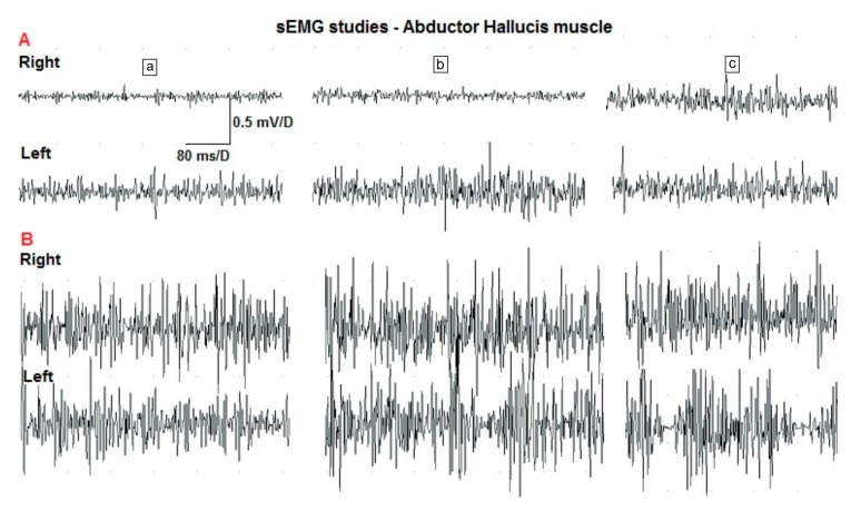
Despite the high availability of surface electromyography (sEMG), it is not widely used for testing the effectiveness of exercises that activate intrinsic muscles of foot in people with hallux valgus. The aim of this study was to assess the effect of the toe-spread-out (TSO) exercise on the outcomes of sEMG recorded from the abductor hallucis muscle (AbdH). An additional objective was the assessment of nerve conduction in electroneurography. The study involved 21 patients with a diagnosed hallux valgus (research group A) and 20 people without the deformation (research group B) who performed a TSO exercise and were examined twice: before and after therapy. The statistical analysis showed significant differences in the third, most important phase of TSO. After the exercises, the frequency of motor units recruitment increased in both groups. There were no significant differences in electroneurography outcomes between the two examinations in both research groups. The TSO exercise helps in the better activation of the AbdH muscle and contributes to the recruitment of a larger number of motor units of this muscle. The TSO exercises did not cause changes in nerve conduction. The sEMG and ENG are good methods for assessing this exercise but a comprehensive assessment should include other tests as well.

Keywords: abductor hallucis muscle; electroneurography; hallux valgus; surface electromyography; toe-spread-out exercise.


Conflict of interest statement

The authors declare no conflict of interest.

Figures

Figure 1
Figure 1
The design of the study (HVA—hallux valgus angle, 1st examination—first examination before exercise, 2nd examination—second examination after exercise, sEMG—surface electromyography, ENG—electroneurography, AbdH muscle—abductor hallucis muscle, TSO—toe spread out exercise, rep.—repetitions).
Figure 2
Figure 2
Surface EMG study of abductor hallucis muscle. (a,b)—KeyPoint System and set of electrodes; (c) application of electrodes on the AbdH muscle (R—recording electrode, Ref—reference electrode) and (d) the third phase of TSO exercises and (e) recording of AbdH muscle activity during maximal contraction.
Figure 3
Figure 3
Placement of the electrodes in the electroneurographic examination of the tibial nerve and sample results of the evaluated parameters (E1—active recording electrode; E2—reference electrode; stimulation points: A—anode, K—cathode; G—ground electrode, LAT—latency; CV—segmental conduction velocity; AMP—amplitude; AREA—area of motor potential; DUR—duration of motor potential).
Figure 4
Figure 4
Sample sEMG results of a selected patient from study group A (a—the first phase of TSO exercises, b—the second phase of TSO exercise, c—the third phase of TSO exercise, right—recording from AbdH in the right foot, left—recording from AbdH in the left foot).
Figure 5
Figure 5
Comparison of the amplitudes in the second phase of toe-spread-out exercise in research group B (mV—millivolts, 2 TSO—the second phase of the toe-spread-out exercise, 0 EMG—examination before therapy, 1 EMG—examination after therapy).

References

    1. Lisiński P., Huber J., Samborski W., Witkowska A. Neurophysiological assessment of the electrostimulation procedures used in stroke patients during rehabilitation. Int. J. Artif. Organs. 2008;31:76–86. doi: 10.1177/039139880803100111. - DOI - PubMed
    1. Jóźwiak M., Idzior M., Huber J., Szulc A., Grottel K. Podpajęczynówkowe podawanie baklofenu w leczeniu spastyczności u chorych z mózgowym porażeniem dziecięcym—doniesienie wstępne. Chirur. Narz. Ruch. Ortop. Pol. 2003;68:253–259. - PubMed
    1. Moon D., Esquenazi A. Instrumented gait analysis: A tool in the treatment of spastic gait dysfunction. JBJS Rev. 2016:4. doi: 10.2106/JBJS.RVW.15.00076. - DOI - PubMed
    1. Strazza A., Mengarelli A., Fioretti S., Burattini L., Agostini V., Knaflitz M., Di Nardo F. Surface-EMG analysis for the quantification of thigh muscle dynamic co-contractions during normal gait. Gait Posture. 2017;51:228–233. doi: 10.1016/j.gaitpost.2016.11.003. - DOI - PubMed
    1. Giggins O.M., Persson U.M., Caulfield B. Biofeedback in rehabilitation. J. Neuroeng. Rehabil. 2013;10:1–11. doi: 10.1186/1743-0003-10-60. - DOI - PMC - PubMed

LinkOut - more resources