. 2020 Apr 12;12(4):953.
doi: 10.3390/cancers12040953.

High Galectin-7 and Low Galectin-8 Expression and the Combination of both are Negative Prognosticators for Breast Cancer Patients

Affiliations

High Galectin-7 and Low Galectin-8 Expression and the Combination of both are Negative Prognosticators for Breast Cancer Patients

Anna Trebo et al. Cancers (Basel). .

Abstract

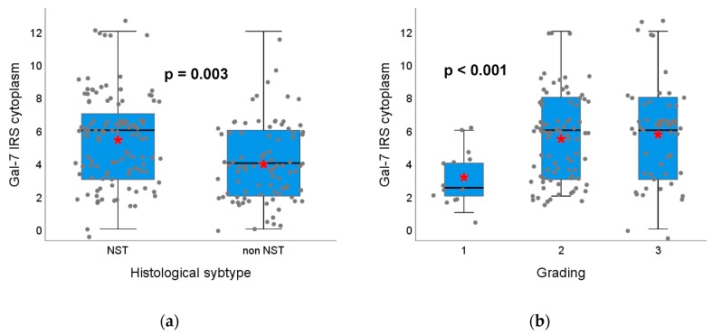
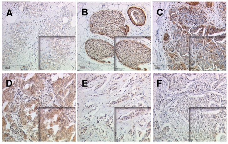
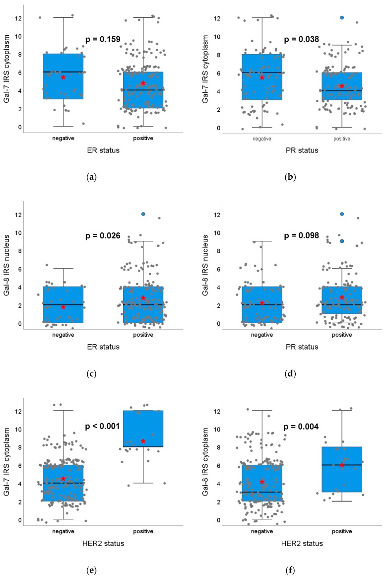
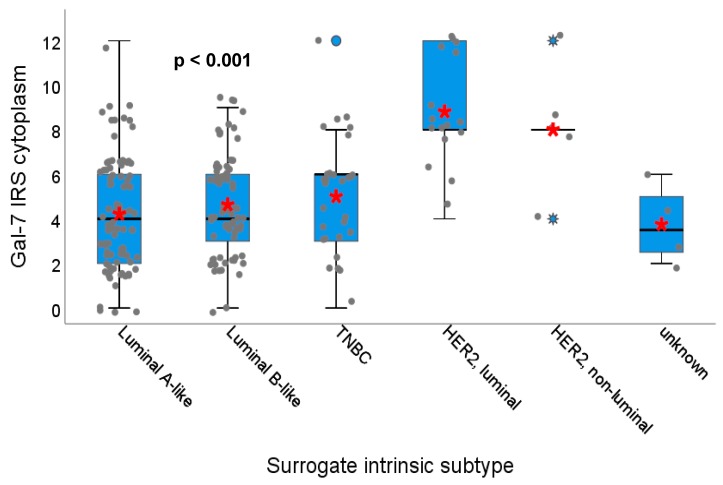
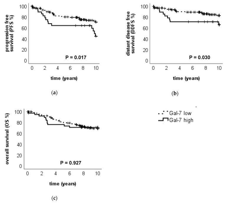
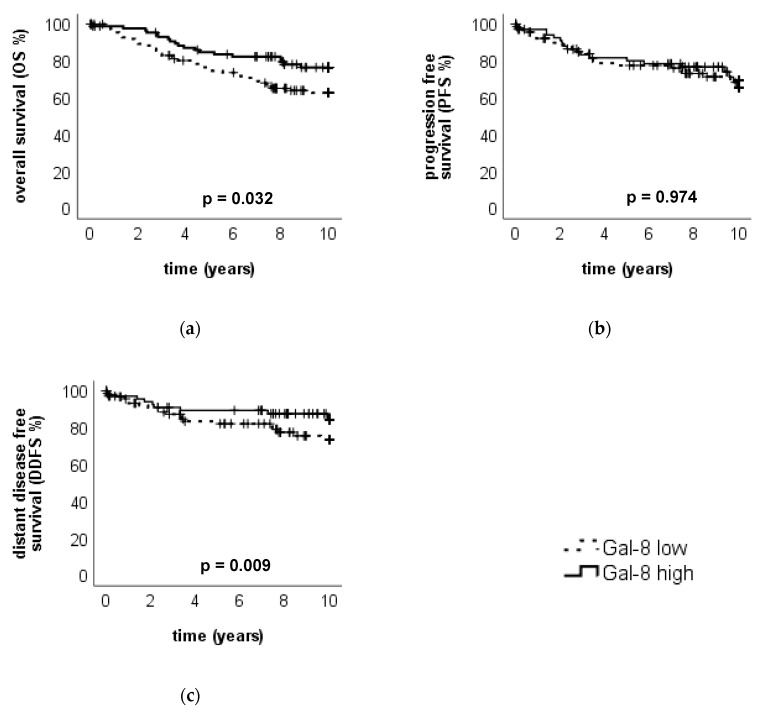
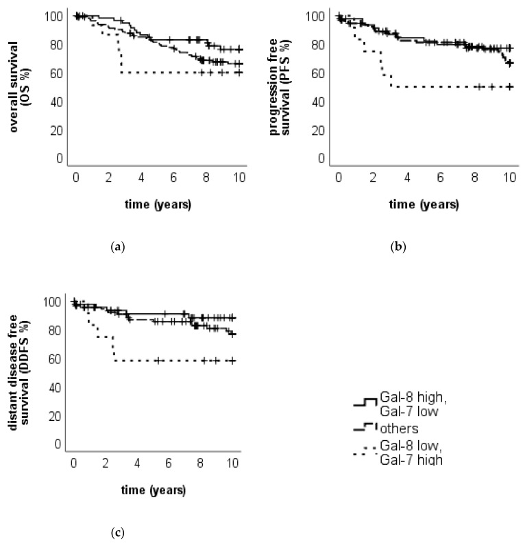
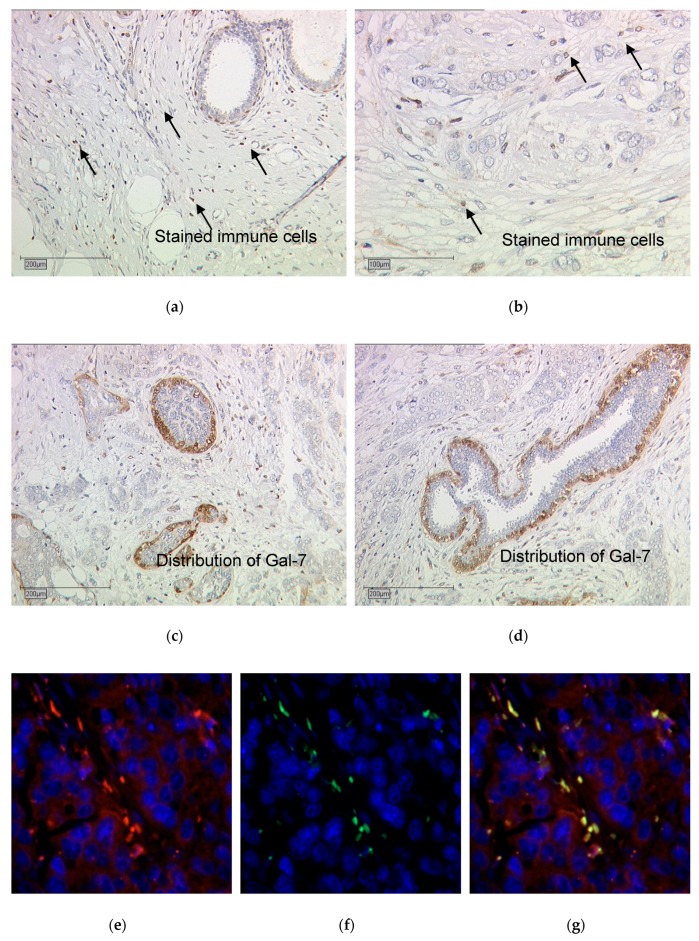
Galectins are commonly overexpressed in cancer cells and their expression pattern is often associated with the aggressiveness and metastatic phenotype of the tumor. This study investigates the prognostic influence of the expression of galectin7 (Gal-7) and galectin8 (Gal-8) in tumor cell cytoplasm, nucleus and on surrounding immune cells. Primary breast cancer tissue of 235 patients was analyzed for the expression of Gal-7 and Gal-8 and correlated with clinical and pathological data and the outcome. To identify immune cell subpopulations, immunofluorescence double staining was performed. Significant correlations of Gal-7 expression in the cytoplasm with HER2-status, PR status, patient age and grading, and of Gal-8 expression in the cytoplasm with HER2-status and patient age and of both galectins between each other were found. A high Gal7 expression in the cytoplasm was a significant independent prognosticator for an impaired progression free survival (PFS) (p = 0.017) and distant disease-free survival (DDFS) (p = 0.030). Gal-7 was also expressed by tumor-infiltrating macrophages. High Gal-8 expression in the cytoplasm was associated with a significantly improved overall survival (OS) (p = 0.032). Clinical outcome in patients showing both high Gal-7 and with low Gal-8 expression was very poor. Further understanding of the role of galectins in the regulation and interaction of tumor cells and macrophages is essential for finding new therapeutic targets.

Keywords: Galectins; breast cancer; galectin-7; galectin-8; prognostic markers; tumor infiltrating macrophages.


Conflict of interest statement

Thomas Kolben holds stock of Roche AG and his relative is employed at Roche AG. Anna Hester has received a research grant from the “Walter Schulz” foundation and advisory board, speech honoraria and travel expenses from Roche and Pfizer. Research support, advisory board, honoraria, and travel expenses from AstraZeneca, Clovis, Medac, MSD, Novartis, PharmaMar, Roche, Sensor Kinesis, Tesaro, Teva have been received by Sven Mahner. All other authors declare no conflict of interest. The funders had no role in the design of the study; in the collection, analyses, or interpretation of data; in the writing of the manuscript, or in the decision to publish the results.

Figures

Figure 1
Figure 1
Association of Gal-7 expression with histological subtype and tumor grading. Boxplots of the median IRS of Gal-7 staining in the cytoplasm dependent on histological subtype (a) and tumor grading (b) of the tumor are shown. (a) In non-NST tumors, Gal-7 expression in the cytoplasm is significantly lower than in NST tumors. (b) Tumors with G2/3 grading show a significantly higher Gal-7 expression in the cytoplasm compared to G1 tumors. Red asterisks indicate means. Please note that individual datapoints have been jittered to avoid overlap.
Figure 2
Figure 2
Gal-7 and Gal-8 expression dependent on tumor grading. Exemplary immunohistochemical staining of Gal-7 in grade 1 (A), 2 (B), and 3 (C) tumors and of Gal-8 in grade 1 (D), 2 (E), 3 (F) tumors are shown. Magnification: main images x10, image sections x25.
Figure 3
Figure 3
Association of Gal-7 and Gal-8 expression with the ER-, PR- and HER2-status. Boxplots of the median IRS of Gal-7 staining in the cytoplasm dependent on ER-status (a), PR-status (b) and HER2-status (e) and of Gal-8 staining in the nucleus dependent on ER-status (c) and PR-status (d) as well as Gal-8 staining in the cytoplasm dependent on HER2-status (f) are shown. ER-positive tumors do not differ concerning Gal-7 staining but show a higher Gal-8 staining. PR-positive tumors show lower Gal-7 staining and a trend towards higher Gal-8 staining. HER2-positive tumors show a significantly higher Gal-7 and Gal 8 expression in the cytoplasm compared to HER2-negative tumors. Staining in the nucleus does not show significant differences. Red asterisks indicate means. Please note that individual data points have been jittered to avoid overlap.
Figure 4
Figure 4
Association of Gal-7 expression with different surrogate intrinsic subtype. Boxplot of the median IRS of Gal-7 staining in the cytoplasm dependent on the surrogate intrinsic subtype of the tumor is shown. HER2-positive, both luminal and non-luminal tumors show a significantly higher Gal-7 expression in the cytoplasm compared to the other subtypes. Red asterisks indicate means. Please note that individual datapoints have been jittered to avoid overlap.
Figure 5
Figure 5
Association of Gal-7 expression in the cytoplasm to the clinical outcome. Kaplan–Meier analysis of PFS (a), DDFS (b) and OS (c) in Gal-7 high- and low-expressing tumors (in the cytoplasm) is shown. Tumors with high Gal-7 expression in the cytoplasm showed a significantly impaired PFS and DDFS.
Figure 6
Figure 6
Association of Gal-8 expression in the cytoplasm to the clinical outcome. Kaplan–Meier analysis of OS (a), PFS (b) and DDFS (c) in Gal-8 high- and low-expressing tumors (in the cytoplasm) is shown. Tumors with high Gal-8 expression in the cytoplasm showed a significantly improved OS.
Figure 7
Figure 7
OS, PFS and DDFS for the two groups. Kaplan–Meier analysis of OS (a), PFS (b) and DDFS (c) in tumors with combined high or low Gal-7 expression with low or high Gal-8 expression (in the cytoplasm), respectively, are shown. Tumors with high Gal-7 and low Gal-8 expression in the cytoplasm show the trend of an impaired OS, while tumors with high Gal-8 and low Gal 7 expression show the trend of an improved OS. Tumors with high Gal-7 and low Gal-8 expression in the cytoplasm show the trend of an impaired PFS and a significantly reduced DDFS, while tumors with high Gal-8 and low Gal-7 expression show the trend of an improved PFS and a DDFS.
Figure 8
Figure 8
Gal-7-staining results (ad) and double immunofluorescence staining with Gal 7 and CD68 (eg). Exemplary pictures of Gal 7-staining results are shown, revealing the distribution of Gal 7-staining in tumor cells (a,b). Stained immune cells in Gal 7-expressing tumors are shown in (c) and (d). Immunofluorescence results: Gal-7 is shown in red (e), CD68 in green (f) and the nucleus is stained with DAPI in blue. Picture (g) shows that Gal 7- and CD8-staining is overlapping in some cells, which appear yellow, showing that these are macrophages. Magnification: Images 8a, 8c and 8d x10, and images 8b, 8e, 8f and 8g x25.

References

    1. Harbeck N., Gnant M. Breast cancer. Lancet. 2017;389:1134–1150. doi: 10.1016/S0140-6736(16)31891-8. - DOI - PubMed
    1. Bray F., Ferlay J., Soerjomataram I., Siegel R.L., Torre L.A., Jemal A. Global cancer statistics 2018: GLOBOCAN estimates of incidence and mortality worldwide for 36 cancers in 185 countries. CA A Cancer J. Clin. 2018;68:394–424. doi: 10.3322/caac.21492. - DOI - PubMed
    1. Sorlie T., Tibshirani R., Parker J., Hastie T., Marron J.S., Nobel A., Deng S., Johnsen H., Pesich R., Geisler S., et al. Repeated observation of breast tumor subtypes in independent gene expression data sets. Proc. Natl. Acad. Sci. USA. 2003;100:8418–8423. doi: 10.1073/pnas.0932692100. - DOI - PMC - PubMed
    1. Perou C.M., Sorlie T., Eisen M.B., van de Rijn M., Jeffrey S.S., Rees C.A., Pollack J.R., Ross D.T., Johnsen H., Akslen L.A., et al. Molecular portraits of human breast tumours. Nature. 2000;406:747–752. doi: 10.1038/35021093. - DOI - PubMed
    1. Cho S.H., Jeon J., Kim S.I. Personalized medicine in breast cancer: A systematic review. J. Breast Cancer. 2012;15:265–272. doi: 10.4048/jbc.2012.15.3.265. - DOI - PMC - PubMed