. 2020 Apr 12;13(8):1815.
doi: 10.3390/ma13081815.

Description of a Digital Work-Flow for CBCT-Guided Construction of Micro-Implant Supported Maxillary Skeletal Expander

Affiliations

Description of a Digital Work-Flow for CBCT-Guided Construction of Micro-Implant Supported Maxillary Skeletal Expander

Antonino Lo Giudice et al. Materials (Basel). .

Abstract

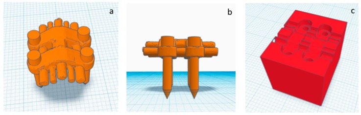
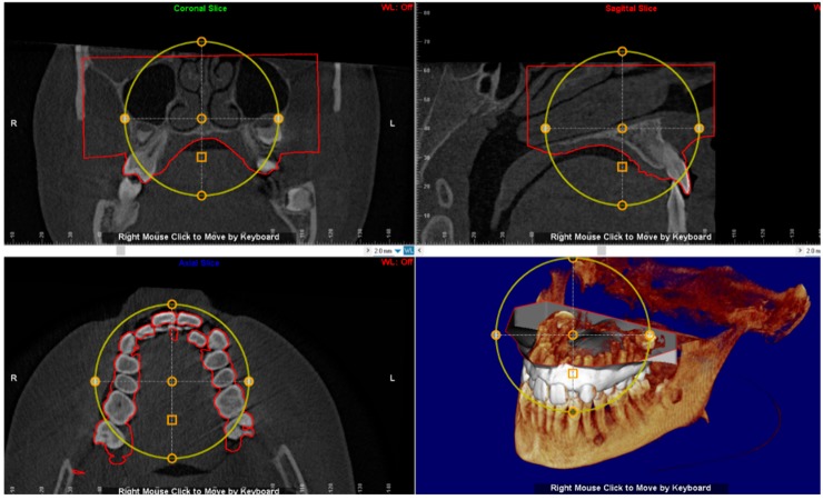
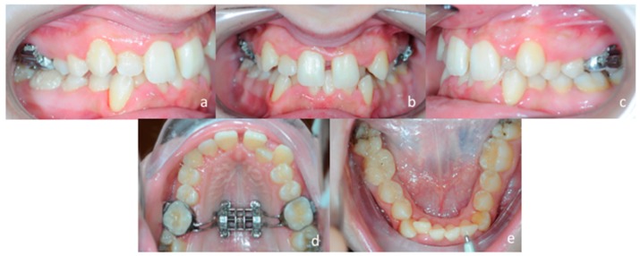
The introduction of miniscrew-assisted rapid palatal expansion (MARPE) has widened the boundaries of orthodontic skeletal correction of maxillary transversal deficiency to late adolescence and adult patients. In this respect, Maxillary Skeletal Expander (MSE) is a particular device characterized by the engagement of four miniscrews in the palatal and nasal cortical bone layers. Thus, the availability of sufficient supporting bone and the perforation of both cortical laminas (bi-corticalism) are two mandatory parameters for mini-screw stability, especially when orthopedic forces are used. Virtual planning and construction of MSE based on cone-beam computed tomography (CBCT)-derived stereolithography (.stl) files have been recently described in the literature. In this manuscript we described: (a) a user-friendly digital workflow which can provide a predictable placement of maxillary skeletal expander (MSE) appliance according to the patient's anatomical characteristics, (b) the construction of a positional template of the MSE that allows lab technician to construct the MSE appliance in a reliable and accurate position, according to the virtual project planned by the orthodontist on the patient CBCT scans. We also described a case report of an adult female patient affected by skeletal transversal maxillary deficiency treated with MSE appliance that was projected according to the described workflow.

Keywords: digital dentistry; digital orthodontics; maxillary skeletal expander; miniscrews-assisted maxillary expansion; rapid maxillary expansion.


Conflict of interest statement

The authors declare no conflict of interest.

Figures

Figure 1
Figure 1
(a) Conventional hybrid skeletal anchored expander with bands on first molars and two miniscrews placed in the anterior palate; (b) MSE expander placed barely anterior to the soft palate with bands on first molars and four miniscrews inserted via four slots within the expander itself.
Figure 2
Figure 2
(a) Printed template of the expander connected to a handle which facilitates the test of adaptability of the MSE avoiding discomfort to the patient; (b) digital version of the 3D template.
Figure 3
Figure 3
(a,b) upper and lateral view of the .stl file of the MSE palatal expander with the four miniscrews, (c) negative template of the MSE with miniscrews
Figure 4
Figure 4
Superimposition of the digital model of maxillary arch onto the DICOM file for identification of the most suitable vertical and anteroposterior placement of the MSE.
Figure 5
Figure 5
The position of the negative template of MSE is determined in sagittal, coronal and axial views. See the design of the expander with the four miniscrews that are engaged in the cortical bone of the palate and nasal floor.
Figure 6
Figure 6
(a) .stl file of the negative template of the MSE merged with the maxillary 3d model; (b) 3d printed maxillary model includes a template that accurately represents the position of the MSE virtually projected by the clinician; (c) this template aid the lab technician in stabilizing the position of the screw during the realization of supporting structures such as the arms connected to anchored teeth.
Figure 7
Figure 7
Extra-oral patient’s facial examination. (a) frontal view at rest, (b) frontal view while smiling, (c) right lateral profile at rest.
Figure 8
Figure 8
Intra-oral patient’s examination. (a) Right lateral occlusion, (b) front view, (c) left lateral occlusion, (d) occlusal view of the maxillary arch, (e) occlusal view of the mandibular arch.
Figure 9
Figure 9
Panorex.
Figure 10
Figure 10
(a) Lateral cephalogram, (b) pre-treatment cephalometric tracing.
Figure 11
Figure 11
Procedure of miniscrews insertion. Four self-drilling mini-screws (1.8 mm in diameter, 11 mm in length) were used to fix the MSE expander to the palate.
Figure 12
Figure 12
Post expansion intra-oral examination. (a) right lateral occlusion, (b) front view, (c) left lateral occlusion, (d) occlusal view of the maxillary arch, (e) occlusal view of the mandibular arch.

References

    1. Lo Giudice A., Barbato E., Cosentino L., Ferraro C.M., Leonardi R. Alveolar bone changes after rapid maxillary expansion with tooth-born appliances: A systematic review. Eur. J. Orthod. 2018;40:296–303. doi: 10.1093/ejo/cjx057. - DOI - PubMed
    1. Bucci R., D’Anto V., Rongo R., Valletta R., Martina R., Michelotti A. Dental and skeletal effects of palatal expansion techniques: A systematic review of the current evidence from systematic reviews and meta-analyses. J. Oral. Rehabil. 2016;43:543–564. doi: 10.1111/joor.12393. - DOI - PubMed
    1. Zhou Y., Long H., Ye N., Xue J., Yang X., Liao L., Lai W. The effectiveness of non-surgical maxillary expansion: A meta-analysis. Eur. J. Orthod. 2014;36:233–242. doi: 10.1093/ejo/cjt044. - DOI - PubMed
    1. Leonardi R., Lo Giudice A., Rugeri M., Muraglie S., Cordasco G., Barbato E. Three-dimensional evaluation on digital casts of maxillary palatal size and morphology in patients with functional posterior crossbite. Eur. J. Orthod. 2018;40:556–562. doi: 10.1093/ejo/cjx103. - DOI - PubMed
    1. Lo Giudice A., Fastuca R., Portelli M., Militi A., Bellocchio M., Spinuzza P., Briguglio F., Caprioglio A., Nucera R. Effects of rapid vs slow maxillary expansion on nasal cavity dimensions in growing subjects: A methodological and reproducibility study. Eur. J. Paediatr. Dent. 2017;18:299–304. - PubMed

LinkOut - more resources