. 2020 Apr 14;21(1):240.
doi: 10.1186/s12891-020-03252-y.

Imaging algorithm and multimodality evaluation of spinal osteoblastoma

Affiliations

Imaging algorithm and multimodality evaluation of spinal osteoblastoma

Zihuan Huang et al. BMC Musculoskelet Disord. .

Abstract

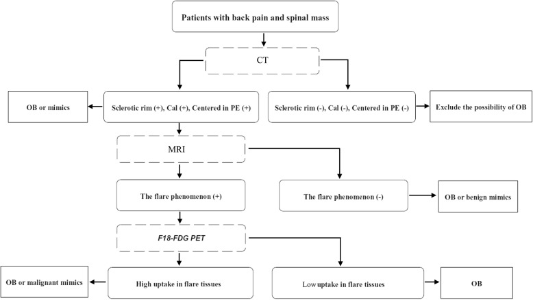
Background: To analyze the features of CT, MRI and PET/CT and their diagnostic value for spinal osteoblastomas (OBs).

Methods: The radiological and clinical data of 21 patients with histopathologically-confirmed spinal OBs were analyzed retrospectively.

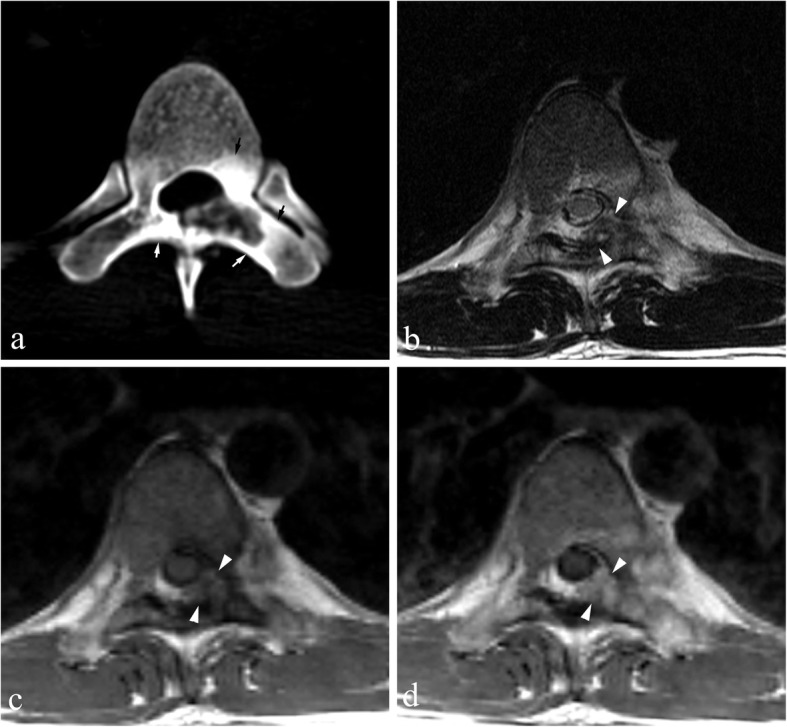
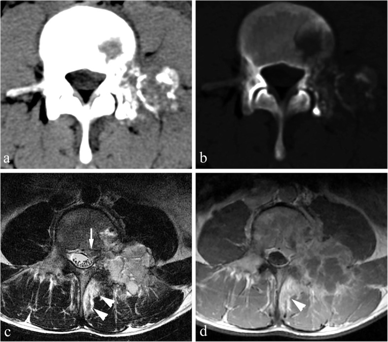
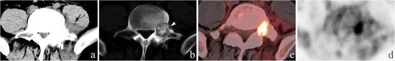
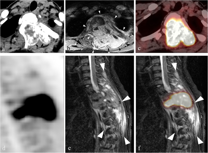
Results: Sixteen of the 21 cases were benign and 5 were aggressive OBs. Tumors were located in the lumbar (n = 11), cervical (n = 4), thoracic (n = 5), and sacral (n = 1) spinal regions. Nineteen cases were centered in the posterior elements of the spine, 13 of which extended into the vertebral body. Punctate or nodular calcifications were found in all cases on CT with a complete sclerotic rim (n = 12) or incomplete sclerotic rim (n = 8). The flare phenomenon (indicative of surrounding tissue inflammation) was found in 17/21 cases on CT, thin in 11 cases and thick in 6 cases, and in 19/19 cases on MRI, thin in 1 case and thick in 18 cases. On 18F-FDG PET/CT, all cases (8/8) were metabolically active with the SUVmax of 12.3-16.0; the flare sign was observed in 8 cases, including 7 cases of hypometabolism and 1 case of coexistence of hypermetabolism and hypometabolism. Based on CT, 3, 12, and 6 cases were classified as Enneking stage 1, 2 and 3, respectively. Of 19 cases with MRI, 1 and 18 cases were classified as Enneking stage 2 and 3, respectively.

Conclusions: Spinal OB has multiple unique characteristic radiological features. Although a larger sample size is needed, combining CT, MRI and PET may be beneficial to optimize preoperative diagnosis and care of patients with OBs.

Keywords: CT, MRI, PET; Osteoblastoma; Radiography; Spine.


Conflict of interest statement

The authors declare that they have no competing interests.

Figures

Fig. 1
Fig. 1
Osteoblastoma in a 25-year-old male, Enneking stage 3 on both CT and MRI. a and b Axial CT soft tissue and bone windows show a lesion causing extensive osteolytic destruction in L4 (which mainly affects the left pedicle, traverse process and lamina) with an incomplete sclerotic rim, unclear borders, and spotted calcifications. Note the thin flares in the epidural space (arrow). c and d Axial MRI T2WI and contrast-enhanced T1WI demonstrate the flare phenomenon adjacent to the tumor, i.e., abnormal swollen soft tissues in the spinal canal compressing the dural sac (arrow) lateral to the spinous process (arrowheads), showing hyperintensity on T2WI and marked enhancement with contrast
Fig. 2
Fig. 2
Osteoblastoma in a 29-year-old male, Enneking stage 2 on CT and stage 3 on MRI. a Axial CT bone window shows expansile bone destruction in the left lamina of L7 with a clear sclerotic rim and speckled calcifications. Note sclerotic changes (arrows) in the surrounding areas. Axial MRI T2WI (b), T1WI (c), and contrast-enhanced T1WI (d) show a soft tissue mass in the epidural space (arrowheads)
Fig. 3
Fig. 3
Osteoblastoma in a 28-year-old male, Enneking stage 2 on CT. a Axial CT soft tissue window shows a hypo-dense soft tissue mass (arrow) medial to the left psoas major muscle, which demonstrates the flare phenomenon. b Axial CT bone window shows expansile bone destruction in the lamina of L4 with a large quantity of matrix calcifications and sclerotic changes in the surrounding areas. Note the cortical breakthrough in the left pedicle (arrowhead). c and d F18-FDG PET/CT images show high uptake of FDG (SUVmax: 14.3) in the area of bone destruction, without FDG uptake abnormalities of the surrounding reactive sclerosis or inflamed tissues
Fig. 4
Fig. 4
Aggressive osteoblastoma in a 16-year-old female, Enneking stage 3 on both CT and MRI. This case was diagnosed as Ewing’s sarcoma or osteosarcoma before needle biopsy. a Axial CT soft tissue window shows extensive osteolytic destruction in the vertebral body, left pedicle and lamina of T2 vertebrae, with scattered internal nodular calcifications and a sclerotic rim (arrows). Note that the structures in the spinal canal are not clearly depicted. b Axial MRI contrast-enhanced T1WI shows a diffuse mass (arrowheads) with avid enhancement greater than that observed on CT, involving the T2 vertebral body and anterior soft tissues, bilateral lamina, spinous process, left pedicle, head and neck of the left 2nd rib and their surroundings, as well as soft tissues inside the spinal canal encapsulating the cord. c and d Axial and sagittal F18-FDG PET/CT images show both the mass and flares with high uptake of FDG (SUVmax: 15.7), which indicates tumor extension into the surrounding soft tissues. Note that PET does not clearly show the spinal cord due to its low spatial resolution. e Sagittal T2WI shows the tumor (asterisk) and extensively swollen tissues surrounding it that display the flare phenomenon (arrowheads). f Co-registered PET and T2WI sagittal image shows that a lesion visible on MRI (arrowheads) is significantly larger than the foci of high uptake on PET-CT, which indicates the existence of inflammatory issues in the flares
Fig. 5
Fig. 5
Proposed diagnostic work-up for patients suspected of having spinal osteoblastoma. OB, osteoblastoma; Cal, calcification; PE, posterior elements

References

    1. Versteeg AL, Dea N, Boriani S, Varga PP, Luzzati A, Fehlings MG, Blisky MH, Rhines LD, Reynolds JJ, Dekutoski MB, Gokaslan ZL, Germscheid NM. Fisher CG surgical management of spinal osteoblastomas. J Neurosurgical Spine. 2017;27:321–327. doi: 10.3171/2017.1.SPINE16788. - DOI - PubMed
    1. Galgano MA, Goulart CR, Iwenofu H, Chin LS, Lavelle W, Mendel E. Osteoblastomas of the spine: a comprehensive review. Neurosurgical Focus. 2016;41:E4. doi: 10.3171/2016.5.FOCUS16122. - DOI - PubMed
    1. Reynolds JJ, Rothenfluh DA, Athanasou N, Wilson S, Kieser DC. Neoadjuvant denosumab for the treatment of a sacral osteoblastoma. Eur Spine J. 2017;27:446–452. doi: 10.1007/s00586-018-5461-z. - DOI - PubMed
    1. Shimizu N, Sakata K, Yamamoto I. Benign osteoblastoma of the temporal bone: case report and review of the literature. Surg Neurol. 2006;66:534–538. doi: 10.1016/j.surneu.2006.02.044. - DOI - PubMed
    1. Tang H, Zou D, Chen W. Imaging diagnosis of aggressive osteoblastoma. J Clin Radiol. 2011;30:76–78.

LinkOut - more resources