Review
. 2020 May;101(5):263-268.
doi: 10.1016/j.diii.2020.03.014. Epub 2020 Apr 3.

COVID-19 pneumonia: A review of typical CT findings and differential diagnosis

Affiliations
Review

COVID-19 pneumonia: A review of typical CT findings and differential diagnosis

C Hani et al. Diagn Interv Imaging. 2020 May.

Abstract

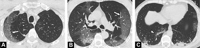
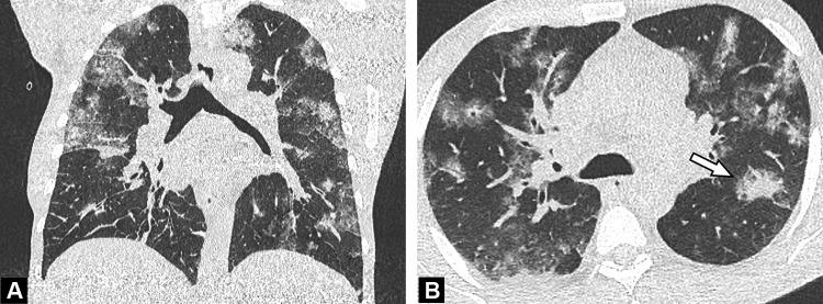
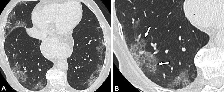
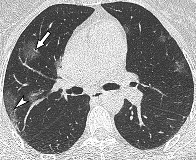
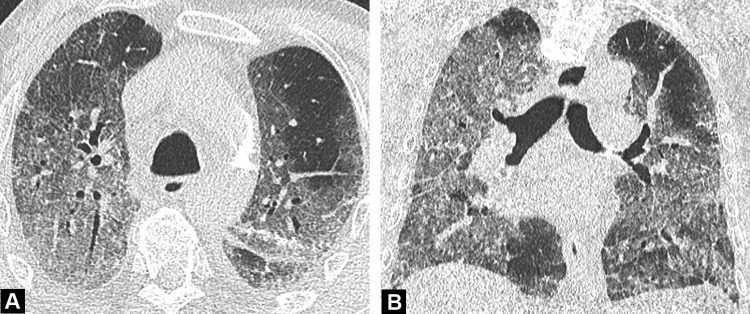
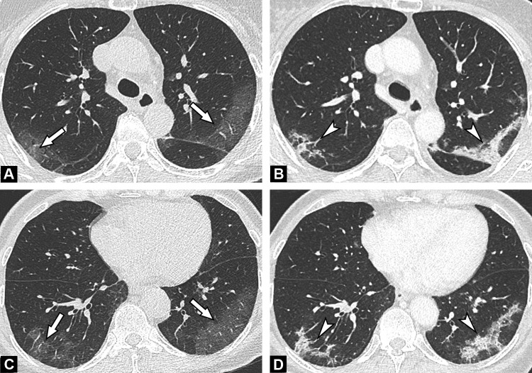
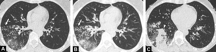
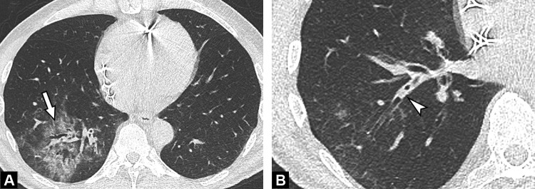
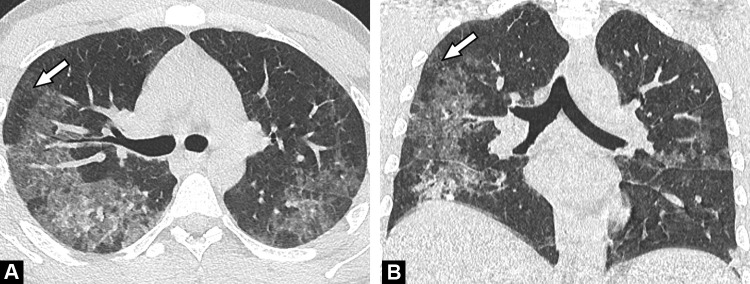
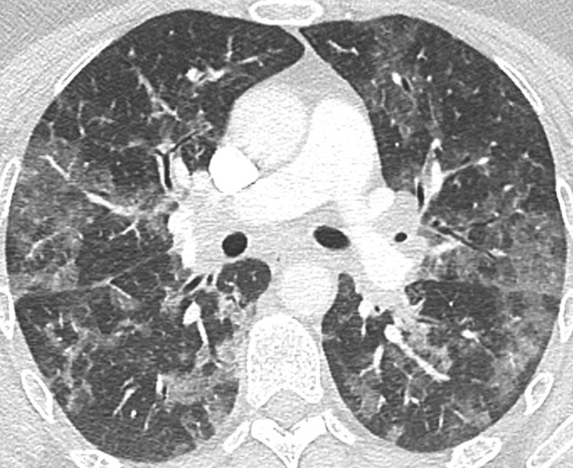
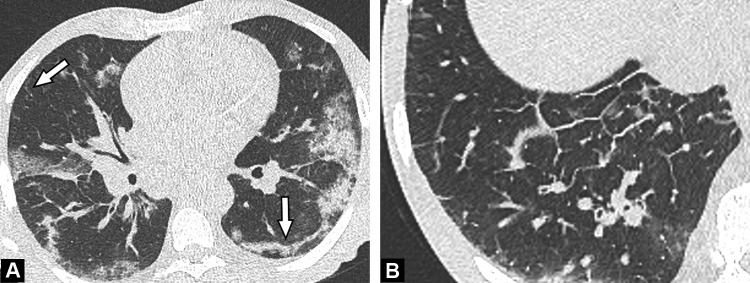
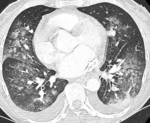
The standard of reference for confirming COVID-19 relies on microbiological tests such as real-time polymerase chain reaction (RT-PCR) or sequencing. However, these tests might not be available in an emergency setting. Computed tomography (CT) can be used as an important complement for the diagnosis of COVID-19 pneumonia in the current epidemic context. In this review, we present the typical CT features of COVID-19 pneumonia and discuss the main differential diagnosis.

Keywords: COVID-19 pneumonia; Cryptogenic Organizing Pneumonia; Pneumonia; Tomography; X-Ray Computed.


Figures

Fig. 1
Fig. 1
Unenhanced CT images show typical findings of COVID-19 pneumonia in a 55-year-old man. Peripheral GGO is seen in the upper portion of both lungs (A, B) (arrows), associated with linear consolidations in the lower lobes (C) (arrowhead). The results of first and second RT-PCRs were negative, with only the third test, repeated in view of CT findings, becoming positive.
Fig. 2
Fig. 2
Unenhanced CT examination performed 6 days after the onset of symptoms in a 64-year-old-man with COVID-19 pneumonia. Axial (A) and coronal (B) CT images demonstrate bilateral ground glass opacities admixed with patchy areas of consolidation (arrow) in the central and peripheral portions of the lung.
Fig. 3
Fig. 3
Unenhanced CT images of an 86-year-old woman with a crazy-paving pattern due to COVID-19 pneumonia. (a) CT examination performed 4 days after symptom onset (dry cough and chest pain) demonstrates moderate disease extent (10–25%). (b) Peripheral ground-glass opacities with superimposed intralobular reticulations (arrows) resulting in a crazy-paving pattern, are seen in both lower lobes.
Fig. 4
Fig. 4
Unenhanced CT image in a 50-year-old woman with a mild form of COVID-19. She has asthma with no respiratory symptoms except fever. Her husband has been recently diagnosed with COVID-19. CT demonstrates rounded ground-glass opacities in both upper lobes, some in the sub pleural region (arrowhead) and others more centrally distributed (arrow).
Fig. 5
Fig. 5
Unenhanced CT images of a “white lung” appearance in an 89-year-old man with respiratory distress due to COVID-19 pneumonia. Axial (A) and coronal (B) CT images, performed before admission in ICU demonstrate extensive ground glass opacities, with more than 75% of the lung involved.
Fig. 6
Fig. 6
Initial and follow-up CT images in a 71-year-old woman with COVID-19 pneumonia. Unenhanced initial CT performed before RT-PCR confirmation (A,C) shows bilateral peripheral ground-glass in the dorsal segment of upper (a) and lower lobes (C) (arrows). Contrast-enhanced CT (B,dD) performed 6 days later to rule out pulmonary embolism demonstrates linear consolidations typical for an organizing pneumonia pattern (arrowheads).
Fig. 7
Fig. 7
Unenhanced CT images in a 55-year-old-patient with bacterial bronchopneumonia. Centrilobular nodules (A) with a tree-in-bud pattern better seen on MIP reformatted images (B) (arrows) are seen in the right lower lobe, together with a segmental consolidation (arrowhead) (C).
Fig. 8
Fig. 8
Unenhanced CT images of a 45-year-old man with bacterial pneumonia. Ground glass opacities (arrow) limited to the posterior and lateral segment of the right lower lobe are demonstrated (A), associated with endobronchial secretions (arrowhead) (B) more proximally.
Fig. 9
Fig. 9
Unenhanced CT images of a 30-year-old man with Pneumocystis Jiroveci infection. Bilateral ground glass opacities with right lung predominance are demonstrated. Note the relative subpleural sparing (arrows) on both axial (A) and coronal (B) CT images.
Fig. 10
Fig. 10
Unenhanced CT image in a 45-year-old-patient with Influenza virus-associated pneumonia. Bilateral diffuse ground glass opacities are demonstrated. Differential diagnosis with COVID-19 pneumonia is not possible and relies on RT-PCR results, even though peripheral predominance is less common. RT-PCR result for SARS-Cov-2 was negative but positive for influenza A.
Fig. 11
Fig. 11
Unenhanced CT images of a 64-year-old man with COVID-19 and pulmonary edema. Ground glass opacities admixed with patchy consolidation are seen in both lungs and associated with linear consolidation in the subpleural region of the left lower lobe (arrow) (A). The associated smooth thickening of the interlobular septa (B), indicating pulmonary edema, suggests left ventricular failure and potentially associated myocarditis.
Fig. 12
Fig. 12
Unenhanced CT performed in a 48-year-old woman with Goodpasture syndrome. CT image shows bilateral ground glass opacities with central predominance, associated with pleural effusion, which is very uncommon in COVID-19 pneumonia. The patient presented with mild hemoptysis related to intra alveolar hemorrhage and acute renal failure. Histopathological analysis of tissues samples obtained from renal biopsy confirmed Goodpasture syndrome.

Comment in

  • Lessons learned from chest CT in COVID-19.
    Soyer P. Soyer P. Diagn Interv Imaging. 2020 May;101(5):261-262. doi: 10.1016/j.diii.2020.04.006. Diagn Interv Imaging. 2020. PMID: 32362428 Free PMC article. No abstract available.

References

    1. WHO Director-General's opening remarks at the media briefing on COVID-19-11 March 2020 n.d. https://www.who.int/dg/speeches/detail/who-director-general-s-opening-re... March 22, 2020).
    1. Coronavirus Update (Live): 629,450 Cases and 28,963 Deaths from COVID-19 Virus Outbreak - Worldometer n.d. https://www.worldometers.info/coronavirus/.(accessed March 28, 2020).
    1. Corman V.M., Landt O., Kaiser M., Molenkamp R., Meijer A., Chu D.K. Detection of 2019 novel coronavirus (2019-nCoV) by real-time RT-PCR. Eurosurveillance. 2020;25:2000045. - PMC - PubMed
    1. Fang Y., Zhang H., Xie J., Lin M., Ying L., Pang P. Sensitivity of chest CT for COVID-19: comparison to RT-PCR. Radiology. 2020 doi: 10.1148/radiol.20202004.32. - DOI - PMC - PubMed
    1. Ai T., Yang Z., Hou H., Zhan C., Chen C., Lv W. Correlation of chest CT and RT-PCR testing in coronavirus disease 2019 (COVID-19) in China: a report of 1014 cases. Radiology. 2020 doi: 10.1148/radiol.2020200642. - DOI - PMC - PubMed