. 2020 Sep;30(9):4865-4873.
doi: 10.1007/s00330-020-06823-8. Epub 2020 Apr 15.

Evolution of CT findings in patients with mild COVID-19 pneumonia

Affiliations

Evolution of CT findings in patients with mild COVID-19 pneumonia

Ting Liang et al. Eur Radiol. 2020 Sep.

Abstract

Objectives: To delineate the evolution of CT findings in patients with mild COVID-19 pneumonia.

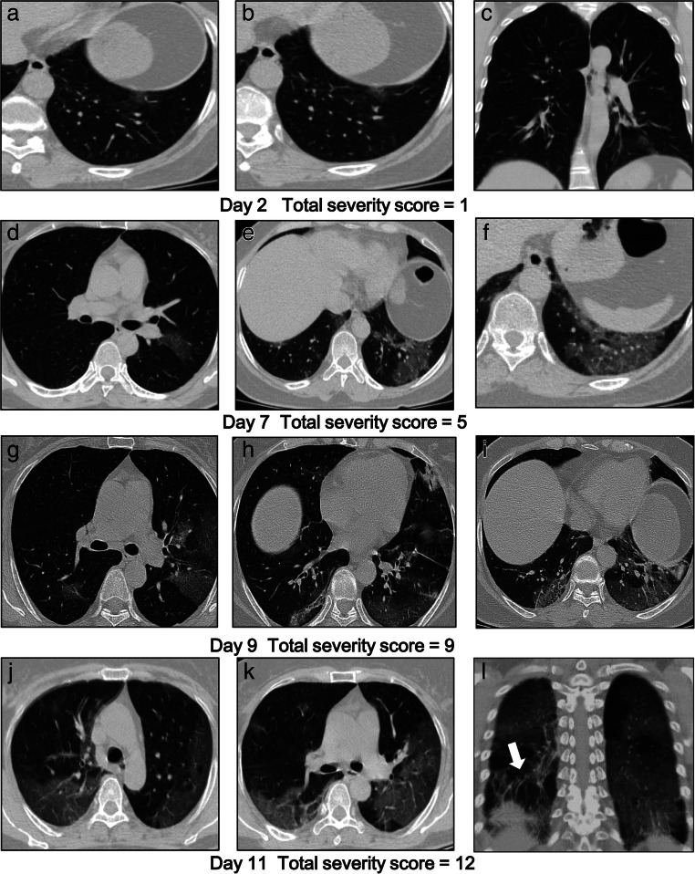
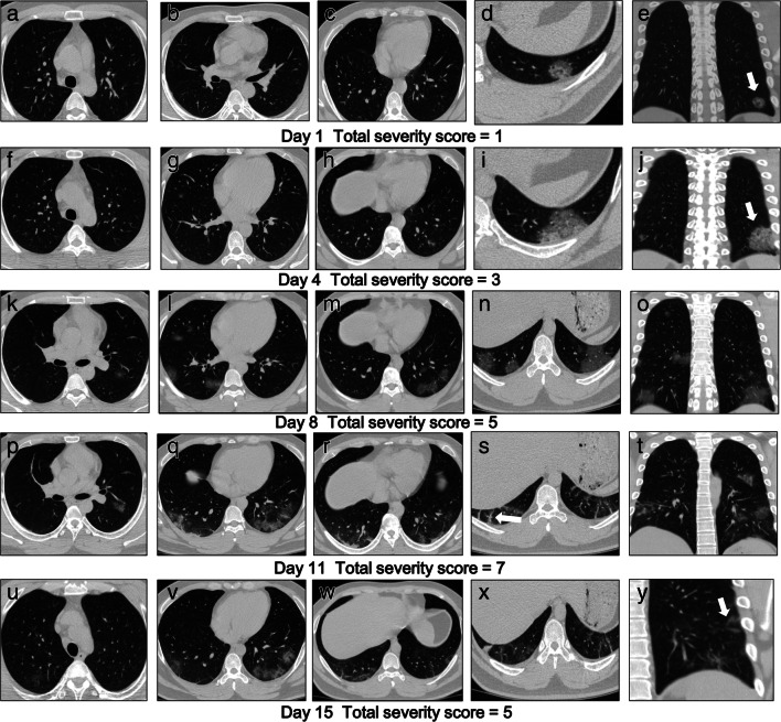
Methods: CT images and medical records of 88 patients with confirmed mild COVID-19 pneumonia, a baseline CT, and at least one follow-up CT were retrospectively reviewed. CT features including lobar distribution and presence of ground glass opacities (GGO), consolidation, and linear opacities were analyzed on per-patient basis during each of five time intervals spanning the 3 weeks after disease onset. Total severity scores were calculated.

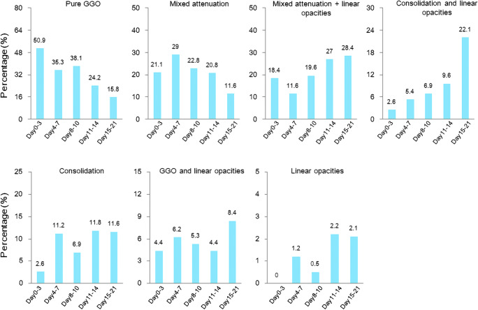
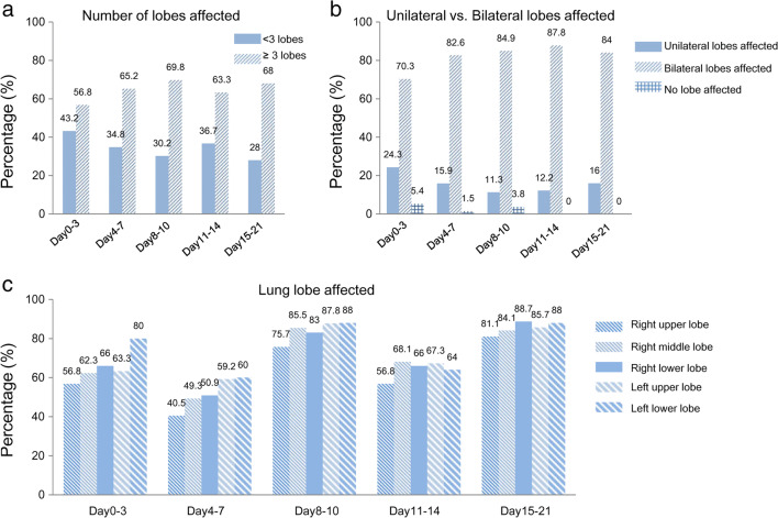
Results: Of patients, 85.2% had travel history to Wuhan or known contact with infected individuals. The most common symptoms were fever (84.1%) and cough (56.8%). The baseline CT was obtained on average 5 days from symptom onset. Four patients (4.5%) had negative initial CT. Significant differences were found among the time intervals in the proportion of pulmonary lesions that are (1) pure GGO, (2) mixed attenuation, (3) mixed attenuation with linear opacities, (4) consolidation with linear opacities, and (5) pure consolidation. The majority of patients had involvement of ≥ 3 lobes. Bilateral involvement was more prevalent than unilateral involvement. The proportions of patients observed to have pure GGO or GGO and consolidation decreased over time while the proportion of patients with GGO and linear opacities increased. Total severity score showed an increasing trend in the first 2 weeks.

Conclusions: While bilateral GGO are predominant features, CT findings changed during different time intervals in the 3 weeks after symptom onset in patients with COVID-19.

Key points: • Four of 88 (4.5%) patients with COVID-19 had negative initial CT. • Majority of COVID-19 patients had abnormal CT findings in ≥ 3 lobes. • A proportion of patients with pure ground glass opacities decreased over the 3 weeks after symptom onset.

Keywords: Coronavirus; Pneumonia; Tomography.


Conflict of interest statement

The authors declare that they have no conflict of interest.

Figures

Fig. 1
Fig. 1
Evolution of CT characteristics of pulmonary lesions in patients with COVID-19. The time intervals after disease onset (days) were divided into 5 groups, i.e., days 0–3 (lesion number = 114), days 4–7 (lesion number = 241), days 8–10 (lesion number = 189), days 11–14 (lesion number = 178), and days 15–21 (lesion number = 95). GGO, ground glass opacities; mixed attenuation = ground glass opacities + consolidation
Fig. 2
Fig. 2
Evolution of lobar distribution of the (a) number of lobe affected, (b) unilateral and bilateral lobes affected, and (c) lung lobe affected in patients with COVID-19 within 21 days after disease onset. The time after disease onset (day) is categorized as 5 groups, i.e., days 0–3 (patient number = 37), days 4–7 (patient number = 69), days 8–10 (patient number = 53), days 11–14 (patient number = 49), and days 15–21 (patient number = 25)
Fig. 3
Fig. 3
A 54-year-old woman with exposure to a COVID-infected patient presented with fever, cough, and low back pain for 2 days and remains hospitalized. ac CT shows left lower lobe GGO on day 2. The total severity score is 1. df CT on day 7 shows a significant increase in GGO in both lungs. The severity score of the left lower lobe is 2. The total severity score is 5. gi CT on day 9 shows bilateral GGO and subpleural consolidation. The severity score of the left lower lobe is 3. The total severity score is 9. jl CT on day 11 shows diffuse GGO and linear opacities (thick arrow). The severity score of the left lower lobe is 4. The total severity score is 12
Fig. 4
Fig. 4
A 24-year-old man with exposure to a COVID-infected patient presented with fever and cough for 1 day and remains hospitalized currently. ae CT scan shows GGO in the subpleural left lower lobe on day 1 (thick arrow). The overall total severity score is 1. fj CT on day 4 shows enlarging GGO in left lower lobes (thick arrow). The overall total severity score is 3. ko CT on day 8 shows increased GGO in bilateral lower lobes. The total severity score was 5. pt CT obtained on day 11 shows bilateral ground glass opacities and linear opacities (thick arrow). The overall total severity score was 7. uy CT obtained on day 15 shows improvement of bilateral lower lobe GGO and atoll sign (thick arrow). The overall total severity score is 5

Similar articles

Cited by

References

    1. Huang C, Wang Y, Li X, et al. Clinical features of patients infected with 2019 novel coronavirus in Wuhan, China. Lancet. 2020;395:497–506. - PMC - PubMed
    1. Chen N, Zhou M, Dong X, et al. Epidemiological and clinical characteristics of 99 cases of 2019 novel coronavirus pneumonia in Wuhan, China: a descriptive study. Lancet. 2020;395:507–513. - PMC - PubMed
    1. Zhu N, Zhang D, Wang W et al (2020) A novel coronavirus from patients with pneumonia in China, 2019. N Engl J Med 382:727–733 - PMC - PubMed
    1. Coronavirus toll update: cases and deaths by country and territory. https://www.worldometers.info/coronavirus/. Accessed 12 Feb 2020
    1. Chung M, Bernheim A, Mei X et al (2020) CT imaging features of 2019 novel coronavirus (2019-nCoV). Radiology 295:202–207 - PMC - PubMed

LinkOut - more resources