. 2020 May;53(5):e12802.
doi: 10.1111/cpr.12802. Epub 2020 Apr 14.

Placental endovascular extravillous trophoblasts (enEVTs) educate maternal T-cell differentiation along the maternal-placental circulation

Affiliations

Placental endovascular extravillous trophoblasts (enEVTs) educate maternal T-cell differentiation along the maternal-placental circulation

Yeling Ma et al. Cell Prolif. 2020 May.

Abstract

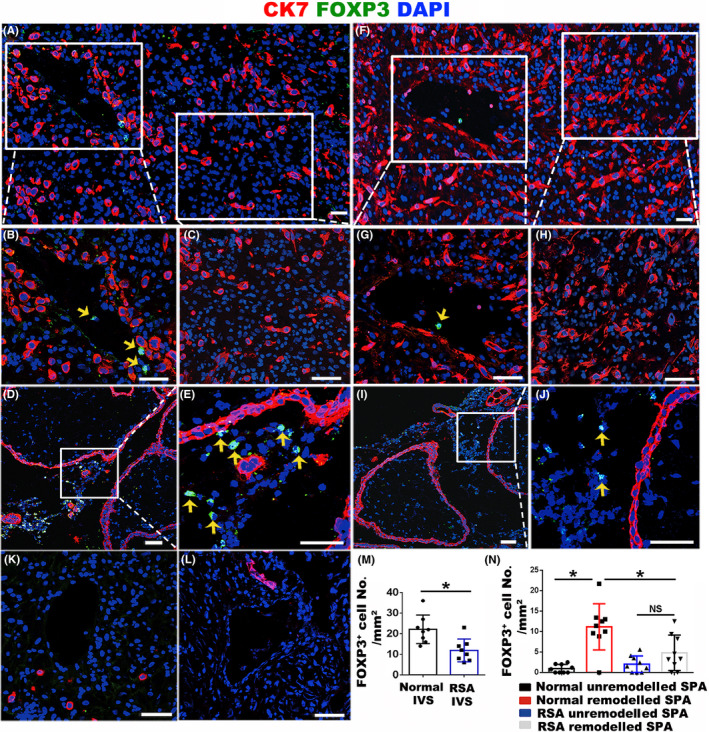
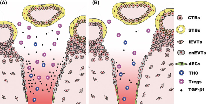
Objectives: During human pregnancy, the endothelial cells of the uterine spiral arteries (SPA) are extensively replaced by a subtype of placental trophoblasts, endovascular extravillous trophoblasts (enEVTs), thus establishing a placental-maternal circulation. On this pathway, foetus-derived placental villi and enEVTs bath into the maternal blood that perfuses along SPA being not attacked by the maternal lymphocytes. We aimed to reveal the underlying mechanism of such immune tolerance.

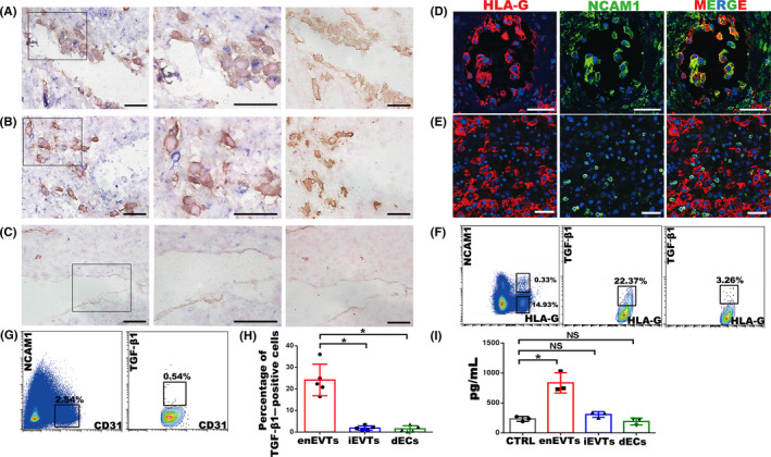
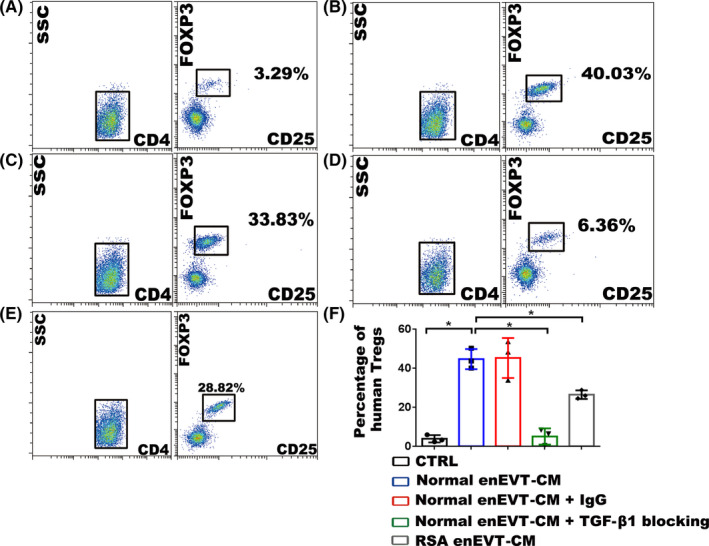
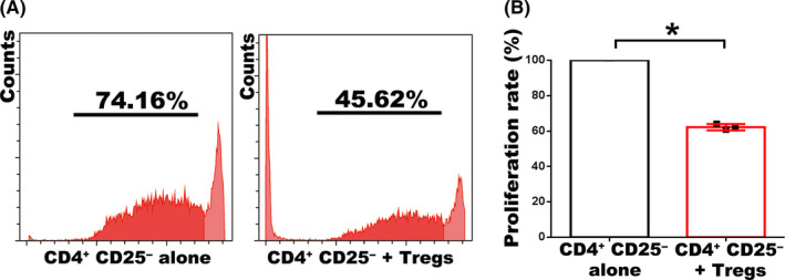
Methods: In situ hybridization, immunofluorescence, ELISA and FCM assay were performed to examine TGF-β1 expression and distribution of regulatory T cells (Tregs) along the placental-maternal circulation route. The primary enEVTs, interstitial extravillous trophoblasts (iEVTs) and decidual endothelial cells (dECs) were purified by FACS, and their conditioned media were collected to treat naïve CD4+ T cells. Treg differentiation was measured by FLOW and CFSE assays.

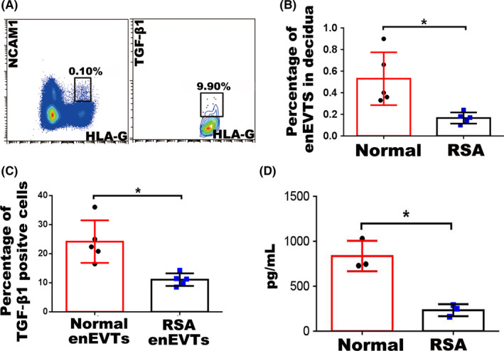
Results: We found that enEVTs but not iEVTs or dECs actively produced TGF-β1. The primary enEVTs significantly promoted naïve CD4+ T-cell differentiation into immunosuppressive FOXP3+ Tregs, and this effect was dependent on TGF-β1. In recurrent spontaneous abortion (RSA) patients, an evidently reduced proportion of TGF-β1-producing enEVTs and their ability to educate Tregs differentiation were observed.

Conclusions: Our findings demonstrate a unique immune-regulatory characteristic of placental enEVTs to develop immune tolerance along the placental-maternal circulation. New insights into the pathogenesis of RSA are also suggested.

Keywords: RSA; TGF-β1; Tregs; enEVTs; placental-maternal circulation.


Conflict of interest statement

The authors declare no competing interests.

Figures

FIGURE 1
FIGURE 1
Distribution and proportion of Tregs at the maternal‐foetal interface in healthy and RSA pregnancies at gestational weeks 7‐8. A, Immunofluorescent staining of CK7 (red) and FOXP3 (green) in normal pregnant decidua. B, C, Enlargement of the areas as indicated in panel a, showing remodelled SPA (B) and the area nearby the remodelled SPA (C). D, E, Immunofluorescent staining of CK7 (red) and FOXP3 (green) in placental villi of normal pregnancy and the enlargement of the IVS area are shown in panel E. F, Immunofluorescent staining of CK7 (red) and FOXP3 (green) in RSA decidua. G, H, Enlargement of the areas as indicated in panel F, showing remodelled SPA (G) and the area nearby the remodelled SPA (H). I, J, Immunofluorescent staining of CK7 (red) and FOXP3 (green) in placental villi of RSA pregnancy and the enlargement of the IVS area are shown in panel J. K, L, Immunofluorescent staining of CK7 (red) and FOXP3 (green) in unremodelled SPA of normal pregnancy (K) and RSA pregnancy (L). M, N, The statistical analysis of FOXP3+ Treg number in a unit area of IVS (M) and SPA (N) in normal and RSA pregnancies. Three random views from each case were counted, and results from 3 pairs of normal and RSA cases were statistically analysed using ANOVA. Data are presented as mean ± SD. *P < .05. Scale bars indicate 100 μm. Yellow arrows indicate FOXP3+ Tregs
FIGURE 2
FIGURE 2
Identification of TGF‐β1–producing cells at the maternal‐foetal interface in normal pregnancy at gestational weeks 7‐9. A‐C, In situ hybridization of TGF‐β1 (blue) and immunohistochemistry staining of HLA‐G (yellow in A and B) or CD31 (yellow in C) in normal pregnant decidua. Middle panels are enlargement of the rectangular areas in left panels, showing the enEVTs in remodelled SPA (A), iEVTs in the area nearby the remodelled SPA (B) and dECs in unremodelled SPA (C). Right panels are negative control of in situ hybridization, with the immunohistochemistry signals of HLA‐G (A, B) or CD31 (C). D, E, Immunofluorescence of HLA‐G (red) and NCAM1 (green) in remodelled SPA (D) and the area nearby remodelled SPA (E). F, Flow cytometry of TGF‐β1 expression in EVTs of normal pregnancy. Left panel, FACS isolation of enEVTs and iEVTs with antibodies against HLA‐G and NCAM1. Middle panel, flow cytometry of TGF‐β1–positive enEVTs that are gated from the left panel as HLA‐G+NCAM1+. Right panel: flow cytometry analysis of TGF‐β1–positive iEVTs that were gated from the left panel as HLA‐G+NCAM1. G, FACS isolation of CD31+NCAM1 dECs in normal pregnancy (left panel), and flow cytometry of TGF‐β1–positive dECs gated from the left panel as CD31+NCAM1 (right panel). H, The statistical analysis of TGF‐β1–positive primary cells based on the results from 5 normal pregnant cases. I, ELISA for TGF‐β1 in supernatants of the FACS‐sorted enEVTs, iEVTs and dECs (n = 3). The foetal bovine serum in cell‐free media also contains TGF‐β1, so the cell‐free media was set as a control group (CTRL). Data are presented as mean ± SD, and comparison between groups was performed with Student's t test. *P < .05. NS, no significance. Scale bars indicate 100 μm
FIGURE 3
FIGURE 3
The number of enEVTs and TGF‐β1–producing enEVTs in RSA pregnancy. A, Flow cytometry of enEVTs with antibodies against HLA‐G and NCAM1 in RSA pregnancy (left panel). Flow cytometry of TGF‐β1–positive enEVTs that are gated from the left panel as HLA‐G+NCAM1+ in RSA pregnancy (right panel). B, The statistical analysis of the number of enEVTs in normal (n = 5) and RSA (n = 5) pregnancy. C, The statistical analysis of the proportion of TGF‐β1–producing enEVTs in normal (n = 5) and RSA (n = 5) pregnancy. D, ELISA for TGF‐β1 in supernatants of the normal enEVTs (n = 3) and RSA enEVTs (n = 3). Data are presented as mean ± SD, and comparison between groups was performed with Student's t test. *, P < .05
FIGURE 4
FIGURE 4
Effect of the conditioned media from enEVTs on the differentiation of Tregs. (A‐E) The results of flow cytometry showing the influence of the conditioned media from enEVTs (enEVT‐CM) on the differentiation of human Tregs. Human peripheral naïve CD4+ T cells are isolated and cultured in cell‐free RPMI‐1640 medium (CTRL; A), 50% RPMI‐1640 medium + 50% normal enEVT‐CM (B), 50% complete RPMI‐1640 medium + 50% normal enEVT‐CM + 20 μg/mL normal mouse IgG (C), 50% complete RPMI‐1640 medium + 50% normal enEVT‐CM + 20 μg/mL blocking antibody against TGF‐β1 (D), or 50% RPMI‐1640 medium + 50% RSA enEVT‐CM (E). The proportions of CD4+ CD25+ FOXP3+ Tregs were analysed after culture. F, Statistical analysis of flow cytometry showing the proportions of human CD4+ CD25+ FOXP3+ Tregs upon various treatments. The statistical analysis was performed based on the results from three independently repeated experiments using different batches of enEVT and naïve CD4+ T cells. Data are presented as mean ± SD, and the comparisons between groups were finally accomplished with Student's t test. *P < .05
FIGURE 5
FIGURE 5
enEVT‐primed Tregs inhibit CD4+ CD25 T‐cell proliferation. A, The results of CFSE assay to measure the proliferation rate of CD4+ CD25 T cells that were co‐cultured with (right panel) or without (left panel) the enEVT‐primed Tregs. B, Statistical analysis of CFSE assay based on the results from three independently repeated experiments. Data are presented as Mean ± SD, and the comparison between groups was performed with Student's t test. *P < .05
FIGURE 6
FIGURE 6
A scheme of the suggested model based on the results revealed in this study. During normal pregnancy (A), enEVTs replace the dECs and produce TGF‐β1 to educate maternal CD4+ T‐cell differentiation to Tregs when maternal blood perfuses through the remodelled SPA. The expanding Tregs along the placental‐maternal circulation contribute substantially to form a safe microenvironment in the remodelled SPA and IVS, which protects enEVTs and villous trophoblasts from maternal immune attack. However, in RSA patients (B), the total number of enEVTs and the ability to produce TGF‐β1 in enEVTs decline sharply. The proportion of Tregs decreases accordingly in SPA and IVS, which may lead to the immune attack of trophoblasts by maternal lymphocytes and therefore the adverse pregnancy outcome

References

    1. De Carolis C, Perricone C, Perricone R. War and peace at the feto‐placental front line: recurrent spontaneous abortion. Isr Med Assoc J. 2014;16:667‐668. - PubMed
    1. Sargent IL, Wilkins T, Redman CW. Maternal immune responses to the fetus in early pregnancy and recurrent miscarriage. Lancet. 1988;2:1099‐1104. - PubMed
    1. Larsen EC, Christiansen OB, Kolte AM, Macklon N. New insights into mechanisms behind miscarriage. BMC Med. 2013;11:154. - PMC - PubMed
    1. Krieg S, Westphal L. Immune function and recurrent pregnancy loss. Semin Reprod Med. 2015;33:305‐312. - PubMed
    1. Clifford K, Rai R, Watson H, Regan L. An informative protocol for the investigation of recurrent miscarriage: preliminary experience of 500 consecutive cases. Hum Reprod. 1994;9:1328‐1332. - PubMed

Substances