. 2020 May 1;318(5):R1014-R1023.
doi: 10.1152/ajpregu.00346.2019. Epub 2020 Apr 15.

Ghrelin signaling contributes to fasting-induced attenuation of hindbrain neural activation and hypophagic responses to systemic cholecystokinin in rats

Affiliations

Ghrelin signaling contributes to fasting-induced attenuation of hindbrain neural activation and hypophagic responses to systemic cholecystokinin in rats

James W Maniscalco et al. Am J Physiol Regul Integr Comp Physiol. .

Abstract

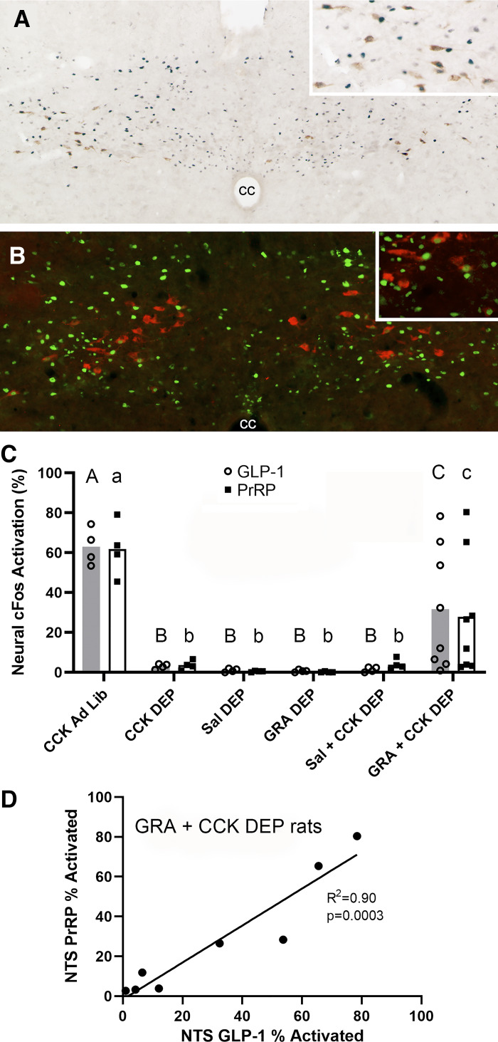
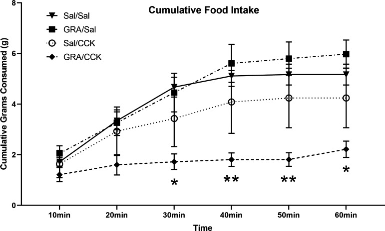
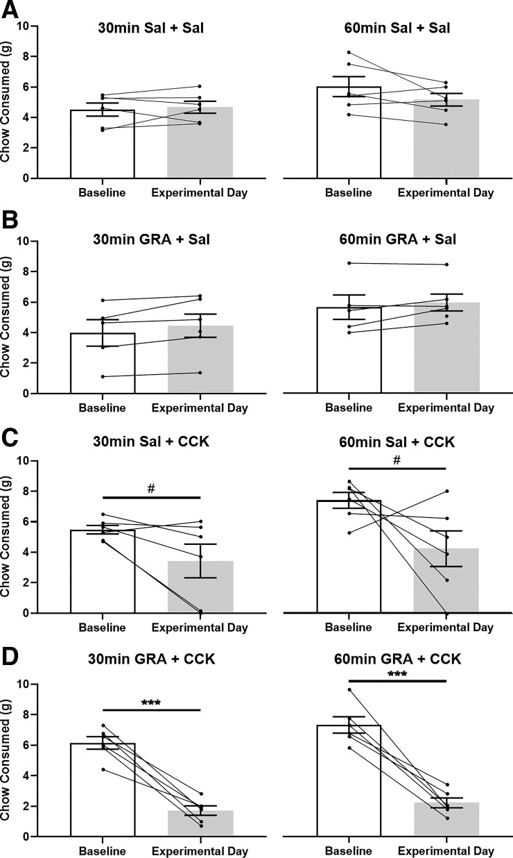
In rats, overnight fasting reduces the ability of systemic cholecystokinin-8 (CCK) to suppress food intake and to activate cFos in the caudal nucleus of the solitary tract (cNTS), specifically within glucagon-like peptide-1 (GLP-1) and noradrenergic (NA) neurons of the A2 cell group. Systemic CCK increases vagal sensory signaling to the cNTS, an effect that is amplified by leptin and reduced by ghrelin. Since fasting reduces plasma leptin and increases plasma ghrelin levels, we hypothesized that peripheral leptin administration and/or antagonism of ghrelin receptors in fasted rats would rescue the ability of CCK to activate GLP-1 neurons and a caudal subset of A2 neurons that coexpress prolactin-releasing peptide (PrRP). To test this, cFos expression was examined in ad libitum-fed and overnight food-deprived (DEP) rats after intraperitoneal CCK, after coadministration of leptin and CCK, or after intraperitoneal injection of a ghrelin receptor antagonist (GRA) before CCK. In fed rats, CCK activated cFos in ~60% of GLP-1 and PrRP neurons. Few or no GLP-1 or PrRP neurons expressed cFos in DEP rats treated with CCK alone, CCK combined with leptin, or GRA alone. However, GRA pretreatment increased the ability of CCK to activate GLP-1 and PrRP neurons and also enhanced the hypophagic effect of CCK in DEP rats. Considered together, these new findings suggest that reduced behavioral sensitivity to CCK in fasted rats is at least partially due to ghrelin-mediated suppression of hindbrain GLP-1 and PrRP neural responsiveness to CCK.

Keywords: cFos; cholecystokinin-8; fasting; hypophagia; satiety.


Conflict of interest statement

No conflicts of interest, financial or otherwise, are declared by the authors.

Figures

Fig. 1.
Fig. 1.
Leptin is insufficient to rescue CCK-induced activation of glucagon-like peptide-1 (GLP-1) or prolactin-releasing peptide (PrRP) neurons in overnight food-deprived (DEP) rats. A: color image depicting neuronal cFos expression (black nuclear label) and GLP-1 immunolabeling (brown cytoplasmic label) in the caudal nucleus of the solitary tract (cNTS) of a DEP rat that received intraperitoneal coinjection of CCK (3 μg/kg body wt) and leptin (800 μg/kg body wt). Despite robust cNTS cFos activation, very few double-labeled neurons (i.e., positive for both cFos and GLP-1) are present. Inset: higher-magnification view of several GLP-1 neurons, none of which express cFos. cc, central canal. B: color image depicting neuronal cFos expression (green nuclear label) and PrRP neurons (red cytoplasmic label) in the cNTS of a DEP rat that received intraperitoneal coinjection of CCK (3 μg/kg body wt) and leptin (800 μg/kg body wt). Despite robust cNTS cFos activation, very few double-labeled neurons (i.e., immunopositive for both cFos and PrRP) are present. Inset: higher-magnification view of several PrRP neurons, none of which express cFos. C: summary data reporting the proportion of cFos-positive hindbrain GLP-1 neurons (shaded bars; open circles are individual data points) or PrRP neurons (open bars; solid squares are individual data points) in ad libitum-fed (Ad Lib) vs. fasted (DEP) rats (n = 4/treatment group). Within the same neuronal population (i.e., GLP-1 or PrRP), bar values with different letters are significantly different (P < 0.05).
Fig. 2.
Fig. 2.
Ghrelin receptor antagonism enhances CCK-induced activation of glucagon-like peptide-1 (GLP-1) and prolactin-releasing peptide (PrRP) neurons in overnight food-deprived (DEP) rats. A: color image depicting neuronal cFos expression (black nuclear label) within GLP-1 neurons (brown cytoplasmic label) in the caudal nucleus of the solitary tract (cNTS) of a DEP rat that received intraperitoneal pretreatment with ghrelin receptor antagonist (GRA; 3.3 mg/kg body wt) before intraperitoneal CCK (3 μg/kg body wt). Inset: higher-magnification view of several GLP-1 neurons, many of which express cFos. cc, central canal. B: color image depicting neuronal cFos expression (green nuclear label) within PrRP neurons (red cytoplasmic label) in the cNTS of a DEP rat that received intraperitoneal pretreatment with GRA (3.3 mg/kg body wt) before intraperitoneal CCK (3 μg/kg body wt). Inset: higher-magnification view of several PrRP neurons, some of which express cFos. C: summary data illustrating the proportion of cFos-positive hindbrain GLP-1 neurons (shaded bars; open circles are individual data points) or PrRP neurons (open bars; solid squares are individual data points) in ad libitum-fed (Ad Lib) vs. fasted (DEP) rats (n = 4/group, except n = 8 for GRA + CCK). Within the same neuronal population (i.e., GLP-1 or PrRP), bar values with different letters are significantly different (P < 0.05). D: proportions of GLP-1- and PrRP-positive neurons activated to express cFos are highly correlated in individual DEP rats within the GRA + CCK treatment group (n = 8).
Fig. 3.
Fig. 3.
One-hour cumulative food intake (means ± SE) in overnight food-deprived (DEP) rats. DEP rats (n = 6/group) received intraperitoneal injection of ghrelin receptor antagonist (GRA; 3.3 mg/kg body wt) or saline vehicle (Sal), followed 30 min later by intraperitoneal injection of Sal or CCK (1 μg/kg body wt) just before food access. Compared with food intake by Sal/Sal control rats, feeding was suppressed only in rats that received GRA before CCK (*P < 0.05, **P < 0.01, GRA/Sal vs. Sal/Sal). Other between-group differences in food intake at each time point were not statistically significant.
Fig. 4.
Fig. 4.
Within-subjects comparison of fasting-induced food intake by overnight food-deprived (DEP) rats within each treatment group. Open bars depict cumulative 30-min (left) and 60-min (right) chow intake (means ± SE) by DEP rats under baseline (i.e., noninjected) conditions. Shaded bars depict fasting-induced chow intake by the same rats after intraperitoneal injection of saline (Sal) or ghrelin receptor antagonist (GRA; 3.3 mg/kg body wt), followed 30 min later by intraperitoneal injection of saline or CCK (1 μg/kg body wt) just before food access (n = 6/group). Within-subject data points are connected by lines. Compared with baseline intake, 30- and 60-min food intakes were suppressed only in GRA + CCK-treated rats (D: ***P < 0.001). Conversely, rats treated with Sal + CCK displayed only a nonsignificant trend (C: #P = 0.075 at 30 min, #P = 0.109 at 60 min) toward suppression of fasting-induced food intake.

References

    1. Akana SF, Strack AM, Hanson ES, Dallman MF. Regulation of activity in the hypothalamo-pituitary-adrenal axis is integral to a larger hypothalamic system that determines caloric flow. Endocrinology 135: 1125–1134, 1994. doi: 10.1210/endo.135.3.8070356. - DOI - PubMed
    1. Akieda-Asai S, Poleni PE, Date Y. Coinjection of CCK and leptin reduces food intake via increased CART/TRH and reduced AMPK phosphorylation in the hypothalamus. Am J Physiol Endocrinol Metab 306: E1284–E1291, 2014. doi: 10.1152/ajpendo.00664.2013. - DOI - PubMed
    1. Appleyard SM, Marks D, Kobayashi K, Okano H, Low MJ, Andresen MC. Visceral afferents directly activate catecholamine neurons in the solitary tract nucleus. J Neurosci 27: 13292–13302, 2007. doi: 10.1523/JNEUROSCI.3502-07.2007. - DOI - PMC - PubMed
    1. Asakawa A, Inui A, Kaga T, Katsuura G, Fujimiya M, Fujino MA, Kasuga M. Antagonism of ghrelin receptor reduces food intake and body weight gain in mice. Gut 52: 947–952, 2003. doi: 10.1136/gut.52.7.947. - DOI - PMC - PubMed
    1. Bado A, Levasseur S, Attoub S, Kermorgant S, Laigneau JP, Bortoluzzi MN, Moizo L, Lehy T, Guerre-Millo M, Le Marchand-Brustel Y, Lewin MJ. The stomach is a source of leptin. Nature 394: 790–793, 1998. doi: 10.1038/29547. - DOI - PubMed

Publication types

MeSH terms

LinkOut - more resources