

Multiple Intracranial Tuberculomas with an Intra-medullary Spinal Cord Tuberculoma in a Pediatric Patient
- PMID: 32292663
- PMCID: PMC7152571
- DOI: 10.7759/cureus.7248
Multiple Intracranial Tuberculomas with an Intra-medullary Spinal Cord Tuberculoma in a Pediatric Patient
Abstract
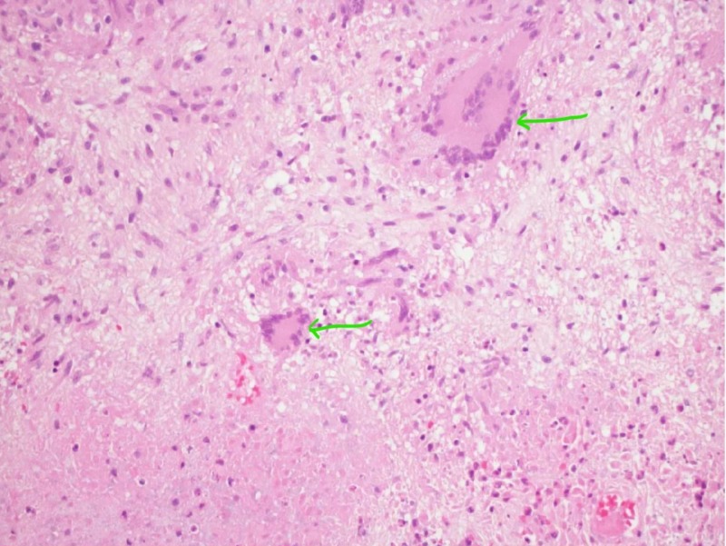
Central nervous system (CNS) tuberculosis (TB), caused by Mycobacterium tuberculosis (MT), is a severe form of TB, which presents as meningitis, cerebritis, abscesses, spinal tuberculous arachnoiditis, and rarely tuberculomas. CNS TB is prevalent in the underdeveloped or developing world and is common in malnourished, alcoholics, children, young adults, immunocompromised, and cancer patients. Intracranial tuberculomas (ICT) can present with symptoms and signs of focal neurological deficits with or without systemic manifestations. ICT is the least common presentation of CNS TB. Medical management with anti-TB drugs and steroids is the mainstay of treatment, while surgical intervention is usually reserved for refractory cases. Here, we present the case of a 10-year-old Indian American girl with headaches, diplopia, fever, and neck pain diagnosed with ICT and intramedullary spinal cord tuberculoma.
Keywords: central nervous system tuberculosis; cerebral tuberculomas; disseminated tuberculosis; intra-medullary spinal cord tuberculoma; intracranial tuberculoma.
Copyright © 2020, Meegada et al.
Conflict of interest statement
The authors have declared that no competing interests exist.
Figures


References
-
- Intracranial tuberculoma:comparison of MR with pathologic findings. Kim TK, Chang KH, Kim CJ, Goo JM, Kook MC, Han MH. http://www.ajnr.org/content/16/9/1903.long. Am J Neuroradiol. 1995;16:1903–1908. - PMC - PubMed
-
- WHO Global Tuberculosis Report. [Feb;2019 ];https://www.who.int/tb/publications/global_report/tb19_Exec_Sum_12Nov201... 2019
-
- Central nervous system tuberculosis. Cherian A, Thomas SV. https://www.ajol.info/index.php/ahs/article/view/65007. Afr Health Sci. 2011;11:116–127. - PMC - PubMed
-
- Rom WN, Garay SM. Philadelphia: Lippincott Williams & Wilkins; 2004. Tuberculosis.
Publication types
LinkOut - more resources
Full Text Sources