. 2020 Jun;34(6):7915-7926.
doi: 10.1096/fj.201902179R. Epub 2020 Apr 15.

Modulation of radiation-induced damage of human glomerular endothelial cells by SMPDL3B

Affiliations

Modulation of radiation-induced damage of human glomerular endothelial cells by SMPDL3B

Alaa Abou Daher et al. FASEB J. 2020 Jun.

Abstract

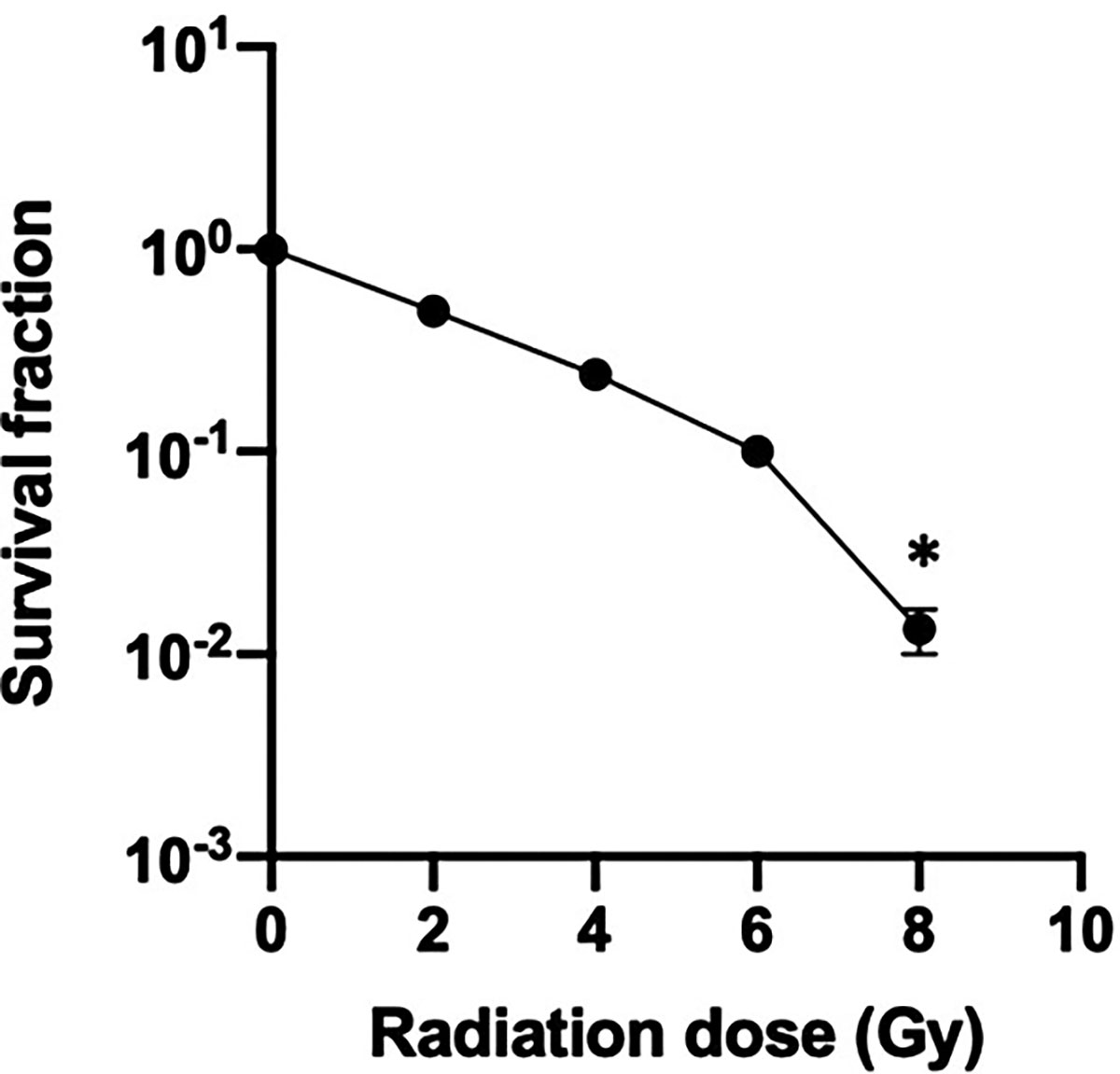
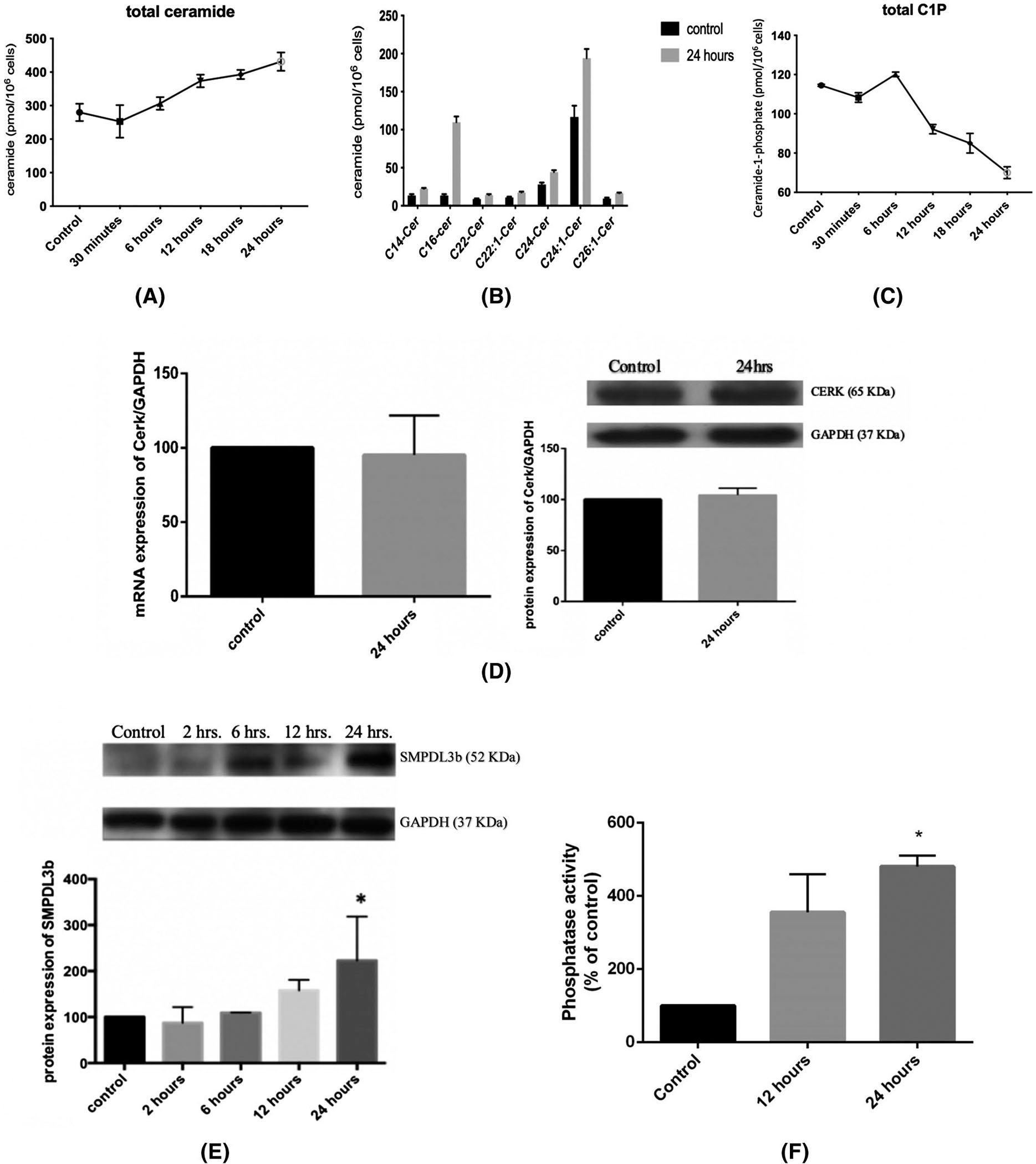
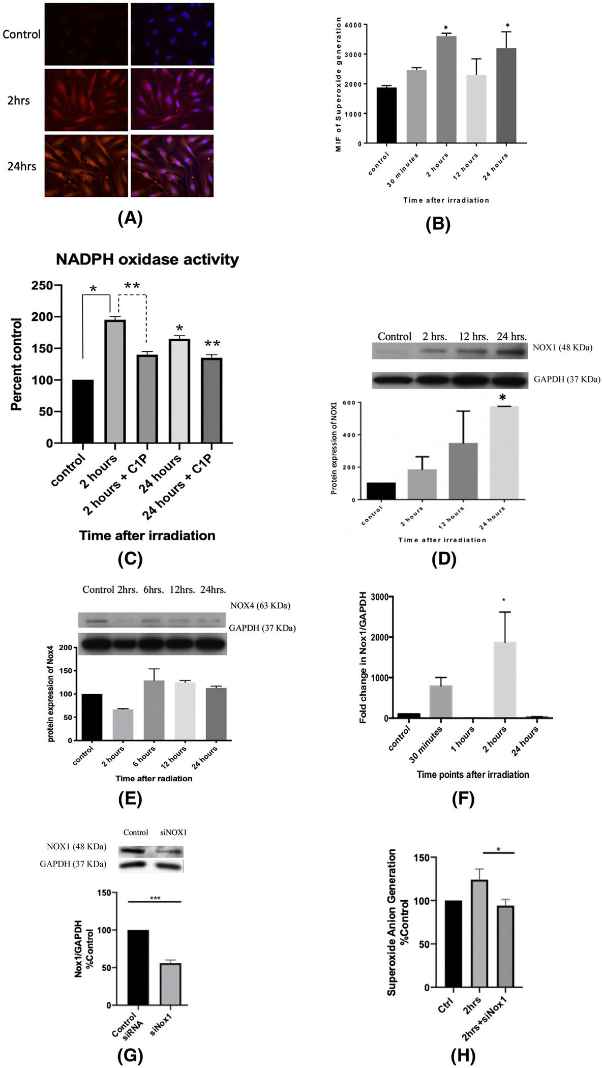
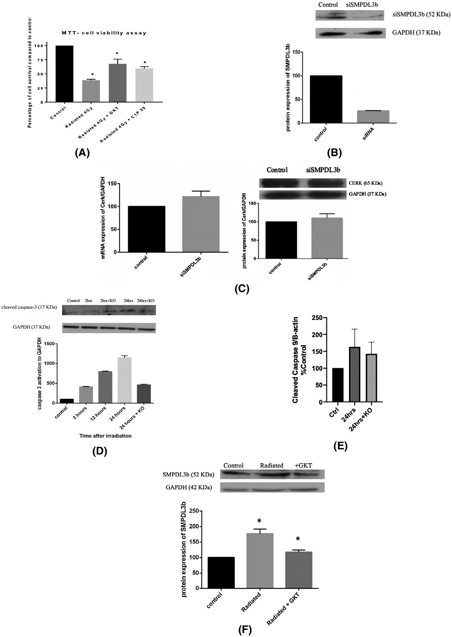
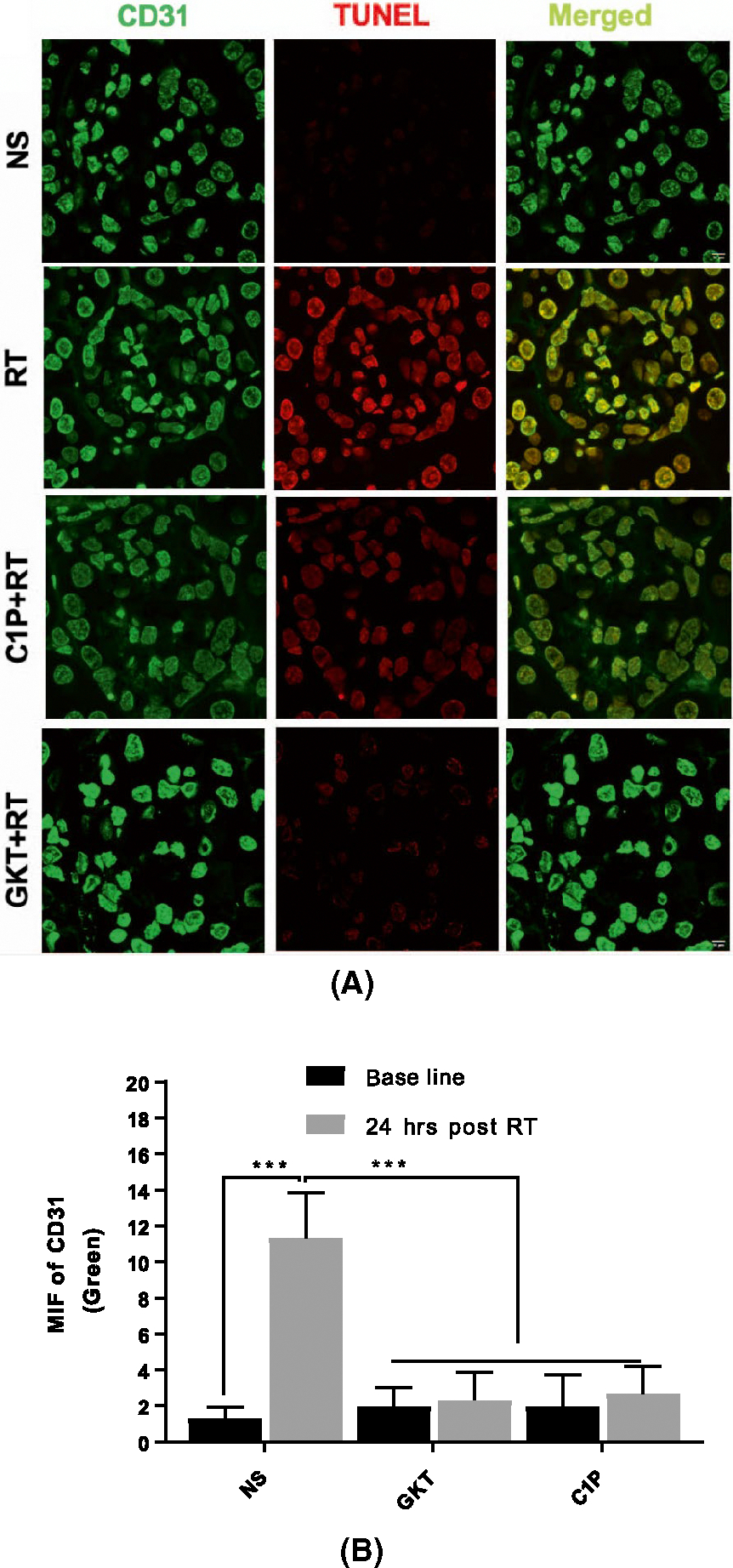
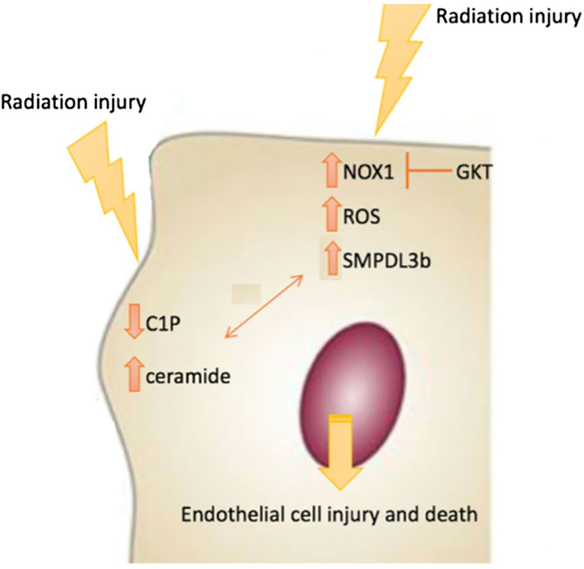
The intracellular molecular pathways involved in radiation-induced nephropathy are still poorly understood. Glomerular endothelial cells are key components of the structure and function of the glomerular filtration barrier but little is known about the mechanisms implicated in their injury and repair. The current study establishes the response of immortalized human glomerular endothelial cells (GEnC) to ionizing radiation (IR). We investigated the role of sphingolipids and the lipid-modifying enzyme sphingomyelin phosphodiesterase acid-like 3b (SMPDL3b) in radiation-induced GEnC damage. After delivering a single dose of radiation, long and very-long-chain ceramide species, and the expression levels of SMPDL3b were elevated. In contrast, levels of ceramide-1-phosphate (C1P) dropped in a time-dependent manner although mRNA and protein levels of ceramide kinase (CERK) remained stable. Treatment with C1P or knocking down SMPDL3b partially restored cell survival and conferred radioprotection. We also report a novel role for the NADPH oxidase enzymes (NOXs), namely NOX1, and NOX-derived reactive oxygen species (ROS) in radiation-induced GEnC damage. Subjecting cultured endothelial cells to radiation was associated with increased NOX activity and superoxide anion generation. Silencing NOX1 using NOX1-specific siRNA mitigated radiation-induced oxidative stress and cellular injury. In addition, we report a novel connection between NOX and SMPDL3b. Treatment with the NOX inhibitor, GKT, decreased radiation-induced cellular injury and restored SMPDL3b basal levels of expression. Our findings indicate the importance of SMPDL3b as a potential therapeutic target in radiation-induced kidney damage.

Keywords: SMPDL3b; cancer; ceramide; glomerular endothelial cells; nephropathy; radioprotection; reactive oxygen species; sphingolipids.


Conflict of interest statement

DISCLOSURES

Alessia Fornoni is consultant for Hoffman-La Roche, Alexion, and Mesoblast on subject matters that are unrelated to this publication. The authors declare no conflict of interest.

Figures

FIGURE 1
FIGURE 1
Human glomerular endothelial cell logarithmic survival curve. Cultured endothelial cells were subjected to increasing doses of radiation, incubated at 37 degrees and the colony forming units (CFU) were counted under the microscope after staining with crystal violet. CFU were reported as percentage of control (nonirradiated cells). The results are representative of at least five independent experiments. *P < .05
FIGURE 2
FIGURE 2
Radiation leads to a significant alteration of the sphingolipid profile in GEnC. Cells were irradiated with 0 Gy (control) or 4 Gy, and pellets containing 106 cells were collected after 30 minutes, 6, 12, 18, and 24 hours. After extraction of lipids, the treatment groups were subjected to liquid chromatography-mass spectrometric analysis to determine the levels of total ceramide (A), the different ceramide sub-species (B), and the total level of C1P (C). Results represent the average of three independent experiments. D, Changes in the mRNA and protein levels of CERK at 24 hours post-irradiation. E, Changes in the level of protein expression of SMPDL3b post-irradiation. All blots were quantified by densitometry using Image J software, normalized to the house-keeping gene GAPDH and expressed as percentage of control. F, C1P Phosphatase in vitro assay after radiation at 4 Gy using NBD-labeled C1P at 12- and 24-hours post-irradiation. Results shown are the mean values of at least four independent experiments. *P < .05
FIGURE 3
FIGURE 3
Radiation increases superoxide anion generation by increasing the transcription and translation of NOX1. A, Immunofluorescence staining with DHE and DAPI of endothelial cells radiation at 0 Gy (control) and at 4 Gy, 2 and 24 hours post-irradiation. B, Quantification of mean immunofluorescence at baseline (control) and at 30 minutes, 2, 24, and 24 hours post-irradiation at 4 Gy. Cells were plated in T-25 flasks until 80% confluency, differentiated and then irradiated at 4 Gy. The pellets were collected at various time points. C, NADPH oxidase activity was assessed via a lucigenin-based assay with and without C1P administration. Photon emission was quantified as percentage of control (0 Gy) after subtracting blank. D, NOX1 and (E) NOX4 protein levels post-IR were assessed using western blot. F, NOX1 gene expression profile at 30 minutes, 1, 2, and 24 hours post-irradiation. G, siRNA was used to knockdown NOX1 and the protein expression levels of NOX1 levels were analyzed after 24 hours of incubation. H, NOX1-knockout GEnC show a significant decrease in superoxide anion generation at 2 hours post-irradiation. Results shown are the mean values of three independent experiments. *P < .05 compared to control and **P < .05 compared to the treated condition in the NADPH oxidase assay
FIGURE 4
FIGURE 4
SMPDL3b mediates IR-induced cell injury in a caspase-3 dependent mechanism. Endothelial cells were cultured in plates, differentiated, and treated either with siRNA, GKT, or C1P before irradiation at 4 Gy. A, Cell survival was assessed using MTT. Results were obtained by spectrophotometry. The readings were subtracted from blank and expressed as percentage relative to control. B, siRNA was used to knockdown SMPDL3b and the protein expression levels of SMPDL3b were analyzed after 24 hours of incubation. C, Changes in the mRNA and protein levels of CERK with siSMPDL3b. D, SMPDL3b-knockout GEnC show improved survival post-irradiation as demonstrated by caspase-3 activation. Results are normalized to GAPDH and expressed as percentage to control. E, GEnC show no significant change in radiation-induced caspase-9 cleavage after SMPDL3b silencing (F) GKT treatment downregulates SMPDL3b protein expression post-radiation. Results were quantified by densitometry using Image J software, normalized to the house-keeping gene and expressed as percentage of control. Results are the mean of at least three independent experiments each done in triplicates. *P < .05
FIGURE 5
FIGURE 5
Treatment with GKT or C1P improves glomerular endothelial cell viability in vivo. Murine kidneys were sectioned and processed for immunofluorescence co-staining of TUNEL with CD31. A, Images were captured using a Zeiss confocal microscope via a 63x oil objective lens in different planes using a Z-series pattern with a step size of 0.5 μm, scale bar 0.5 inches. B, Quantification of green fluorescence for CD31 and TUNEL (Red). Endothelial cell death increased significantly with IR. Treatment of mice with GKTor C1P restored cell survival to post-irradiation. Scale bar is 10 μm
FIGURE 6
FIGURE 6
Model of radiation-induced endothelial cell damage. The model depicts induction of NOX1 and NOX1-mediated reactive oxygen species (ROS) downstream radiation injury. The increase in SMPDL3b downstream of NOX1 is a key event in radiation injury of GEnC. This triggers changes in sphingolipid metabolism, including a drop in C1P and an increase in ceramide, which contributes to the damage phenotype

References

    1. Cassady JR. Clinical radiation nephropathy. Int J Radiat Oncol Biol Phys. 1995;31(5):1249–1256. - PubMed
    1. Dawson LA, Kavanagh BD, Pauliono AC, et al. Radiation-associated kidney injury. Int J Radiat Oncol Biol Phys. 2010;76(Suppl. 3):S108–S115. - PubMed
    1. Cohen EP, Robbins ME. Radiation nephropathy. Semin Nephrol. 2003;23(5):486–499. - PubMed
    1. Luxton RW. Radiation nephritis. A long-term study of 54 patients. Lancet. 1961;2(7214):1221–1224. - PubMed
    1. Yang GY, May KS, Iyer RV, et al. Renal atrophy secondary to chemoradiotherapy of abdominal malignancies. Int J Radiat Oncol Biol Phys. 2010;78(2):539–546. - PubMed

Publication types

LinkOut - more resources