

DCE-MRI of locally-advanced carcinoma of the uterine cervix: Tofts analysis versus non-model-based analyses
- PMID: 32293487
- PMCID: PMC7158049
- DOI: 10.1186/s13014-020-01526-2
DCE-MRI of locally-advanced carcinoma of the uterine cervix: Tofts analysis versus non-model-based analyses
Abstract
Background: Dynamic contrast-enhanced magnetic resonance imaging (DCE-MRI) may provide biomarkers of the outcome of locally-advanced cervical carcinoma (LACC). There is, however, no agreement on how DCE-MR recordings should be analyzed. Previously, we have analyzed DCE-MRI data of LACC using non-model-based strategies. In the current study, we analyzed DCE-MRI data of LACC using the Tofts pharmacokinetic model, and the biomarkers derived from this analysis were compared with those derived from the non-model-based analyses.
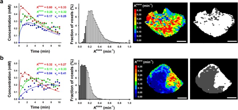
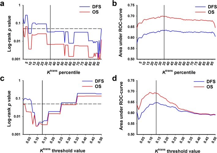
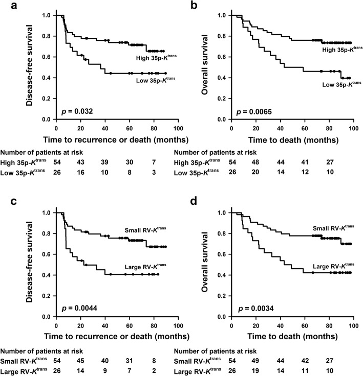
Methods: Eighty LACC patients given cisplatin-based chemoradiotherapy with curative intent were included in the study. Treatment outcome was recorded as disease-free survival (DFS) and overall survival (OS). DCE-MRI series were analyzed voxelwise to produce Ktrans and ve frequency distributions, and ROC analysis was used to identify the parameters of the frequency distributions having the greatest potential as biomarkers. The prognostic power of these parameters was compared with that of the non-model-based parameters LETV (low-enhancing tumor volume) and TVIS (tumor volume with increasing signal).
Results: Poor DFS and OS were associated with low values of Ktrans, whereas there was no association between treatment outcome and ve. The Ktrans parameters having the greatest prognostic value were p35-Ktrans (the Ktrans value at the 35 percentile of a frequency distribution) and RV-Ktrans (the tumor subvolume with Ktrans values below 0.13 min- 1). Multivariate analysis including clinical parameters and p35-Ktrans or RV-Ktrans revealed that RV-Ktrans was the only independent prognostic factor of DFS and OS. There were significant correlations between RV-Ktrans and LETV and between RV-Ktrans and TVIS, and the prognostic power of RV-Ktrans was similar to that of LETV and TVIS.
Conclusions: Biomarkers of the outcome of LACC can be provided by analyzing DCE-MRI series using the Tofts pharmacokinetic model. However, these biomarkers do not appear to have greater prognostic value than biomarkers determined by non-model-based analyses.
Keywords: Biomarkers; Cervical carcinoma; DCE-MRI; Tofts pharmacokinetic model.
Conflict of interest statement
The authors declare that they have no competing interests.
Figures


Similar articles
-
DCE-MRI of Tumor Hypoxia and Hypoxia-Associated Aggressiveness.Cancers (Basel). 2020 Jul 20;12(7):1979. doi: 10.3390/cancers12071979. Cancers (Basel). 2020. PMID: 32698525 Free PMC article. Review.
-
Pharmacokinetic analysis of DCE-MRI data of locally advanced cervical carcinoma with the Brix model.Acta Oncol. 2019 Jun;58(6):828-837. doi: 10.1080/0284186X.2019.1580386. Epub 2019 Feb 27. Acta Oncol. 2019. PMID: 30810443
-
Pharmacokinetic parameters derived from dynamic contrast enhanced MRI of cervical cancers predict chemoradiotherapy outcome.Radiother Oncol. 2013 Apr;107(1):117-22. doi: 10.1016/j.radonc.2012.11.007. Epub 2013 Jan 17. Radiother Oncol. 2013. PMID: 23333024
-
Voxelwise comparison of perfusion parameters estimated using dynamic contrast enhanced (DCE) computed tomography and DCE-magnetic resonance imaging in locally advanced cervical cancer.Acta Oncol. 2013 Oct;52(7):1360-8. doi: 10.3109/0284186X.2013.813637. Epub 2013 Sep 5. Acta Oncol. 2013. PMID: 24003852
-
Tracer-kinetic modeling of dynamic contrast-enhanced MRI and CT: a primer.J Pharmacokinet Pharmacodyn. 2013 Jun;40(3):281-300. doi: 10.1007/s10928-013-9315-3. Epub 2013 Apr 6. J Pharmacokinet Pharmacodyn. 2013. PMID: 23563847 Review.
Cited by
-
Role of MRI-Based Functional Imaging in Improving the Therapeutic Index of Radiotherapy in Cancer Treatment.Front Oncol. 2021 Aug 27;11:645177. doi: 10.3389/fonc.2021.645177. eCollection 2021. Front Oncol. 2021. PMID: 34513659 Free PMC article. Review.
-
Correlation study of functional magnetic resonance index and clinicopathological features of rectal cancer.Abdom Radiol (NY). 2024 Jul;49(7):2368-2386. doi: 10.1007/s00261-024-04375-9. Epub 2024 Jun 13. Abdom Radiol (NY). 2024. PMID: 38872052
-
Assessment of Hypoxic Tissue Fraction and Prediction of Survival in Cervical Carcinoma by Dynamic Contrast-Enhanced MRI.Front Oncol. 2021 May 20;11:668916. doi: 10.3389/fonc.2021.668916. eCollection 2021. Front Oncol. 2021. PMID: 34094964 Free PMC article.
-
DCE-MRI of Tumor Hypoxia and Hypoxia-Associated Aggressiveness.Cancers (Basel). 2020 Jul 20;12(7):1979. doi: 10.3390/cancers12071979. Cancers (Basel). 2020. PMID: 32698525 Free PMC article. Review.
-
The Utility of Contrast-Enhanced Magnetic Resonance Imaging in Uterine Cervical Cancer: A Systematic Review.Life (Basel). 2023 Jun 12;13(6):1368. doi: 10.3390/life13061368. Life (Basel). 2023. PMID: 37374150 Free PMC article. Review.
