. 2020 Apr 15;16(4):e1008440.
doi: 10.1371/journal.ppat.1008440. eCollection 2020 Apr.

A refined model of how Yersinia pestis produces a transmissible infection in its flea vector

Affiliations

A refined model of how Yersinia pestis produces a transmissible infection in its flea vector

Amélie Dewitte et al. PLoS Pathog. .

Abstract

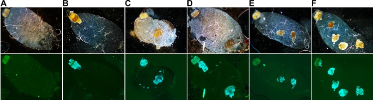
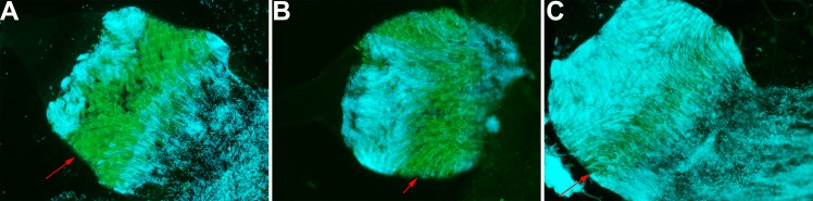
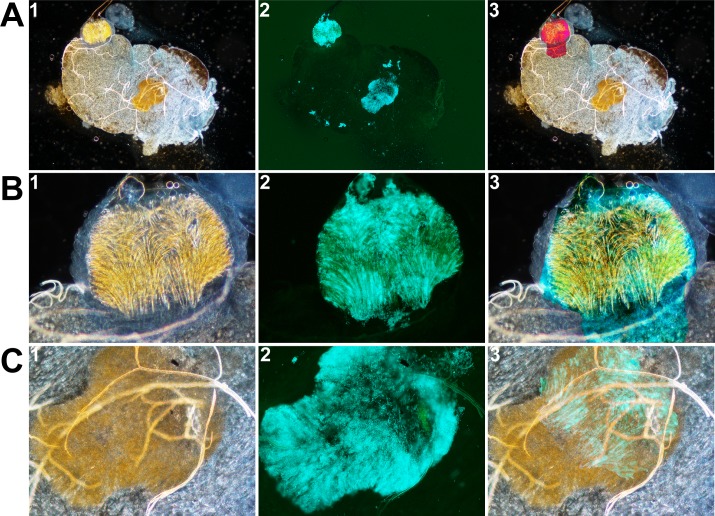
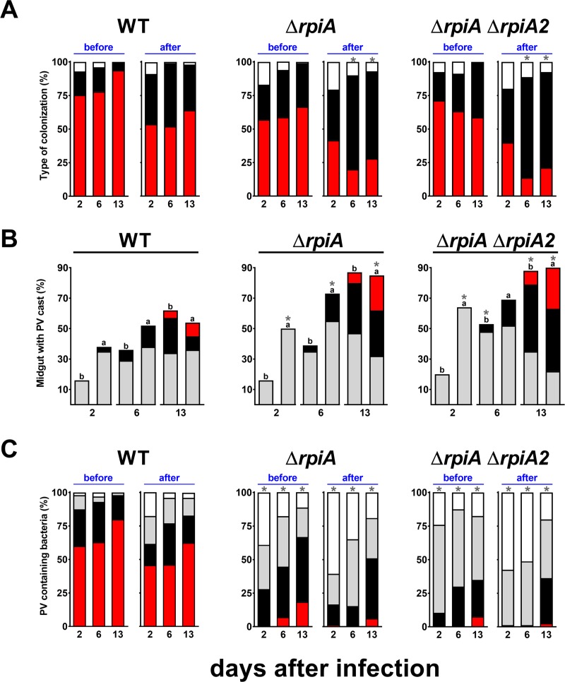
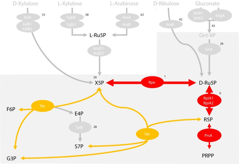
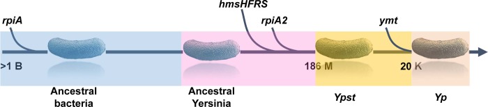
In flea-borne plague, blockage of the flea's foregut by Yersinia pestis hastens transmission to the mammalian host. Based on microscopy observations, we first suggest that flea blockage results from primary infection of the foregut and not from midgut colonization. In this model, flea infection is characterized by the recurrent production of a mass that fills the lumen of the proventriculus and encompasses a large number of Y. pestis. This recurrence phase ends when the proventricular cast is hard enough to block blood ingestion. We further showed that ymt (known to be essential for flea infection) is crucial for cast production, whereas the hmsHFRS operon (known to be essential for the formation of the biofilm that blocks the gut) is needed for cast consolidation. By screening a library of mutants (each lacking a locus previously known to be upregulated in the flea gut) for biofilm formation, we found that rpiA is important for flea blockage but not for colonization of the midgut. This locus may initially be required to resist toxic compounds within the proventricular cast. However, once the bacterium has adapted to the flea, rpiA helps to form the biofilm that consolidates the proventricular cast. Lastly, we used genetic techniques to demonstrate that ribose-5-phosphate isomerase activity (due to the recent gain of a second copy of rpiA (y2892)) accentuated blockage but not midgut colonization. It is noteworthy that rpiA is an ancestral gene, hmsHFRS and rpiA2 were acquired by the recent ancestor of Y. pestis, and ymt was acquired by Y. pestis itself. Our present results (i) highlight the physiopathological and molecular mechanisms leading to flea blockage, (ii) show that the role of a gene like rpiA changes in space and in time during an infection, and (iii) emphasize that evolution is a gradual process punctuated by sudden jumps.


Conflict of interest statement

The authors declare that no competing interests exist.

Figures

Fig 1
Fig 1. The flea gut infected with Y. pestis contains zero, one or several brownish masses.
Images were acquired using a bright-field microscope (upper images) and a fluorescence microscope (lower images). Using Adobe Photoshop, the fluorescence microscopy images were modified to highlight bacteria (in blue) that were attached (or not) to the proventriculus (in green). After an infection, the flea gut may (B and D) or may not (A) feature a brownish, bacteria-containing mass anchored within the proventriculus. In some cases, one or more free-floating masses (C to F) are observed in the midgut. The images shown here are representative of experiments on WT and mutant strains. The photos shown in panels A to E are representative of what can be seen in the gut of fleas collected 2 and 6 days post-infection, whereas all the panels are representative of what can be seen in the gut of fleas collected 13 days after infection.
Fig 2
Fig 2. The three-zone colonization of the proventriculus.
Fluorescence microscopy images of infected proventriculi taken one day post-infection. The images were modified using Adobe Photoshop to highlight bacteria (in blue) and the proventriculus (in green). The proventriculus contained Y. pestis in the anterior and posterior spine-bearing regions, and the posterior spineless region of the proventriculus—the so-called stomodaeum valve that telescopes with the midgut (A and C). The red arrowheads indicate a “green buffer zone”; i.e. a central spine-bearing region with no (A), few (B) or some (C) bacteria.
Fig 3
Fig 3. The free-floating mass observed in the midgut is a proventricular cast containing Y. pestis.
Images of the whole flea gut (A), the proventriculus (B) and the free-floating mass (C) contained within the midgut were taken using bright-field (left) and fluorescence (middle) microscopes. The images on the right are merged images generated after appropriate rotation and blending mode (using Photoshop) to show that the free-floating mass is a proventricular cast. Notably, image A3 was produced by merging image A1 with the free-floating mass extracted from A1, and the “difference” blending mode. Images B3 and C3 were produced by merging a bright-field image with the corresponding fluorescence image (i.e. B3 = B1 merged with C2; C3 = C1 merged with B2) and the “lighten” blending mode. Fluorescence images were also modified (using Photoshop) to highlight the bacteria in blue.
Fig 4
Fig 4. List of Y. pestis mutants affected in biofilm formation.
In the figure, a, "-" means to; b, shown is the biofilm production relative to the parental strain. White squares mean that there was no difference between the mutant and the parental strain. For Congo red plate assay, black, blue and red squares mean that the bacterial colony is white, less red, and redder compared to the parental strain respectively.; *, the gene between the bracket is that responsible for the phenotype. NT, not tested; NA, not applicable.
Fig 5
Fig 5. rep, hdfR, glpD and ail are required for biofilm formation in vitro.
Biofilm formation of selected mutants (relative to the WT strain) was measured after a 24-hour incubation at 21°C with shaking in LB supplemented with Ca2+ and Mg2+ (see Fig 4). The mean ± SEM values from at least three independent experiments are shown. *: mutants producing significantly less biofilm than the WT strain (p<0.01 in a one-way analysis of variance).
Fig 6
Fig 6. RpiA and RpiA2 are redundant in the production of a transmissible infection in fleas.
We measured the ability of ΔrpiA (in yellow), ΔrpiA2 (in green) and ΔrpiA ΔrpiA2 (ΔrpiA-A2; in violet) mutants complemented or not with a high-copy-number plasmid harboring rpiA (yellow hatches) or rpiA2 (green hatches) to (A) form biofilms in vitro, (B) block fleas and (C) colonize fleas, relative to the WT strain (in grey). (A) The bars represent the mean ± SEM value from at least three independent experiments, except for the ΔrpiA ΔrpiA2 mutant complemented with rpiA2 (two experiments only). The ΔrpiA and the ΔrpiA ΔrpiA2 mutants produced significantly less biofilm than the WT and the complemented strains (*: p<0.01 in a one-way analysis of variance). (B) The bars represent the mean ± SEM from three independent experiments (ΔrpiA and ΔrpiA ΔrpiA2 mutants), two independent experiments (ΔrpiA2 and ΔrpiA ΔrpiA2 + rpiA) or one experiment (ΔrpiA ΔrpiA2 + rpiA2 mutant). The ΔrpiA and ΔrpiA ΔrpiA2 mutants blocked significantly fewer fleas than the WT strain (*: p<0.01 in a one-way analysis of variance). (C) Box-and-whisker plots (5–95% percentiles) show the bacterial loads determined from up to 20 fleas in each experiment (i.e. a total of 50 to 259 fleas). The result of one experiment (ΔrpiA2 D6 and ΔrpiA ΔrpiA2 + rpiA2 at all time points) and the cumulative results of two experiments (ΔrpiA D27) and ≥ three experiments (all other strains and time points) are shown. Symbols indicate outliers. X: not determined. The bacterial loads for the ΔrpiA strain were significantly lower than for the WT strain at D1, whereas those for the ΔrpiA ΔrpiA2 strain were significantly lower throughout the experiment (*: p<0.01 in a one-way analysis of variance). The ΔrpiA2 mutant and the complemented mutants behaved like the WT strain.
Fig 7
Fig 7. rpiA (but not hms) is needed to maintain a normal bacillary shape during the early (but not late) colonization of the mass anchored within the proventriculus.
Fluorescence microscopy images show the presence of WT, Δhms or ΔrpiA bacteria (in blue) in the brownish mass anchored within the proventriculus (PV, green spines) on day (D)1 and D13 post-infection. Images were post-processed using the curve adjustment tool in Adobe Photoshop, in order to highlight the bacteria (in blue) and the PV’s spines (in green).
Fig 8
Fig 8. The proventricular casts contain WT, ΔrpiA and ΔrpiA ΔrpiA2 Y. pestis.
Images of the flea guts and proventricular casts were acquired using a bright-field microscope (top) and a fluorescence microscope (center and bottom). The fluorescence images were modified (using Photoshop) to highlight the bacteria in blue. The proventriculus autofluoresces in green.
Fig 9
Fig 9. RpiA activity is needed for the production of a thick Y. pestis biofilm that blocks the flea proventriculus.
(A) the percentage of fleas showing no masses (white), a brownish mass anchored within the proventriculus (red), or only a free-floating mass within the midgut (black) [see Fig 1], (B) the percentage of fleas containing one (grey), two (black) or more than 2 (red) proventricular casts in the midgut (regardless of the presence of a mass associated with the proventriculus), and (C) proventriculi containing no (white), very few (grey), few (black) or many (red) bacteria were determined at 2, 6 and 13 days post-infection, before (b) and immediately after (a) feeding. The stacked data from 4 independent experiments (the ΔrpiA ΔrpiA2 mutant and WT, D13, after feeding) and 5 independent experiments (WT and ΔrpiA mutant) using >15 to 20 fleas are presented (see S7 Fig). *, p <0.05 using 2-way analysis of variance with Tukey's multiple comparisons test.
Fig 10
Fig 10. RpiA activity is needed for the production of a thick mass in the flea gut.
A scanning electron micrograph of the biofilms produced by the WT (upper photos) and the ΔrpiA mutant (lower photos) in the flea at days 13 post-infection and taken at different magnifications.
Fig 11
Fig 11. Pentose-related metabolic pathways that might have a role in flea blockage.
The figure shows the flea blockage rate for mutants lacking D-xylulose (ΔxylAB1), L-xylulose (ΔsgbK-sgbU), arabinose/D-ribulose (ΔaraAB), D- and L-xylulose and arabinose/D-ribulose (ΔaraD1 ΔaraD2 ΔxylB1 ΔxylB2) pathways with or without Rpe (ΔaraD1 ΔaraD2 ΔxylB1 ΔxylB2 Δrpe), gluconate import (ΔidnK1 Δindk2 ΔkdgK) or use via the upper part of the pentose phosphate pathway (Δgnd), ΔtalB, Δrpe or ΔrpiA ΔrpiA2). Grey arrows indicate pathways that are not important for flea blockage. The other reactions are considered to be important for flea blockage because the mutant either (i) does not block fleas (Δrpe and ΔrpiA ΔrpiA2 mutants), (ii) was highly deficient for growth in vitrotktA) (see supplementary text) or (iii) could not be deleted (ΔprsA), presumably because it is essential (as reported for E. coli). The grey area encloses the enzyme reactions of the pentose phosphate pathway.
Fig 12
Fig 12. A physical and molecular model of the processes leading to flea blockage by Y. pestis.
(A) Blockage of the flea’s foregut results from recurrent production of a soft bactericidal proventricular mass (a cast), and then the repeated colonization and [partial] decolonization of the proventriculus and the gradual consolidation of the cast until it is strong enough to withstand the incoming blood flow. The occasional eviction of the mass by the flea itself (i.e. without the need to ingest blood) is not shown. (B) The induction of, resistance against and consolidation of the soft bactericidal proventricular cast (the flea’s response to infection) respectively involve the phospholipase D Ymt, the ribose phosphate isomerases A and A2, and the biofilm synthesis complex HmsHFRS. It should be noted that following Y. pestis' entry into the flea, RpiA is initially required to resist toxic compounds within the proventricular cast. Once the bacterium has adapted to its new host, the enzyme is no longer required for toxicity resistance but is then needed to produce the biofilm that consolidates the proventricular cast.
Fig 13
Fig 13. Slow, gradual genetic accretion punctuated by sudden jumps led to the emergence of plague transmission via the flea vector.

References

    1. Butler T. Yersinia infections: centennial of the discovery of the plague bacillus. Clin Infect Dis. 1994;19(4):655–61; quiz 62–3. 10.1093/clinids/19.4.655 . - DOI - PubMed
    1. Hinnebusch BJ, Rudolph AE, Cherepanov P, Dixon JE, Schwan TG, Forsberg A. Role of Yersinia murine toxin in survival of Yersinia pestis in the midgut of the flea vector. Science. 2002;296(5568):733–5. 10.1126/science.1069972 . - DOI - PubMed
    1. Hinnebusch BJ, Jarrett CO, Bland DM. "Fleaing" the Plague: Adaptations of Yersinia pestis to Its Insect Vector That Lead to Transmission. Annual review of microbiology. 2017;71:215–32. 10.1146/annurev-micro-090816-093521 . - DOI - PubMed
    1. Vadyvaloo V, Jarrett C, Sturdevant DE, Sebbane F, Hinnebusch BJ. Transit through the flea vector induces a pretransmission innate immunity resistance phenotype in Yersinia pestis. PLoS pathogens. 2010;6(2):e1000783 10.1371/journal.ppat.1000783 . - DOI - PMC - PubMed
    1. Zhou W, Russell CW, Johnson KL, Mortensen RD, Erickson DL. Gene expression analysis of Xenopsylla cheopis (Siphonaptera: Pulicidae) suggests a role for reactive oxygen species in response to Yersinia pestis infection. Journal of medical entomology. 2012;49(2):364–70. 10.1603/me11172 . - DOI - PubMed

Publication types

Substances