

Generation and Characterization of Patient-Derived Head and Neck, Oral, and Esophageal Cancer Organoids
- PMID: 32294323
- PMCID: PMC7350550
- DOI: 10.1002/cpsc.109
Generation and Characterization of Patient-Derived Head and Neck, Oral, and Esophageal Cancer Organoids
Abstract
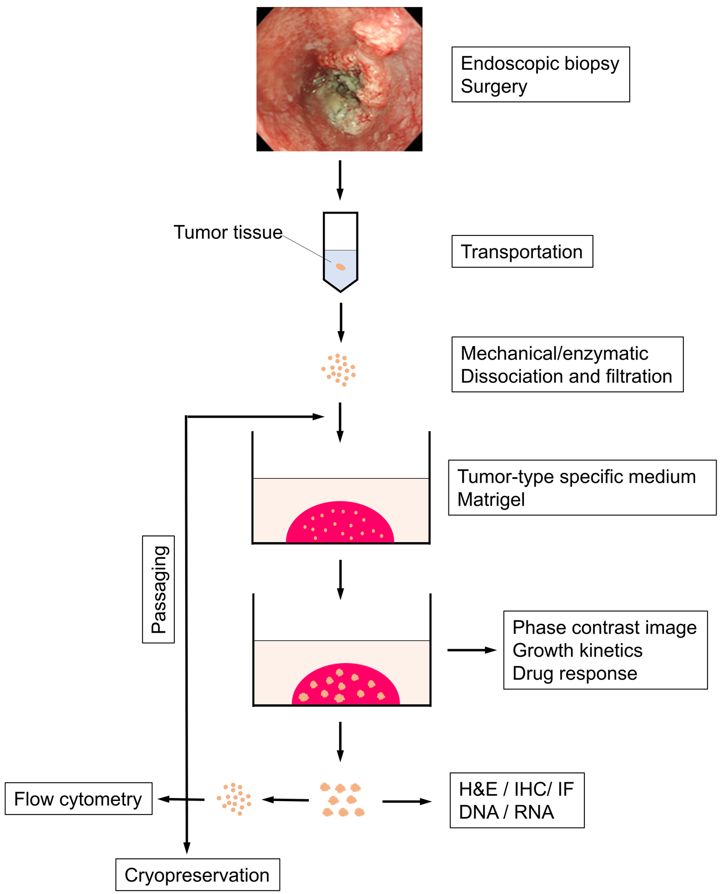
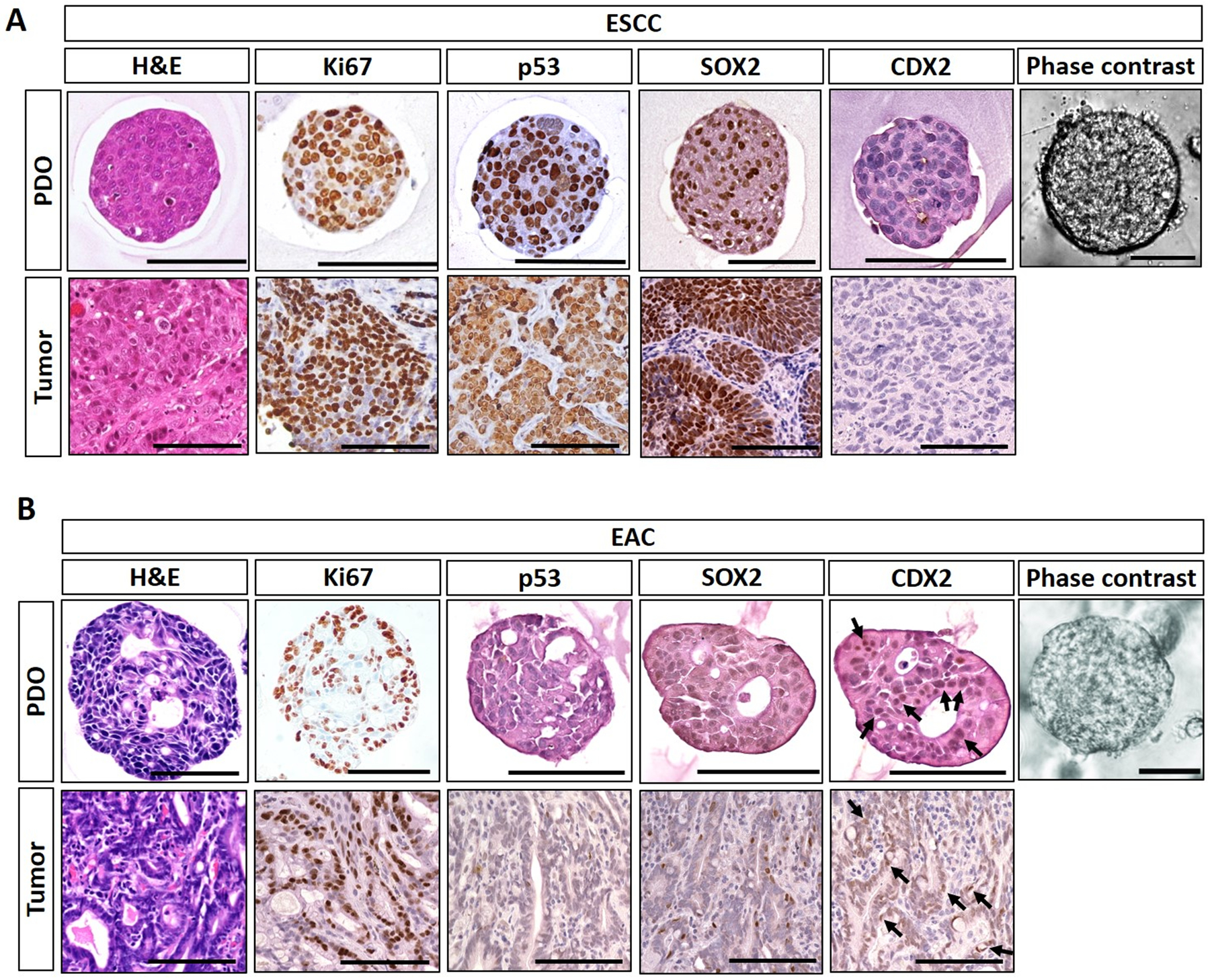
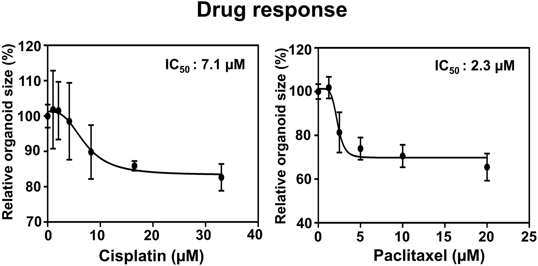
Esophageal cancers comprise adenocarcinoma and squamous cell carcinoma, two distinct histologic subtypes. Both are difficult to treat and among the deadliest human malignancies. We describe protocols to initiate, grow, passage, and characterize patient-derived organoids (PDO) of esophageal cancers, as well as squamous cell carcinomas of oral/head-and-neck and anal origin. Formed rapidly (<14 days) from a single-cell suspension embedded in basement membrane matrix, esophageal cancer PDO recapitulate the histology of the original tumors. Additionally, we provide guidelines for morphological analyses and drug testing coupled with functional assessment of cell response to conventional chemotherapeutics and other pharmacological agents in concert with emerging automated imaging platforms. Predicting drug sensitivity and potential therapy resistance mechanisms in a moderate-to-high throughput manner, esophageal cancer PDO are highly translatable in personalized medicine for customized esophageal cancer treatments. © 2020 by John Wiley & Sons, Inc. Basic Protocol 1: Generation of esophageal cancer PDO Basic Protocol 2: Propagation and cryopreservation of esophageal cancer PDO Basic Protocol 3: Imaged-based monitoring of organoid size and growth kinetics Basic Protocol 4: Harvesting esophageal cancer PDO for histological analyses Basic Protocol 5: PDO content analysis by flow cytometry Basic Protocol 6: Evaluation of drug response with determination of the half-inhibitory concentration (IC50 ) Support Protocol: Production of RN in HEK293T cell conditioned medium.
Keywords: esophageal cancer; patient-derived organoids; personalized medicine.
© 2020 John Wiley & Sons, Inc.
Figures
References
-
- Driehuis E, Kolders S, Spelier S, Lohmussaar K, Willems SM, Devriese LA, … Clevers H (2019). Oral Mucosal Organoids as a Potential Platform for Personalized Cancer Therapy. Cancer Discov, 9(7), 852–871. doi:10.1158/2159-8290.CD-18-1522 - DOI - PubMed
-
- Driehuis E, Spelier S, Beltran Hernandez I, de Bree R, S MW, Clevers H, & Oliveira S (2019). Patient-Derived Head and Neck Cancer Organoids Recapitulate EGFR Expression Levels of Respective Tissues and Are Responsive to EGFR-Targeted Photodynamic Therapy. J Clin Med, 8(11). doi:10.3390/jcm8111880 - DOI - PMC - PubMed
Publication types
MeSH terms
Grants and funding
- K01 DK103953/DK/NIDDK NIH HHS/United States
- R01 DK100342/DK/NIDDK NIH HHS/United States
- R03 DK118310/DK/NIDDK NIH HHS/United States
- R01 DE027185/DE/NIDCR NIH HHS/United States
- R03 DK114463/DK/NIDDK NIH HHS/United States
- P30 ES013508/ES/NIEHS NIH HHS/United States
- P01 CA098101/CA/NCI NIH HHS/United States
- K01 DK100485/DK/NIDDK NIH HHS/United States
- R01 DK120650/DK/NIDDK NIH HHS/United States
- T32 ES019851/ES/NIEHS NIH HHS/United States
- R01 AA026297/AA/NIAAA NIH HHS/United States
- R01 DK113144/DK/NIDDK NIH HHS/United States
- R01 DE026801/DE/NIDCR NIH HHS/United States
- R01 DK121159/DK/NIDDK NIH HHS/United States
- U54 CA163004/CA/NCI NIH HHS/United States
- P30 DK050306/DK/NIDDK NIH HHS/United States
- R01 DK114436/DK/NIDDK NIH HHS/United States
- K08 DK106444/DK/NIDDK NIH HHS/United States
- P30 CA013696/CA/NCI NIH HHS/United States
LinkOut - more resources
Full Text Sources
Other Literature Sources
Medical
