. 2020 Jul;81(1):e33-e39.
doi: 10.1016/j.jinf.2020.04.004. Epub 2020 Apr 12.

CT imaging and clinical course of asymptomatic cases with COVID-19 pneumonia at admission in Wuhan, China

Affiliations

CT imaging and clinical course of asymptomatic cases with COVID-19 pneumonia at admission in Wuhan, China

Heng Meng et al. J Infect. 2020 Jul.

Abstract

Purpose: Aimed to characterize the CT imaging and clinical course of asymptomatic cases with COVID-19 pneumonia.

Methods: Asymptomatic cases with COVID-19 pneumonia confirmed by SARS-COV-2 nucleic acid testing in Renmin Hospital of Wuhan University were retrospectively enrolled. The characteristics of CT imaging and clinical feature were collected and analyzed.

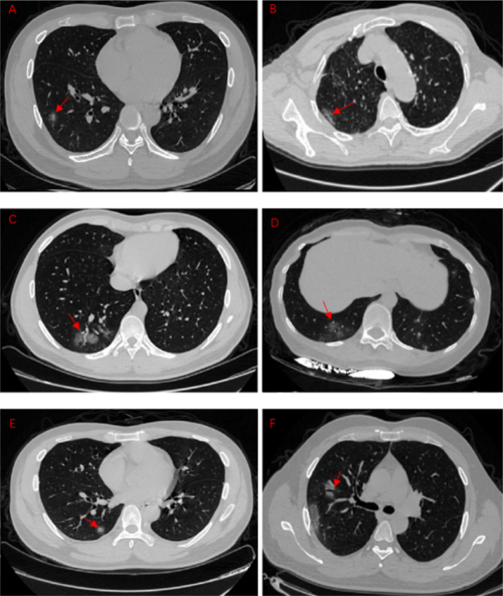
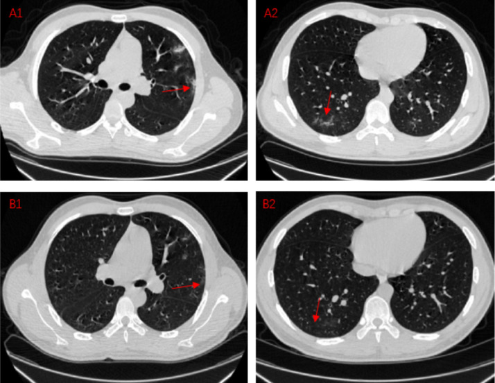
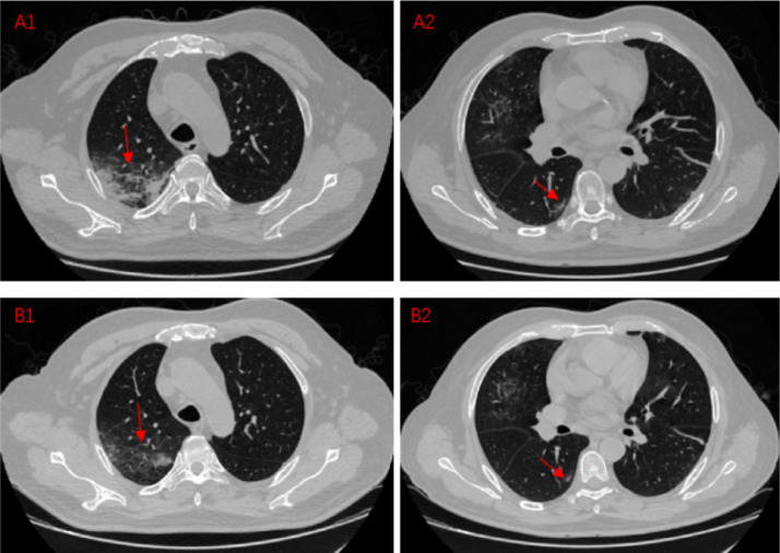
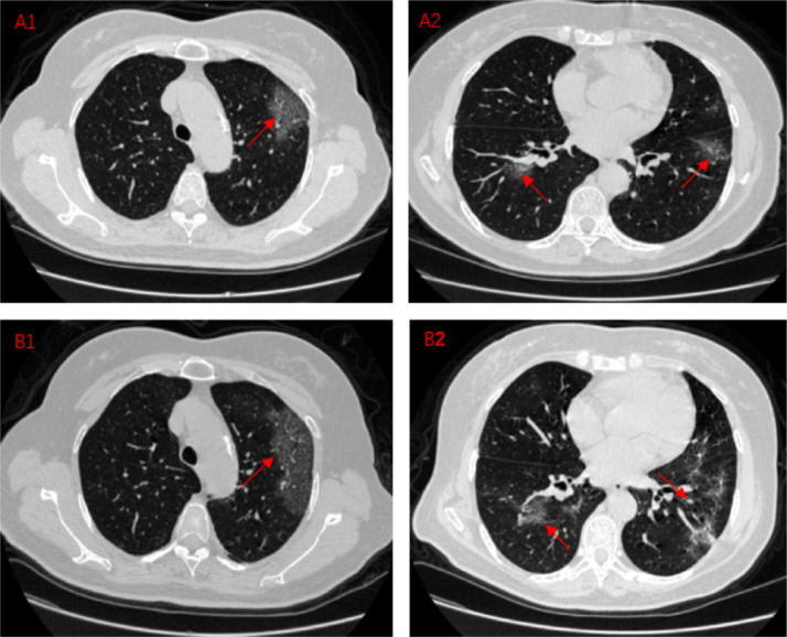
Results: 58 asymptomatic cases with COVID-19 pneumonia admitted to our hospital between Jan 1, 2020 and Feb 23, 2020 were enrolled. All patients had history of exposure to SARS-CoV-2. On admission, patients had no symptoms and laboratory findings were normal. The predominant feature of CT findings in this cohort was ground glass opacity (GGO) (55, 94.8%) with peripheral (44, 75.9%) distribution, unilateral location (34, 58.6%) and mostly involving one or two lobes (38, 65.5%), often accompanied by characteristic signs. After short-term follow-up, 16 patients (27.6%) presented symptoms with lower lymphocyte count and higher CRP, mainly including fever, cough and fatigue. The evolution of lesions on CT imaging were observed in 10 patients (17.2%). The average days of hospitalization was19.80±10.82 days, and was significantly longer in progression patients (28.60±7.55 day).

Conclusion: CT imaging of asymptomatic cases with COVID-19 pneumonia has definite characteristics. Since asymptomatic infections as "covert transmitter", and some patients can progress rapidly in the short term. It is essential to pay attention to the surveillance of asymptomatic patients with COVID-19. CT scan has great value in screening and detecting patients with COVID-19 pneumonia, especially in the highly suspicious, asymptomatic cases with negative nucleic acid testing.

Keywords: Asymptomatic; Computed Tomography; Coronavirus Disease 2019(COVID-19); Ground Glass Opacity; SARS-CoV-2.


Conflict of interest statement

Declaration of Competing Interest The authors declare no conflicts of interest.

Figures

Fig 1
Fig. 1
The different GGO manifestations in COVID-19 pneumonia patient. A: single, pure GGO. B: Pleural parallel sign. C: Vascular thickening sign. D: Fine reticulation. E: Halo sign. F: Air bronchogram.
Fig 2
Fig. 2
Comparison between recheck chest CT results and first CT results in COVID-19 pneumonia patients. Situation1:Lesion absorption A1,A2:A COVID-19 pneumonia patient, CT images on Feb 20,2020 B1,B2:A COVID-19 pneumonia patient, CT images on Feb 25,2020 These four CT images mainly shows the absorption of the lesion during the course of the disease. A1 shows the lesions parallel to the pleura under the pleura of the patient's left lung. A2 shows an ill-defined, irregularly shaped lesion. After 5 days, we recheck chest CT and find out the lesion at the original location is reduced or even disappeared. The lung tissue in the original position is gradually restored and returns to normal. Situation2:Lesion improvement A1,A2:A COVID-19 pneumonia patient, CT images on Feb 13,2020 B1,B2:A COVID-19 pneumonia patient, CT images on Feb 19,2020 These four CT images mainly shows the improvement of the lesion during the course of the disease. A1 shows a large, fused lesion in the patient's right lung, and the lesions are extensive. A2 shows a small lesion under the pleura in right lung. After 6 days, we recheck chest CT and find out the lesion at the original location is reduced. The expansion of the lesion is contained. The scope of the lesion is gradually reduced. Lung lesions are getting better after treatment. Situation3:Lesion progression A1,A2:A COVID-19 pneumonia patient, CT images on Feb 9,2020 B1,B2:A COVID-19 pneumonia patient, CT images on Feb 16,2020 These four CT images mainly shows the improvement of the lesion during the course of the disease. A1 shows a large, fused lesion in the patient's left lung. A2 shows multiple lesions in both lungs. After 7 days, we recheck chest CT and find out the lesion at the original location is expanded. The expansion of the lesion is larger than before. The scope of the lesion is out of control. Diffuse or scattered ground glass-like shadows, superimposed with thin grid-like shadows, named as paving stone sign. This patient gradually showed clinical symptoms during this period.

Similar articles

Cited by

References

    1. Lu R., Zhao X., Li J., Niu P., Yang B., Wu H. Genomic characterisation and epidemiology of 2019 novel coronavirus: implications for virus origins and receptor binding. The Lancet. 2020;395(10224):565–574. doi: 10.1016/S0140-6736(20)30251-8. - DOI - PMC - PubMed
    1. Wang D., Hu B., Hu C., Zhu F., Liu X., Zhang J. Clinical characteristics of 138 hospitalized patients with 2019 novel coronavirus-infected pneumonia in wuhan, china. JAMA. 2020 doi: 10.1001/jama.2020.1585. [Epub ahead of print] - DOI - PMC - PubMed
    1. Gorbalenya A.E., Baker S.C., Baric R.S. Severe acute respiratory syndrome-related coronavirus: the species and its viruses-a statement of the coronavirus study group. bioRxiv. 2020 doi: 10.1101/2020.02.07.937862. 02.07.937862. - DOI
    1. World Health Organization. WHO director-general's remarks at the media briefing on 2019-nCoV on 11Feb 11, 2020. https://www.who.int/dg/speeches/detail/who-director-general-s-remarks-at... (Accessed 1 March 2020).
    1. Chinese Center for Disease Control and Prevention. Coronavirus disease (COVID-2019) situation reports. March 30, 2020. http://2019ncov.chinacdc.cn/2019-nCoV/global.html (Accessed 30 March 2020).

Publication types