Review
. 2020 Apr 13;10(4):216.
doi: 10.3390/diagnostics10040216.

Imaging of Adverse Events Related to Checkpoint Inhibitor Therapy

Affiliations
Review

Imaging of Adverse Events Related to Checkpoint Inhibitor Therapy

Vanina Vani et al. Diagnostics (Basel). .

Abstract

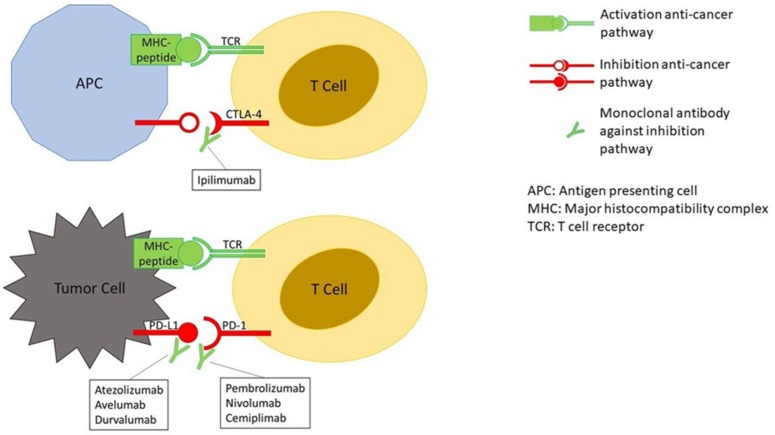
Immunotherapy with checkpoint inhibitors (ICIs) is becoming standard of practice for an increasing number of cancer types. ICIs enhance T-cell action against the cancer cells. By unbalancing the immune system ICIs may cause dysimmune toxicities, a series of disorders broadly defined immune-related adverse events (irAEs). IrAEs may affect any organ or apparatus and most frequently involve skin, colon, endocrine organs, liver, and lungs. Early identification and appropriate treatment of irAEs can improve patient outcome. The paper aims at reviewing mechanisms of the occurrence of irAEs, the importance of a proper diagnosis and the main pillars of therapy. To provide effective guidance to the comprehension of major irAEs imaging findings will be reviewed.

Keywords: immune checkpoint inhibitors; oncologic imaging; therapy; toxicity.


Conflict of interest statement

The authors declare no conflict of interest.

Figures

Figure 1
Figure 1
Mechanism of action of immune checkpoint inhibitors (ICIs). The binding together of MHC-peptide (peptide = tumor antigen) and TCR activates the anti-cancer pathway of T-cell–mediated immune response. The binding of CTLA-4 to its ligand and PD-1 to PD-L1, on the contrary, inhibit the anti-cancer pathway. The binding of monoclonal antibodies, such as ICIs, to CTLA-4, PD-1 or PD-L1 prevent the deactivation of the anti-cancer pathway.
Figure 2
Figure 2
Complicated immune-related colitis in a 54-year-old man with metastatic melanoma treated first with ipilimumab then with Pembrolizumab. CT image shows mesenteric vessel engorgement (a, yellow arrow); CT performed two months later demonstrates free intraperitoneal air (b,c, red arrow) due to intestinal perforation. © Department of Radiology, Candiolo Cancer Institute—IRCCS. Turin.
Figure 3
Figure 3
Complicated immune-related segmental colitis with diverticulosis pattern in a 76-year-old man with angiosarcoma and abdominal pain during Nivolumab treatment. CT images of two different slices show a segmental and circumferential descending colon wall thickening and pericolic fat stranding. © Department of Radiology, Candiolo Cancer Institute—FPO-IRCCs. Turin.
Figure 4
Figure 4
Immune related cholangitis in a 69-year-old female with metastatic melanoma treated with ipilimumab + pembrolizumab. Ultrasound shows layered gallbladder wall thickening likely representing gallbladder wall edema (a, yellow arrow) and hyperechogenicity of periportal space (a, blue arrows). Computed Tomography image shows mild wall thickness and contrast enhancement of the right hepatic duct (b, green arrow) and mild dilated intrahepatic bile duct (b, red arrow). Computed Tomography performed after steroid therapy demonstrates resolution (c). © Department of Radiology, Candiolo Cancer Institute—IRCCS. Turin.
Figure 5
Figure 5
Immune related pneumonitis (pattern AIP/ARDS) in a 47-year-old woman with metastatic thymic carcinoma during treatment with pembrolizumab (a). CT image after 2 months of steroid therapy shows resolution of ARDS (b) and progressive disease (yellow arrow). © Department of Radiology, University of Pisa.
Figure 6
Figure 6
Sarcoid-like post immunotherapy granulomatosis in a 48-year-old man with metastatic melanoma. CT scan was performed before starting pembrolizumab (a) and two months later (b). The latter shows appearance of disseminated small nodules similar in size, enlarged hilar and mediastinal lymph nodes (red arrow), and progressive disease (yellow arrow). © Department of Radiology, Candiolo Cancer Institute—IRCCS. Turin.
Figure 7
Figure 7
Hypophysitis in a 54-year-old woman with metastatic melanoma treated with ipilimumab. MRI performed at time of headache shows homogeneously enlarged pituitary gland (a, yellow arrow). Ipilimumab was stopped and patient was given hormone replacement therapy. MRI obtained one month later shows decrease in size of pituitary gland (b, red arrow). © Department of Neuroradiology, University of Pisa.
Figure 8
Figure 8
Complicated immune-related pancreatitis in a 61-year-old woman with metastatic melanoma treated with Nivolumab. CT image on arterial phase after 6° Nivolumab showed a normal pancreas (a); after 9° Nivolumab (4 months) CT showed a diffuse pancreatic enlargement associated with peripancreatic fat stranding (b). The patient was asymptomatic, but the laboratory test demonstrated an increase of lipase and amylase enzymes. © Department of Radiology, Candiolo Cancer Institute—FPO-IRCCs. Turin.

Similar articles

Cited by

References

    1. Sosa A., Cadena E.L., Olive C.S., Le Pavec J., Collins M., Lallart A., Cengizalp G., Vozy A., Laparra A., Varga A., et al. Clinical assessment of immune-related adverse events. Ther. Adv. Med. Oncol. 2018;10 doi: 10.1177/1758835918764628. - DOI - PMC - PubMed
    1. Wang G.X., Kurra V., Gainor J.F., Sullivan R.J., Flaherty K.T., Lee S.I., Fintelmann F.J. Immune Checkpoint Inhibitor Cancer Therapy: Spectrum of Imaging Findings. Radiographics. 2017;37:2132–2144. doi: 10.1148/rg.2017170085. - DOI - PubMed
    1. Haanen J., Carbonnel F., Robert C., Kerr K.M., Peters S., Larkin J., Jordan K. ESMO Guidelines Committee. Management of Toxicities from Immunotherapy: ESMO Clinical Practice Guidelines. Ann. Oncol. 2017;28:119–142. doi: 10.1093/annonc/mdx225. - DOI - PubMed
    1. Braschi-Amirfarzan M., Tirumani S.H., Hodi F.S., Nishino M. Immune-Checkpoint Inhibitors in the Era of Precision Medicine: What Radiologists Should Know. Korean J. Radiol. 2017;18:42–53. doi: 10.3348/kjr.2017.18.1.42. - DOI - PMC - PubMed
    1. Kim K.W., Ramaiya N.H., Krajewski K.M., Shinagare A.B., Howard S.A., Jagannathan J.P., Ibrahim N. Ipilimumab-Associated Colitis: CT Findings. Am. J. Roentgenol. 2013;200:468–474. doi: 10.2214/AJR.12.9751. - DOI - PubMed

LinkOut - more resources