Review
. 2020 Apr 13;21(8):2685.
doi: 10.3390/ijms21082685.

Vascular Calcification-New Insights Into Its Mechanism

Affiliations
Review

Vascular Calcification-New Insights Into Its Mechanism

Sun Joo Lee et al. Int J Mol Sci. .

Abstract

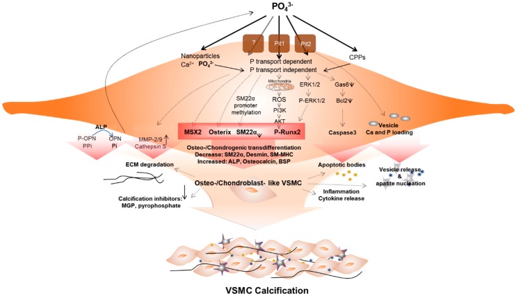
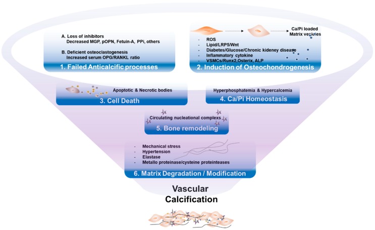
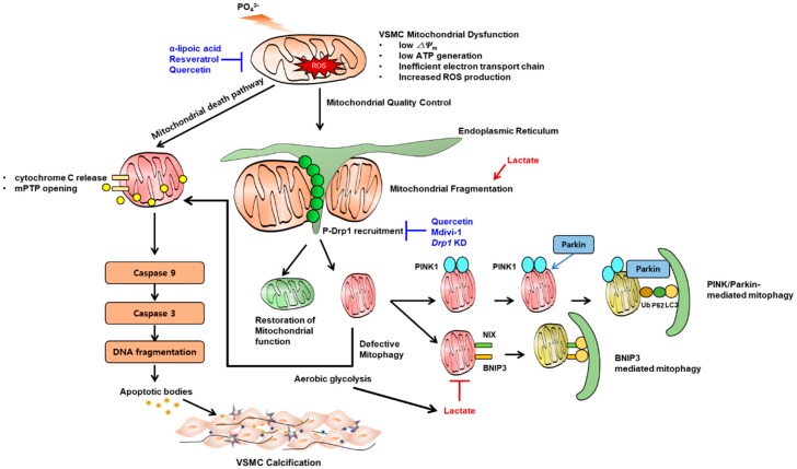
Vascular calcification (VC), which is categorized by intimal and medial calcification, depending on the site(s) involved within the vessel, is closely related to cardiovascular disease. Specifically, medial calcification is prevalent in certain medical situations, including chronic kidney disease and diabetes. The past few decades have seen extensive research into VC, revealing that the mechanism of VC is not merely a consequence of a high-phosphorous and -calcium milieu, but also occurs via delicate and well-organized biologic processes, including an imbalance between osteochondrogenic signaling and anticalcific events. In addition to traditionally established osteogenic signaling, dysfunctional calcium homeostasis is prerequisite in the development of VC. Moreover, loss of defensive mechanisms, by microorganelle dysfunction, including hyper-fragmented mitochondria, mitochondrial oxidative stress, defective autophagy or mitophagy, and endoplasmic reticulum (ER) stress, may all contribute to VC. To facilitate the understanding of vascular calcification, across any number of bioscientific disciplines, we provide this review of a detailed updated molecular mechanism of VC. This encompasses a vascular smooth muscle phenotypic of osteogenic differentiation, and multiple signaling pathways of VC induction, including the roles of inflammation and cellular microorganelle genesis.

Keywords: autophagy; chronic kidney disease; endoplasmic reticulum; hyperphosphatemia; inflammation; matrix vesicle; mitochondrial dysfunction; osteogenic differentiation; vascular calcification; vascular smooth muscle cell.


Conflict of interest statement

The authors declare no conflict of interest.

Figures

Figure 1
Figure 1
Vascular smooth muscle cells (VSMCs) under hyperphosphatemic conditions. Hyperphosphatemic milieu affects VSMC cellular fate by delivering phosphate (P) into VSMC via phosphate transport (Pit1 or Pit2) dependent, independent (as nanoparticles) or as a form of calciprotein particle (CPP). Followed by diverse signaling pathways that enhance sensitivity of VSMCs to calcification, phenotypic differentiation into osteogenic/chondroblast-like VSMCs occurs. This process includes signaling pathways that induce expression of the osteogenic transcription factors Msh Homeobox 2 (MSX2), Osterix, runt-related transcription factor 2 (Runx2), and alkaline phosphatase (ALP). These changes acceleratedly reduce levels of calcification inhibitors. In addition, ROS generated by P-induced mitochondrial dysfunction activates Runx2 via phosphoinositide 3-kinase (PI3K)/protein kinase B (PKB or AKT) signaling and increased apoptosis promote apoptotic bodies or vesicle release. In addition, extracellular matrix (ECM) degradation and inflammatory cytokine releases are increased. These factors create pro-calcifying environment contributing to vascular calcification.
Figure 2
Figure 2
Key mechanisms of vascular calcification. (1) Failure of anti-calcification processes, due to loss of inhibitors and deficiency of constitutively expressed mineralization inhibitors, leads to vascular calcification. (2) Various stressors induce osteogenic transdifferentiation of VSMCs, products of matrix vesicles, which act as a nidus of calcium phosphate deposition. (3) Cell death by apoptosis or necrosis leads to release of apoptotic bodies, or necrotic debris, which may act as nucleation of apatite. (4) Abnormal mineral homeostasis causes deposits calcium phosphate hydroxyapatite. (5) Nucleational complexes formed during bone remodeling, promote ectopic mineralization. (6) Matrix degradation/modifications, caused by environmental stressors, are involved in vascular calcification.
Figure 3
Figure 3
Key mechanism by which mitochondrial dysfunction contributes to vascular calcification. High phosphate induces vascular smooth muscle cell (VSMC) mitochondrial dysfunction represented by low mitochondrial membrane potential, low ATP synthesis and increased reactive oxygen species as a result of inefficient electron transport chain. This leads to release of mitochondrial permeability transition pore (mPTP) and release of cytochrome C which in turn, induces a subsequent cascade of caspase-9 and caspase-3 activation and apoptosis. Mitochondria require quality control in the presence of mitochondrial stress. The representative phenomenon is mitochondrial fission which is mainly mediated by phosphorylated Drp1 recruitment on the site of fission. Once fragmented, dysfunctional mitochondria undergo mitophagy (canonical PINK1-parkin mediated or BCL2-Interacting Protein 3 (BNIP3) mediated). When mitophagic clearance is defective, dysfunctional mitochondria undergo apoptosis. Dysfunctional mitochondria tend to rely on aereobic glycolysis rather than oxidative phosphorylation which might further produce lactate. Lactate promotes mitochondrial fission and blocks mitophagy, both of which promote apoptosis. Some chemicals are preventive in experimental models of vascular calcification (shown in blue text, also see main text).

References

    1. Wu M., Rementer C., Giachelli C.M. Vascular calcification: An update on mechanisms and challenges in treatment. Calcif. Tissue Int. 2013;93:365–373. doi: 10.1007/s00223-013-9712-z. - DOI - PMC - PubMed
    1. Giachelli C.M. Vascular calcification mechanisms. J. Am. Soc. Nephrol. 2004;15:2959–2964. doi: 10.1097/01.ASN.0000145894.57533.C4. - DOI - PubMed
    1. Mizobuchi M., Towler D., Slatopolsky E. Vascular calcification: The killer of patients with chronic kidney disease. J. Am. Soc. Nephrol. 2009;20:1453–1464. doi: 10.1681/ASN.2008070692. - DOI - PubMed
    1. Toussaint N.D., Kerr P.G. Vascular calcification and arterial stiffness in chronic kidney disease: Implications and management. Nephrology (Carlton) 2007;12:500–509. doi: 10.1111/j.1440-1797.2007.00823.x. - DOI - PubMed
    1. Jacob M.P. Extracellular matrix remodeling and matrix metalloproteinases in the vascular wall during aging and in pathological conditions. Biomed. Pharmacother. 2003;57:195–202. doi: 10.1016/S0753-3322(03)00065-9. - DOI - PubMed

LinkOut - more resources