. 2020 Apr 14;11(4):412.
doi: 10.3390/mi11040412.

Differential Sorting of Microparticles Using Spiral Microchannels with Elliptic Configurations

Affiliations

Differential Sorting of Microparticles Using Spiral Microchannels with Elliptic Configurations

Kaan Erdem et al. Micromachines (Basel). .

Abstract

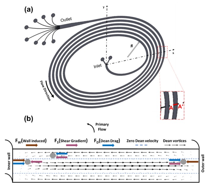
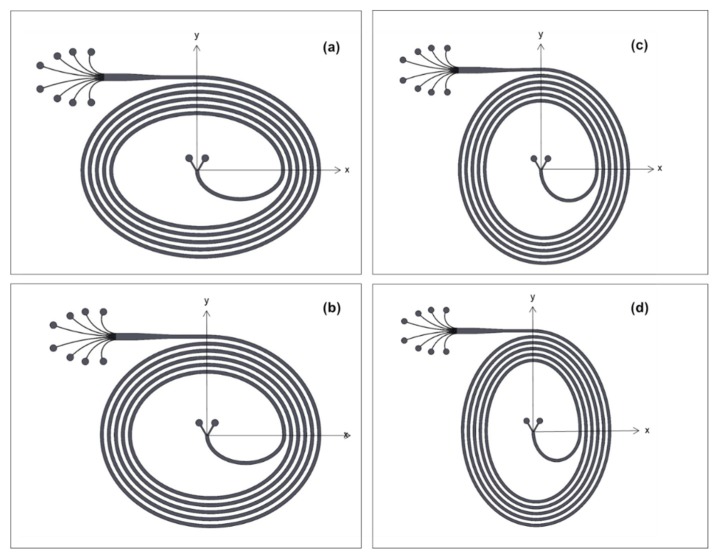
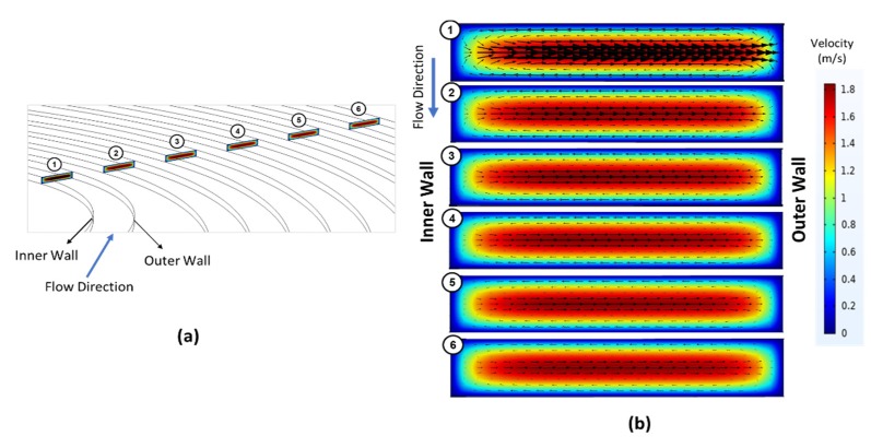
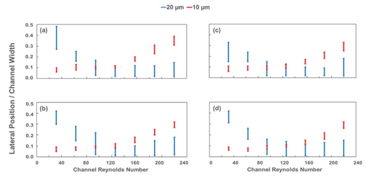
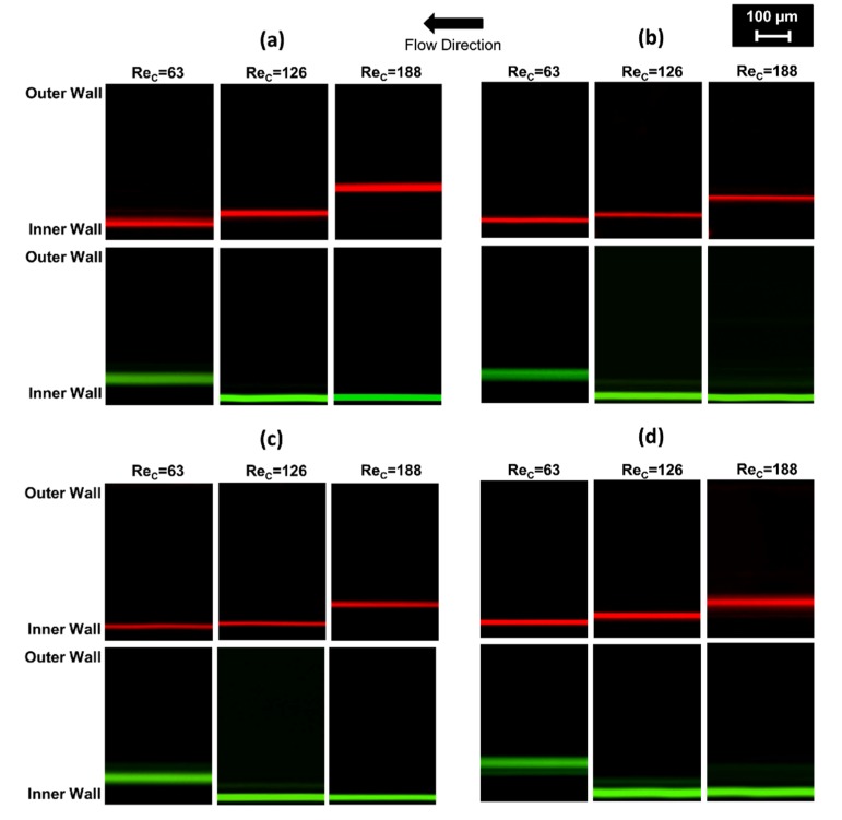
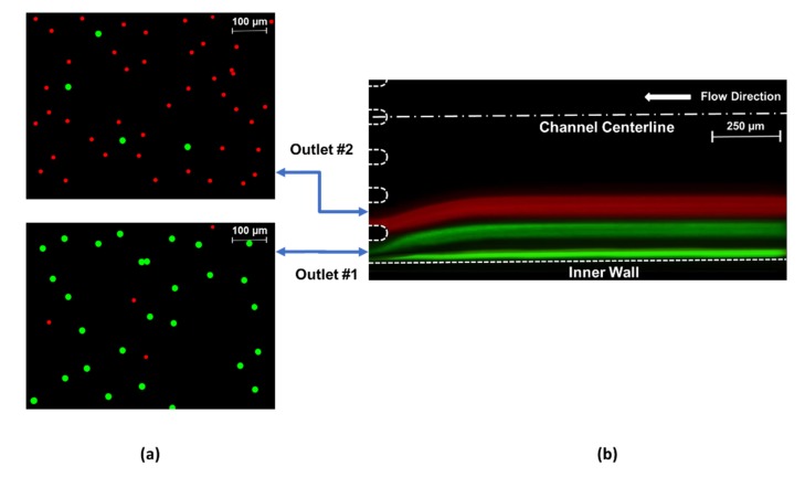
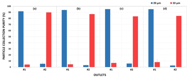
Label-free, size-dependent cell-sorting applications based on inertial focusing phenomena have attracted much interest during the last decade. The separation capability heavily depends on the precision of microparticle focusing. In this study, five-loop spiral microchannels with a height of 90 µm and a width of 500 µm are introduced. Unlike their original spiral counterparts, these channels have elliptic configurations of varying initial aspect ratios, namely major axis to minor axis ratios of 3:2, 11:9, 9:11, and 2:3. Accordingly, the curvature of these configurations increases in a curvilinear manner through the channel. The effects of the alternating curvature and channel Reynolds number on the focusing of fluorescent microparticles with sizes of 10 and 20 µm in the prepared suspensions were investigated. At volumetric flow rates between 0.5 and 3.5 mL/min (allowing separation), each channel was tested to collect samples at the designated outlets. Then, these samples were analyzed by counting the particles. These curved channels were capable of separating 20 and 10 µm particles with total yields up to approximately 95% and 90%, respectively. The results exhibited that the level of enrichment and the focusing behavior of the proposed configurations are promising compared to the existing microfluidic channel configurations.

Keywords: fluorescent particle separation; inertial focusing; microfluidics; spiral microchannels.


Conflict of interest statement

The authors declare no conflict of interest.

Figures

Figure 1
Figure 1
(a) Schematic of an elliptic spiral microchannel shown in a Cartesian plane including a radius of curvature, R at a random position, and the location of the A-A’ cross-section. (b) A close-up view of the A-A’ plane illustrating secondary flow-induced Dean vortices, zero Dean velocity lines, and a force diagram demonstrating the directions of wall-induced lift force (FW), shear gradient lift force (FS), and Dean drag force (FD) acting on a particle at various locations.
Figure 2
Figure 2
Schematics of the elliptic spiral microchannels: (a) Case 1 (initial aspect ratio (IAR): 3:2), (b) Case 2 (IAR: 11:9), (c) Case 3 (IAR: 9:11) and (d) Case 4 (IAR: 2:3).
Figure 3
Figure 3
(a) Six different cut-plane positions labeled starting from the inner loop along the primary flow direction, and (b) a closer look at the vectoral variation of lateral velocity distributions over the colored velocity profile of the primary flow at six different cross-sections throughout the channel. The cross-sectional velocity profiles were retrieved form COMSOL Multiphysics simulation results of Case 1 at volumetric flow rate of 3 mL/min.
Figure 4
Figure 4
The variations of Dean velocity distribution at the beginning and at end of the last quarter loop of the channel for Case 1 (a,b), Case 2 (c,d), Case 3 (e,f), and Case 4 (g,h), respectively. The arrow lengths are proportional to the lateral velocity.
Figure 5
Figure 5
Lateral focusing position from the inside wall normalized by the channel exit width of 1 mm for (a) Case 1, (b) Case 2, (c) Case 3, and (d) Case 4 at the outlet.
Figure 6
Figure 6
The trajectories of the particle streamlines at the end of the last spiral loop, where the channel width is 500 µm for 10 µm (red lines) and 20 µm (green lines) at Rec values of 63, 126, and 188 for (a) Case 1, (b) Case 2, (c) Case 3, and (d) Case 4.
Figure 7
Figure 7
(a) The fluorescent microscopic views of samples utilized for particle counting and (b) the superimposed fluorescent image of two focused streamlines: red (10 µm) and green (20 µm) illustrating particle migration for Case 4 at the optimum flow conditions (approximately Rec 195).
Figure 8
Figure 8
The particle separation purities obtained from the ratio of collected particles at particular outlets to the total collected particles for outlets #1 and #2 in Case 1 (a), Case 2 (b), Case 3 (c), and Case 4 (d).

References

    1. Mach A.J., Di Carlo D. Continuous scalable blood filtration device using inertial microfluidics. Biotechnol. Bioeng. 2010;107:302–311. doi: 10.1002/bit.22833. - DOI - PubMed
    1. Ozbey A., Karimzadehkhouei M., Kocaturk N., Bilir S.E., Kutlu O., Gozuacik D., Koşar A. Inertial focusing of cancer cell lines in curvilinear microchannels. Micro Nano Eng. 2019;2:53–63. doi: 10.1016/j.mne.2019.01.002. - DOI
    1. Su W., Gao X., Jiang L., Qin J. Microfluidic platform towards point-of-care diagnostics in infectious diseases. J. Chromatogr. A. 2015;1377:13–26. doi: 10.1016/j.chroma.2014.12.041. - DOI - PubMed
    1. Martel J.M., Smith K.C., Dlamini M., Pletcher K., Yang J., Karabacak M., Haber D.A., Kapur R., Toner M. Continuous Flow Microfluidic Bioparticle Concentrator. Sci. Rep. 2015;5:11300. doi: 10.1038/srep11300. - DOI - PMC - PubMed
    1. Augustsson P., Magnusson C., Nordin M., Lilja H., Laurell T. Microfluidic, label-Free enrichment of prostate cancer cells in blood based on acoustophoresis. Anal. Chem. 2012;84:7954–7962. doi: 10.1021/ac301723s. - DOI - PMC - PubMed

LinkOut - more resources