. 2020 Apr 14;8(2):184.
doi: 10.3390/vaccines8020184.

The Design and Development of a Multi-HBV Antigen Encoded in Chimpanzee Adenoviral and Modified Vaccinia Ankara Viral Vectors; A Novel Therapeutic Vaccine Strategy against HBV

Affiliations

The Design and Development of a Multi-HBV Antigen Encoded in Chimpanzee Adenoviral and Modified Vaccinia Ankara Viral Vectors; A Novel Therapeutic Vaccine Strategy against HBV

Senthil K Chinnakannan et al. Vaccines (Basel). .

Abstract

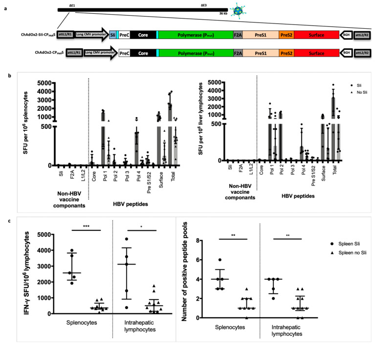
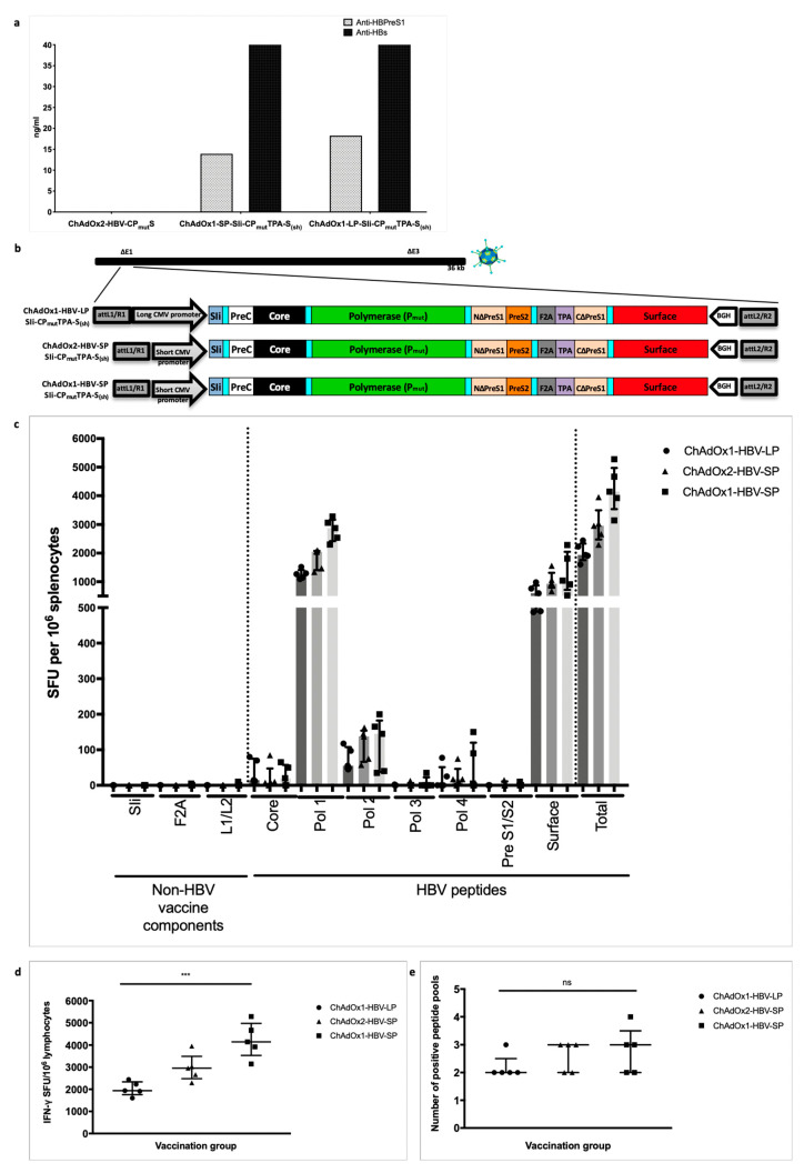
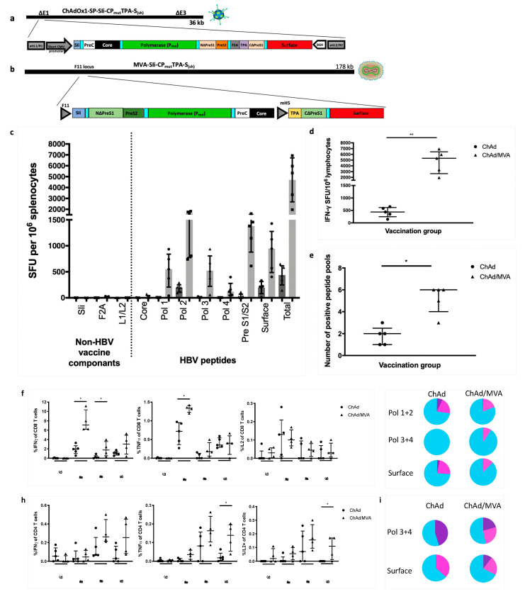
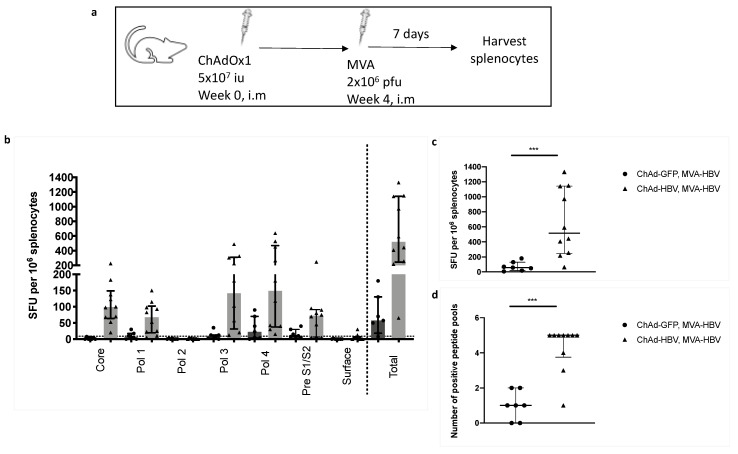
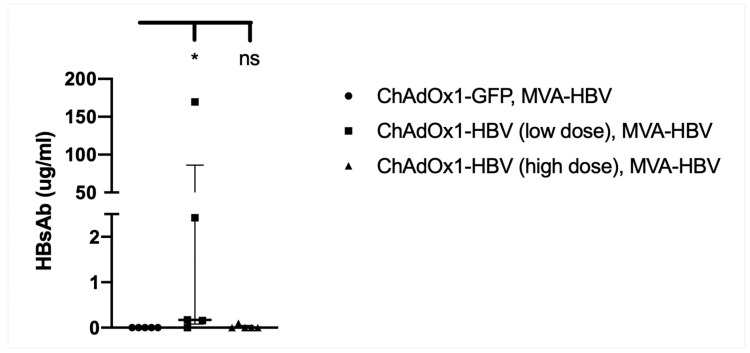
Chronic hepatitis B virus (HBV) infection affects 257 million people globally. Current therapies suppress HBV but viral rebound occurs on cessation of therapy; novel therapeutic strategies are urgently required. To develop a therapeutic HBV vaccine that can induce high magnitude T cells to all major HBV antigens, we have developed a novel HBV vaccine using chimpanzee adenovirus (ChAd) and modified vaccinia Ankara (MVA) viral vectors encoding multiple HBV antigens. ChAd vaccine alone generated very high magnitude HBV specific T cell responses to all HBV major antigens. The inclusion of a shark Invariant (SIi) chain genetic adjuvant significantly enhanced the magnitude of T-cells against HBV antigens. Compared to ChAd alone vaccination, ChAd-prime followed by MVA-boost vaccination further enhanced the magnitude and breadth of the vaccine induced T cell response. Intra-cellular cytokine staining study showed that HBV specific CD8+ and CD4+ T cells were polyfunctional, producing combinations of IFNγ, TNF-α, and IL-2. In summary, we have generated genetically adjuvanted ChAd and MVA vectored HBV vaccines with the potential to induce high-magnitude T cell responses through a prime-boost therapeutic vaccination approach. These pre-clinical studies pave the way for new studies of HBV therapeutic vaccination in humans with chronic hepatitis B infection.

Keywords: ChAd; ChAdOx1; Hepatitis B virus (HBV); MVA; T cell vaccine; chimpanzee adenovirus; modified vaccinia Ankara; therapeutic HBV vaccine.


Conflict of interest statement

The authors declare no conflict of interest.

Figures

Figure 1
Figure 1
Selection of a patient’s hepatitis B virus (HBV) sequence for HBV genotype C immunogen design. 1447 HBV genotype C whole genome sequences downloaded from hepatitis B Virus database (HBVdb), were aligned using MAFFT and used to generate a consensus sequence for HBV genotype C (phylogenetic tree of all sequences shown in (a). The pairwise distance between each sequence in the alignment and the consensus sequence was calculated. The sequence isolated from a person infected with HBV with the closest amino acid sequence to the consensus was selected (accession number: KJ173426 HBV isolate C2). The phylogenetic tree shows the relationship of the chosen sequence (KJ173426 highlighted red) with all downloaded HBV genotype C sequences in (a) and to other closely related sequences (b). Sequence accession number KP017269 is not derived from an infected person (it is a consensus sequence) and so was disregarded. Alignment of precore/core/polymerase/large-surface protein of the consensus and selected KJ173426 sequence (c) showed one amino acid difference, at positions 321 in polymerase protein (leucine (L) in HBV genotype C consensus and phenylalanine (F) in KJ173426 HBV isolate C2), highlighted by grey box with red letters.
Figure 2
Figure 2
HBV immunogen design and in vitro protein expression analysis. (a) HBV viral genome and codon layout (a, i) (illustration from [25]). HBV genome comprise of a partially double stranded circular DNA, of approximately 3.2 kilobase (kb) pairs. It encodes 4 coding regions; precore along with core, polymerase, surface proteins (three forms of the surface proteins, L, M, and S, where the L-form is composed on PreS1, PreS2, and S, the M-form is composed of PreS2 and S) and x-protein. HBV immunogens SIi-CPmutS (a, ii) and SIi-SCPmut (a, iii) were designed to encode HBV precore (PreC), core, polymerase (Pmut), PreS1, PreS2, and surface proteins and non-HBV regions (comprising of a truncated shark Invariant chain (SIi), two linkers and a Furin 2A (F2A) peptide sequence). The preS1/preS2/surface region was positioned at carboxy-terminus of layout 1 and at the amino-terminus of layout 2. Within the mammalian expression cassette, the immunogen sequence was placed in between a long CMV promoter and BGH poly sequence. (b) Plasmids encoding SIi-CPmutS and SIi- SCPmut were transfected into HEK293A cells. 24 h post-transfection, cells were lysed and the lysates were analysed in western blot experiments using mouse anti-HBV-PreS1 and mouse anti-HBV-Polymerase antibodies. Blots probed with mouse anti-GAPDH served as loading controls. Lane 1: cell lysate from un-transfected cells, lane 2 and lane 3: cell lysates from cells transfected with plasmids encoding SIi-CPmutS and SIi-SCPmut, respectively.
Figure 3
Figure 3
Tethering transmembrane region of shark invariant chain to the HBV immunogen enhances the magnitude and breadth of vaccine induced T cell responses. Long-CMV promoter (LP) based mammalian expression cassettes, encoding SIi-CPmutS or CPmutS immunogens, were inserted into E1 locus of replication deficient ChAdOx2 vector and recombinant ChAdOx2-SIi-CPmutS and ChAdOx2-CPmutS viruses were generated in T-REx™-293 cells (a). CD1 mice were vaccinated intramuscularly with 5 × 10⁷ infectious units of ChAdOx2 encoding either SIi-CPmutS (n = 5) or CPmutS (n= 9–10. Splenocytes (left panel) and intrahepatic lymphocytes (right panel) were extracted 14 days after vaccination and plated in interferon-gamma (IFNγ) ELISpot assays with pools of overlapping peptides corresponding to the vaccine immunogen (b). T cell response magnitude was measured by the number of IFNγ spot forming units (SFU) per million lymphocytes (c, left panel) and breath was measured by the number of positive peptide pools (c, right panel, over 100 IFNγ SFU/million cells defined as positive). Median and interquartile ranges are shown. Mann–Whitney tests were used for statistical comparison of medians between groups. * p < 0.05, ** p < 0.01, *** p < 0.001.
Figure 4
Figure 4
Second-generation HBV immunogen SIi-CPmutTPA-S(sh) design and immunogenicity of chimpanzee adenovirus (ChAd) vaccine candidates in inbred Balb/c mice. Supernatants from chimp-adenoviral HBV vaccine infected HEK293A cells were harvested 24 h post-infection and quantified in PreS1 and HBs antigen capture ELISA. PreS1 and S antigen quantities based on commercial standard recombinant HBV surface antigen (PIP002, Biorad) obtained from different chimp-adenoviral HBV vaccines, above background value are shown (a). Long-CMV promoter (LP) or short-CMV promoter (SP) based mammalian expression cassettes, encoding SIi-CPmutTPA-S(sh) immunogen were inserted into E1 locus of replication deficient ChAdOx1 or ChAdOx2 vector and four recombinant ChAdOx1-LP-SIi-CPmutTPA-S(sh), ChAdOx1-SP-SIi-CPmutTPA-S(sh), ChAdOx2-LP-SIi-CPmutTPA-S(sh) and ChAdOx2-SP-SIi-CPmutTPA-S(sh) viruses were generated in T-REx™-293 cells (b). Subsequently, all four recombinant viruses were screened by PCR, for stability analysis. Except ChAdOx2-LP-SIi-CPmutTPA-S(sh), other three viruses showed positive PCR result for the presence of an intact HBV-immunogen cassette. BALB/c mice were vaccinated intramuscularly with 5 × 10⁷ infectious units of ChAdOx1-LP-SIi-CPmutTPA-S(sh), or ChAdOx1-SP-SIi-CPmutTPA-S(sh) or ChAdOx2-SP-SIi-CPmutTPA-S(sh) (n = 5 per group). Splenocytes were extracted 14 days after vaccination and plated in IFNγ ELISpot assays with pools of overlapping peptides corresponding to the vaccine immunogen (c). T cell response magnitude was measured by the number of IFNγ SFU per million lymphocytes (d) and breath was measured by the number of positive peptide pools (e) over 100 IFNγ SFU/million cells defined as positive). Median and interquartile ranges are shown. Kruskal–Wallis tests were used for statistical comparison of the three vaccine groups. *** p < 0.001, ns = not significant.
Figure 5
Figure 5
Mutant HBV polymerase (Pmut) encoded by the chimpanzee-adenovirus and modified vaccinia Ankara (MVA) viral vectored HBV vaccines is non-functional. Schematic representation of wild-type (Pwt-1/FLAG-Pwt-2) and mutant (FLAG-Pmut, FLAG-PΔmut, FLAG-PmutΔYMDD) HBV polymerases used to evaluate the possibility of abolishing the functionality of the HBV polymerase encoded in the vaccine using eight-point mutations (FLAG-Pmut; alanine substitutions at positions Y63, C323, C334, C338, C352, R714, D777, R781, represented by red stars) on its own, or in combination with either 193–326 spacer region deletion (FLAG-PmutΔ193–326) or 549–552 YMDD motif deletion (FLAG-PmutΔYMDD) (a). Mammalian expression plasmids, each encoding a wild-type (Pwt-1 and FLAG-Pwt-2) or a mutant (FLAG-Pmut, FLAG-PmutΔ193–326, FLAG-PmutΔYMDD) HBV polymerase, were generated and tested in a trans-complementation assay that requires co-transfection of a plasmid encoding functional HBV-polymerase, to rescue the replication of polymerase deficient HBV-genome (encoded via plasmid, pCH-9/3091-P11) (b). Plasmid constructs, as indicated in the top of the figure, were co-transfected into Huh7 cells. 4 days post-transfection cells were lysed, capsids were isolated and the levels of capsid protein and capsid DNA from each sample were analyzed in Western blot and Southern blot, using anti-capsid antibody and 32P-labelled HBV specific probe, respectively. Western blot probed with anti-FLAG antibody confirmed equivalent level of expression HBV polymerase in samples receiving FLAG-Pwt-2, FLAG-Pmut, FLAG-PmutΔ193–326, and FLAG-PmutΔYMDD.
Figure 6
Figure 6
Priming with ChAdOx1-SP-SIi-CPmutTPA-S(sh) and boosting with MVA-SIi-CPmutTPA-S(sh) enhances the magnitude of vaccine induced T cell responses. C57BL/6J mice were vaccinated intramuscularly with 5 × 10⁷ IU of ChAdOx1-SP-SIi-CPmutTPA-S(sh) alone (ChAd, n = 5, construct shown in (a)) or followed 7–8 weeks later by 2 × 10⁶ plaque forming units of MVA-SIi-CPmutTPA-S(sh) (MVA, n = 5, construct shown in (b)). Splenocytes extracted 14 days after vaccination and plated in IFNγ ELISpot assays with pools of overlapping peptides corresponding to the vaccine immunogen (c). T cell response magnitude was measured by the number of IFNγ SFU per million lymphocytes (d) and breath was measured by the number of positive peptide pools (e) (over 100 IFNγ SFU/million cells defined as positive). Splenocytes extracted 14 days after vaccination were plated with pools of overlapping peptides corresponding to the vaccine immunogen and stained intracellularly for detection of cytokine production, shown as percentage of CD8+ cells producing IFNγ (left panel), TNFα (middle panel) or IL-2 (right panel) (f), proportion of CD8+ T cells that are single (blue), double (pink) or triple (purple) cytokine producers (g) percentage of CD4+ cells producing IFNγ (left panel), TNFα (middle panel) or IL-2 (right panel) (h) and proportion of CD4+ T cells that are single (blue), double (pink) or triple (purple) cytokine producers (i). Median and interquartile ranges are shown. Mann–Whitney tests were used for statistical comparison of medians between groups. * p < 0.05, ** p < 0.01.
Figure 7
Figure 7
ChAd-MVA prime-boost vaccination in HHD mice induces T-cell response to HBV-core peptides. HHD mice were vaccinated intramuscularly (i.m) with 5 × 10⁷ iu (infectious units) of ChAdOx1-SP-SIi-CPmutTPA-S(sh) or ChAdOx1-GFP as a negative control, followed 4 weeks later by 2 × 10⁶ pfu (plaque forming units) of MVA-SIi-CPmutTPA-S(sh) (a). T cell response magnitude was measured by the number of IFNγ SFU per million lymphocytes 7 days after MVA vaccination and plated in IFNγ ELISpot assays with pools of overlapping peptide corresponding to the vaccine immunogen (b and c) and breath was measured by the number of positive peptide pools (d) (over 20 IFNγ SFU/million cells defined as positive). Median and interquartile ranges are shown. Mann–Whitney tests were used for statistical comparison of medians between groups. *** p < 0.001.
Figure 8
Figure 8
Vaccination with MVA-SIi-CPmutTPA-S(sh) 7–8 weeks after ChAdOx1-SP-SIi-CPmutTPA-S(sh) induces HBs-antibody. C57BL6 mice, n = 5 per group, were given intramuscularly injections with 5 × 10⁵ IU per mice (low dose) or 5 × 10⁷ IU per mice (high dose) of ChAdOx1-SP-SIi-CPmut-TPA-S(sh) at week 0 followed by 2 × 10⁶ pfu per mice of MVA-SIi-HBV-CPmutTPA-Ssh at week 7–8. The third group of C57BL6 mice (n = 5), received 5 × 10⁷ IU per mice of ChAdOx1-GFP at week 0 followed by 2 × 10⁶ pfu per mice of MVA-HBV at week 8. Sera were collected 14 days post MVA vaccination and the levels of anti-HBs induction in response to vaccination was quantified by ELISA. Anti-HBs quantitation based on commercial standard mouse monoclonal antibodies to surface (GeneTex, GTX40707) above background value of naïve un-vaccinated sera are shown. Median and interquartile ranges are shown. The Kruskal–Wallis test and Dunn’s multiple comparisons test were used for statistical comparison of medians between groups. * p < 0.05, ns = not significant.

References

    1. Chan H.L., Chan C.K., Hui A.J., Chan S., Poordad F., Chang T.T., Mathurin P., Flaherty J.F., Lin L., Corsa A., et al. Effects of tenofovir disoproxil fumarate in hepatitis B e antigen-positive patients with normal levels of alanine aminotransferase and high levels of hepatitis B virus DNA. Gastroenterology. 2014;146:1240–1248. doi: 10.1053/j.gastro.2014.01.044. - DOI - PubMed
    1. Li M.R., Xi H.L., Wang Q.H., Hou F.Q., Huo N., Zhang X.X., Li F., Xu X.Y. Kinetics and prediction of HBsAg loss during long-term therapy with nucleos(t)ide analogues of different potency in patients with chronic hepatitis B. PLoS ONE. 2014;9:e98476. doi: 10.1371/journal.pone.0098476. - DOI - PMC - PubMed
    1. Papatheodoridis G.V., Chan H.L., Hansen B.E., Janssen H.L., Lampertico P. Risk of hepatocellular carcinoma in chronic hepatitis B: Assessment and modification with current antiviral therapy. J. Hepatol. 2015;62:956–967. doi: 10.1016/j.jhep.2015.01.002. - DOI - PubMed
    1. Wang J.P., Kao F.Y., Wu C.Y., Hung Y.P., Chao Y., Chou Y.J., Li C.P. Nucleos(t)ide analogues associated with a reduced risk of hepatocellular carcinoma in hepatitis B patients: A population-based cohort study. Cancer. 2015;121:1446–1455. doi: 10.1002/cncr.29159. - DOI - PubMed
    1. Wu C.Y., Lin J.T., Ho H.J., Su C.W., Lee T.Y., Wang S.Y., Wu C., Wu J.C. Association of nucleos(t)ide analogue therapy with reduced risk of hepatocellular carcinoma in patients with chronic hepatitis B: A nationwide cohort study. Gastroenterology. 2014;147:143–151.e145. doi: 10.1053/j.gastro.2014.03.048. - DOI - PubMed

LinkOut - more resources