. 2020 Apr 14;12(4):356.
doi: 10.3390/pharmaceutics12040356.

Characterization and Mathematical Modeling of Alginate/Chitosan-Based Nanoparticles Releasing the Chemokine CXCL12 to Attract Glioblastoma Cells

Affiliations

Characterization and Mathematical Modeling of Alginate/Chitosan-Based Nanoparticles Releasing the Chemokine CXCL12 to Attract Glioblastoma Cells

Suzanne Gascon et al. Pharmaceutics. .

Erratum in

Abstract

Chitosan (Chit) currently used to prepare nanoparticles (NPs) for brain application can be complexed with negatively charged polymers such as alginate (Alg) to better entrap positively charged molecules such as CXCL12. A sustained CXCL12 gradient created by a delivery system can be used, as a therapeutic approach, to control the migration of cancerous cells infiltrated in peri-tumoral tissues similar to those of glioblastoma multiforme (GBM). For this purpose, we prepared Alg/Chit NPs entrapping CXCL12 and characterized them. We demonstrated that Alg/Chit NPs, with an average size of ~250 nm, entrapped CXCL12 with ~98% efficiency for initial mass loadings varying from 0.372 to 1.490 µg/mg NPs. The release kinetic profiles of CXCL12 were dependent on the initial mass loading, and the released chemokine from NPs after seven days reached 12.6%, 32.3%, and 59.9% of cumulative release for initial contents of 0.372, 0.744, and 1.490 µg CXCL12/mg NPs, respectively. Mathematical modeling of released kinetics showed a predominant diffusive process with strong interactions between Alg and CXCL12. The CXCL12-NPs were not toxic and did not promote F98 GBM cell proliferation, while the released CXCL12 kept its chemotaxis effect. Thus, we developed an efficient and tunable CXCL12 delivery system as a promising therapeutic strategy that aims to be injected into a hydrogel used to fill the cavity after surgical tumor resection. This system will be used to attract infiltrated GBM cells prior to their elimination by conventional treatment without affecting a large zone of healthy brain tissue.

Keywords: alginate; cell migration; chemokine; chitosan; delivery system; mathematical modeling; nanoparticles.


Conflict of interest statement

The authors declare no conflict of interest.

Figures

Figure 1
Figure 1
Schematic of the segmentation and counting of cell or nucleus using a U-net convolution neural network.
Figure 2
Figure 2
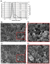
(A) Size distribution in volume % and particle density (number of particle/mL) of Alg/Chit NPs obtained from laser granulometry; 5–10 measurements were taken per experiment and the results are representative of three independent experiments. (B) Representative SEM images of empty NPs (C) and NPs containing CXCL12 (0.744 µg/mg NPs). (D) and (E) are zoom regions highlighted by red squares for NPs and NPs-CXCL12, respectively. White arrows point to individual NPs.
Figure 3
Figure 3
(A) 3D fluorescence scan of CXCL12-AF647 (2 µg/mL). (B) Standard CXCL12-AF647 curve. C) Release kinetics ± SD of CXCL12-AF647 from Alg/Chit NPs for different mass loadings (0.372 µg/mg NPs, 0.744 µg/mg NPs, and 1.490 µg/mg NPs). Results are representative of at least three independent experiments performed in duplicate.
Figure 4
Figure 4
Mathematical modeling of CXCL12 release. (A) Particle size classes used in the modeling process adapted from laser diffraction results. (B) Representative graphic showing the objective function to minimize with respect to the number of generation performed with the evolutionary optimization algorithm. (C) Mathematical modeling of CXCL12 for different initial loading mass. Dashed lines show modeling results. (D) Mathematical modeling results for the first 24 h (zoom from C).
Figure 5
Figure 5
Cytotoxicity of NPs. (A) MTS assay results showing the absorbance measured at 490 nm (±SD), (B) Kinetic of F98 cell proliferation (±SD). (C) Percentage of cell mortality determined from live/dead cell viability assay. Results are representative of three independent experiments performed in duplicate. * p < 0.05, ** p < 0.01, *** p < 0.001.
Figure 5
Figure 5
Cytotoxicity of NPs. (A) MTS assay results showing the absorbance measured at 490 nm (±SD), (B) Kinetic of F98 cell proliferation (±SD). (C) Percentage of cell mortality determined from live/dead cell viability assay. Results are representative of three independent experiments performed in duplicate. * p < 0.05, ** p < 0.01, *** p < 0.001.
Figure 6
Figure 6
Representative Hoechst, segmented Hoechst (Segmented) and live/dead images for F98 cells incubated for 72 h with empty NPs, NPs-CXCL12 (0.744 µg/mg NPs) and CXCL12 (0.8 µg/mL). Bar = 100 µm. Results are representative of three independent experiments performed in duplicate.
Figure 7
Figure 7
(A) Schematic of the invasion chambers used to perform chemotaxis assays. (B) Representative images of crystal violet stained F98 cells located at the porous membrane surface following 6 h of incubation for the following conditions: (1) DMEM 0.1% BSA (CTL); (2) empty NPs; (3) empty NPs 24 h incubation with the extraction of the supernatant + CXCL12 added before seeding the F98 cells; (4) CXCL12 24 h incubation; (5) NPs-CXCL12 24 h incubation and (6) CXCL12 0 h incubation. Each condition always had a theoretical final concentration of 100 ng/mL. (C) Invasion ratio of F98 cells added on the top of a Matrigel layer and allowed to migrate for 6 h for the following conditions: (1) DMEM 0.1% BSA (CTL); (2) empty NPs; (3) NPs-CXCL12 24 h incubation; (4) CXCL12 24 h incubation, and (5) CXCL12 0 h incubation. Results are representative of three to five independent experiments performed in triplicate. *** p < 0.001.

References

    1. Mendes M., Sousa J.J., Pais A., Vitorino C. Targeted Theranostic Nanoparticles for Brain Tumor Treatment. Pharmaceutics. 2018;10:181. doi: 10.3390/pharmaceutics10040181. - DOI - PMC - PubMed
    1. Nagpal K., Singh S.K., Mishra D.N. Optimization of brain targeted chitosan nanoparticles of Rivastigmine for improved efficacy and safety. Int. J. Biol. Macromol. 2013;59:72–83. doi: 10.1016/j.ijbiomac.2013.04.024. - DOI - PubMed
    1. Mohammed M.A., Syeda J.T.M., Wasan K.M., Wasan E.K. An Overview of Chitosan Nanoparticles and Its Application in Non-Parenteral Drug Delivery. Pharmaceutics. 2017;9:53. doi: 10.3390/pharmaceutics9040053. - DOI - PMC - PubMed
    1. Elkadery A.A.S., Elsherif E.A., Ezz Eldin H.M., Fahmy I.A.F., Mohammad O.S. Efficient therapeutic effect of Nigella sativa aqueous extract and chitosan nanoparticles against experimentally induced Acanthamoeba keratitis. Parasitol. Res. 2019;118:2443–2454. doi: 10.1007/s00436-019-06359-x. - DOI - PubMed
    1. Khor E., Lim L.Y. Implantable applications of chitin and chitosan. Biomaterials. 2003;24:2339–2349. doi: 10.1016/S0142-9612(03)00026-7. - DOI - PubMed

LinkOut - more resources