Review
. 2020 Apr 14;21(8):2712.
doi: 10.3390/ijms21082712.

Genotypes and Phenotypes: A Search for Influential Genes in Diabetic Retinopathy

Affiliations
Review

Genotypes and Phenotypes: A Search for Influential Genes in Diabetic Retinopathy

Andrea P Cabrera et al. Int J Mol Sci. .

Abstract

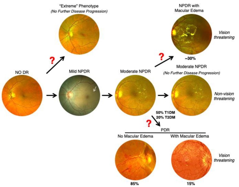
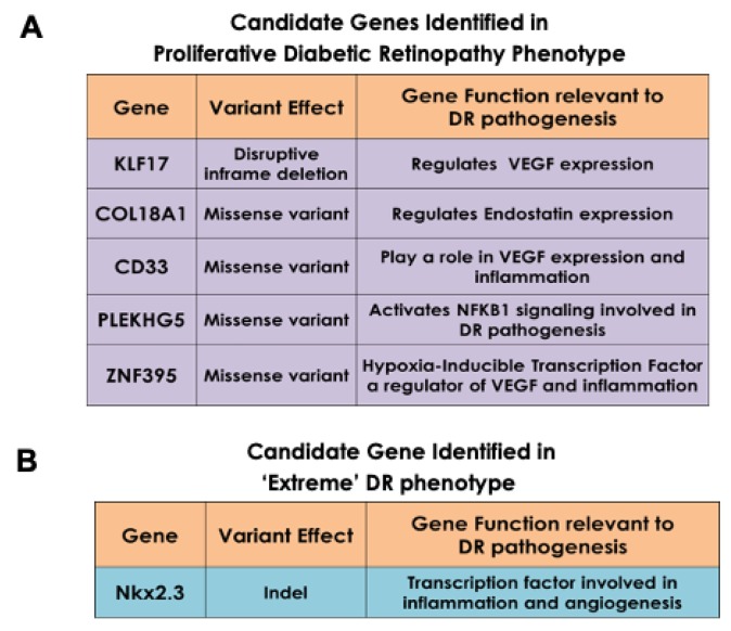
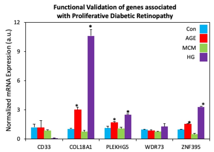
Although gene-environment interactions are known to play an important role in the inheritance of complex traits, it is still unknown how a genotype and the environmental factors result in an observable phenotype. Understanding this complex interaction in the pathogenesis of diabetic retinopathy (DR) remains a big challenge as DR appears to be a disease with heterogenous phenotypes with multifactorial influence. In this review, we examine the natural history and risk factors related to DR, emphasizing distinct clinical phenotypes and their natural course in retinopathy. Although there is strong evidence that duration of diabetes and metabolic factors play a key role in the pathogenesis of DR, accumulating new clinical studies reveal that this disease can develop independently of duration of diabetes and metabolic dysfunction. More recently, studies have emphasized the role of genetic factors in DR. However, linkage analyses, candidate gene studies, and genome-wide association studies (GWAS) have not produced any statistically significant results. Our recently initiated genomics study, the Diabetic Retinopathy Genomics (DRGen) Study, aims to examine the contribution of rare and common variants in the development DR, and how they can contribute to clinical phenotype, rate of progression, and response to available therapies. Our preliminary findings reveal a novel set of genetic variants associated with proangiogenic and inflammatory pathways that may contribute to DR pathogenesis. Further investigation of these variants is necessary and may lead to development of novel biomarkers and new therapeutic targets in DR.

Keywords: diabetes; diabetic retinopathy; genomics; genotype; phenotype.


Conflict of interest statement

The authors declare no conflict of interest.

Figures

Figure 1
Figure 1
Stages of Progressing Nonproliferative Diabetic Retinopathy (NPDR). (A) Fundus photograph of a retina with no retinopathy observed during the first 10 years of diabetes. (B) In Mild NPDR, the first detectible sign of disease consists of a single microaneurysm (arrow, inset). (C) In moderate NPDR, retinal hemorrhages and hard exudates (circled) may be observed. (D) In severe NPDR, >20 intraretinal hemorrhages may be observed throughout the retina. Venous beading and intraretinal microvascular anomalies (IRMA) may be also seen (arrows).
Figure 2
Figure 2
Progressive Stages of Proliferative Diabetic Retinopathy (PDR). (A) The proliferative stage of diabetic retinopathy is marked by neovascularization, visible on the optic nervehead (black arrow). Additionally, preretinal hemorrhage on the superior aspect of the nerve can also be seen (white arrow). (B) A subhyaloid hemorrhage (white arrows) manifests with boat-shaped configuration as it is trapped in the potential space between the posterior hyaloid and the internal limiting membrane. Preretinal hemorrhages can also be observed (black arrows). (C) Another complication occurring in this stage is the formation of a fibrovascular band along the superotemporal arcade, which can contract, causing tractional retinal detachment (white arrows).
Figure 3
Figure 3
Non-center and Center Involving Macular Edema. (A) Optical coherence tomography (OCT) image of the macular region of the retina showing non-center involving macular edema (Central retinal thickness, 244 µm). (B) Corresponding fundus photograph of (A) showing characteristic exudates formed outside of the macular region (marked by dashed circle). (C) OCT image of center involving macular edema with cystic changes within the retina (Central retinal thickness, 453 µm). (D) Corresponding fundus photograph of C showing characteristic hard exudates and edema formed inside the macula (marked by dashed circle).
Figure 4
Figure 4
Coexistence of PDR and DME. (A) Fundus photograph of a PDR patient one day after vitrectomy with endolaser showing the macula without any exudates or edema, (B) Fundus photograph of another PDR patient with laser marks showing absence of any macular edema, (C) Pie chart showing co-existence of proliferative diabetic retinopathy (PDR) and diabetic macular edema (DME). In PDR patients, concurrent DME features were seen in only 15.7% of patients, the remaining 84.3% of PDR patients did not have any concurrent DME. (D) In DME patients, 79.7% of patients did not have any concurrent neovascularization. The remaining 20.3% of DME patients did not exhibit features of PDR. CI; Confidence Interval.
Figure 5
Figure 5
Treatment Algorithm for Diabetic Retinopathy Patients. Flow diagram outlining the various phenotypes of DR which are treated with anti-VEGF injection and laser. Treatment plan is carefully selected based on individual patient. VEGF, Vascular endothelial growth factor; Va, Visual acuity; PRP, Panretinal Photocoagulation VH, Vitreous hemorrhage; TRD, Tractional Retinal Detachment.
Figure 6
Figure 6
Proposed Course of Progression of Diabetic Retinopathy. Based on our clinical observations and large epidemiological studies, we propose that not every DR patient progresses through disease in the same sequence of events. After a period of no diabetes for 10 years, mild NPDR develops and over time advances to moderate NPDR. In some cases, there is no further disease progression. However, 30% of cases develop concurrent DME. About 50% of type 1 diabetics and 20% of type 2 diabetics develop PDR. In PDR patients, 15% develop concurrent macular edema while 85% never develop macular edema. Additionally, about 1–5% diabetic patients never develop retinopathy despite >20 years of diabetes (“Extreme” phenotype). Red question marks indicate divergence points which may be influenced by genetic factors.
Figure 7
Figure 7
Diabetic Nephropathy has a Progressive and Continuous Heterogenous Phenotype. As Stage 1 (normoalbuminuria) of nephropathy progresses to Stage 2 (microalbuminuria), 60% of Stage 2 patients may revert to Stage 1 while 30% of Stage 2 patients progress to Stage 3 macroalbuminuria. The disease further progresses to Stage 4 (renal failure), culminating in Stage 5 (end Stage renal disease with fibrosis). A small number of Stage 1 patients may advance directly to Stage 5 without any signs of albuminuria. This progression may be genetically determined. In diabetic retinopathy, a similar phenomenon may occur where genetic determinates may influence whether patients will advance to PDR and/or DME.
Figure 8
Figure 8
Newly Identified Genetic Variants Associated with Specific Phenotypes of Diabetic Retinopathy. (A) Enrichment of the ‘risk’ allele in five candidate genes in cases of advanced PDR patients revealed a disruptive in-frame deletion and four heterozygous missense variants. (B) A variant in the NKX2.3 gene was shared in the “extreme” DR phenotype (No DR) group.
Figure 9
Figure 9
Functional Validation of genes associated with Proliferative Diabetic Retinopathy. Human retinal endothelial cells treated with advanced glycation end product-BSA (AGE, 500 μg, 3 days; Sigma Aldrich, St. Louis, MO), macrophage conditioned medium (MCM, 1:1 ratio, 24 h), or high glucose medium (HG, 30 mM, 7 days) revealed increased mRNA expression of COL18A1, ZNF395, and PLEKHG5 in AGE and HG-treated cells. * p < 0.05. Bars indicate average ± standard deviation.
Figure 10
Figure 10
NKX2.3 gene associated with ‘extreme’ phenotype leads to mRNA expression changes in angiogenic and inflammatory markers. (A) mRNA expression of NKX2.3 was increased in human retinal endothelial cells (HREC) using varying concentrations of PCDNA plasmid (lo: 5 μg + 3.75 μL, hi: 5 μg + 7.5 μL; 24 h). (B) Overexpression of NKX2.3 in HRECs revealed increased mRNA expression of cell-cell junctional proteins N-cadherin (N-cad) and occludin (OCLN). (C) Changes in pro-angiogenic vascular endothelial growth factor (VEGF), angiopoietin 1 (Ang1) and angiopoietin 2 (Ang2) mRNA expression were also observed. (D) Overexpression of NKX2.3 in HRECs also revealed increased expression of inflammatory markers intracellular adhesion molecule-1 (ICAM-1), vascular cell adhesion molecule-1 (VCAM-1), and cathepsin D (CTS D). * p < 0.05. Bars indicate average ± standard deviation.

References

    1. Paxton W.A., Martin S.R., Tse D., O’Brien T.R., Skurnick J., VanDevanter N.L., Padian N., Braun J.F., Kotler D.P., Wolinsky S.M., et al. Relative resistance to HIV-1 infection of CD4 lymphocytes from persons who remain uninfected despite multiple high-risk sexual exposure. Nat. Med. 1996;2:412–417. doi: 10.1038/nm0496-412. - DOI - PubMed
    1. Liu R., Paxton W.A., Choe S., Ceradini D., Martin S.R., Horuk R., MacDonald M.E., Stuhlmann H., Koup R.A., Landau N.R. Homozygous defect in HIV-1 coreceptor accounts for resistance of some multiply-exposed individuals to HIV-1 infection. Cell. 1996;86:367–377. doi: 10.1016/S0092-8674(00)80110-5. - DOI - PubMed
    1. Vangelista L., Vento S. The Expanding Therapeutic Perspective of CCR5 Blockade. Front. Immunol. 2017;8:1981. doi: 10.3389/fimmu.2017.01981. - DOI - PMC - PubMed
    1. Dobzhansky T. Genetics and the Origin of Species. 1st ed. Columbia University Press; New York, NY, USA: 1937.
    1. Dobzhansky T. Genetics and the Origin of Species. 3rd ed. Columbia University Press; New York, NY, USA: 1951.

MeSH terms