Comparative Study
. 2020 Apr 21;9(8):e015672.
doi: 10.1161/JAHA.119.015672. Epub 2020 Apr 16.

Molecular and Cellular Differences in Cardiac Repair of Male and Female Mice

Affiliations
Comparative Study

Molecular and Cellular Differences in Cardiac Repair of Male and Female Mice

Amanda B Pullen et al. J Am Heart Assoc. .

Abstract

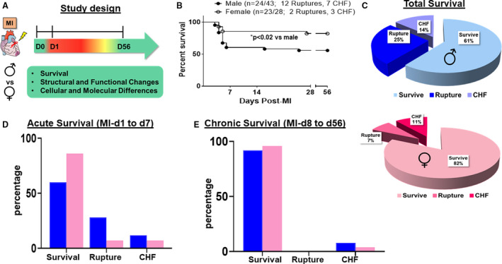
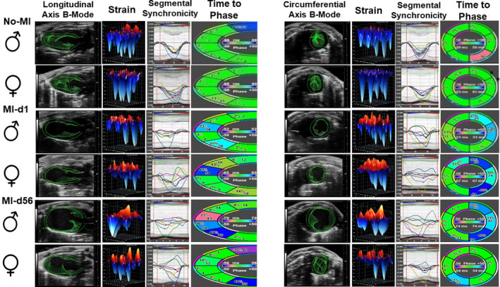
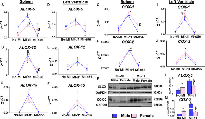
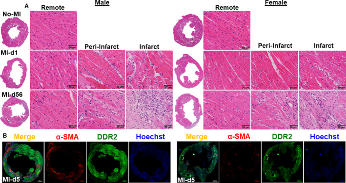
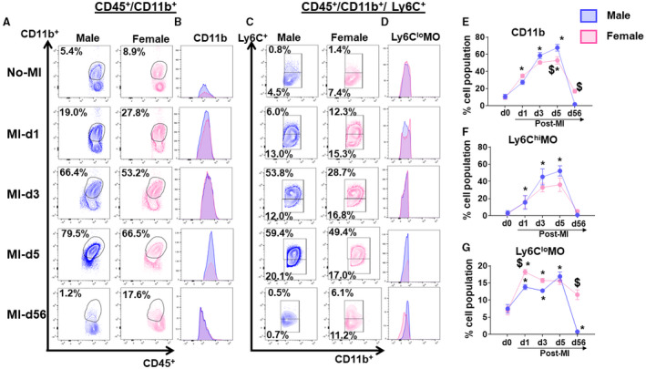
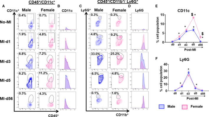
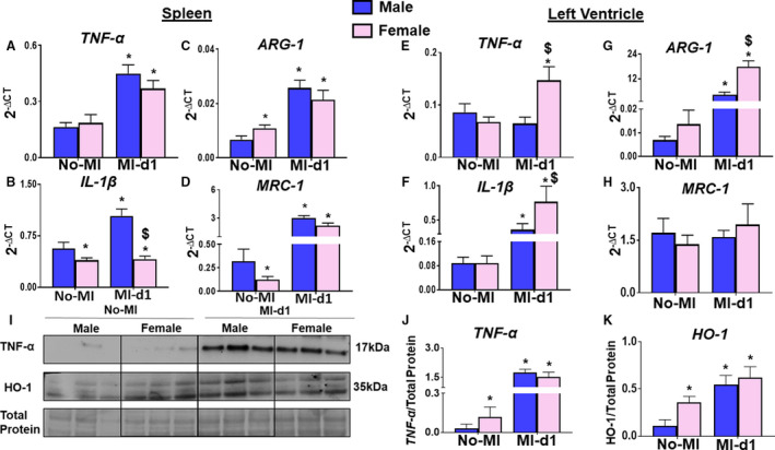
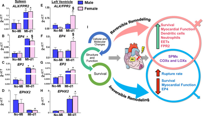
Background Leukocyte-directed biosynthesis of specialized proresolving mediators (SPMs) orchestrates physiological inflammation after myocardial infarction. Deficiency of SPMs drives pathological and nonresolving inflammation, leading to heart failure (HF). Differences in SPMs and inflammatory responses caused by sex-specific differences are of interest. We differentiated leukocyte-directed biosynthesis of lipid mediators in male and female mice, focusing on leukocyte populations, structural remodeling, functional recovery, and survival rates. Methods and Results Risk-free male and female C57BL/6 mice were selected as naïve controls or subjected to myocardial infarction surgery. Molecular and cellular mechanisms that differentiate survival, heart function, and structure and leukocyte-directed lipid mediators were quantified to describe physiological inflammation after myocardial infarction. Female mice show improved survival in acute HF but no statistical difference during chronic HF compared with male mice. Female mice improved survival is marked with functional recovery and limited remodeling compared with male mice. Male and female mice are similarly responsive to arachidonate lipoxygenase (LOX-5, LOX-12, LOX-15) or cyclooxygenase (COX-1, COX-2) in acute HF and particularly male infarcted heart had overall increased SPMs. Female cardiac healing is marked with the biosynthesis of differential p450-derived product, particularly 11,12 epoxyeicosatrienoic acid in acute HF. A sex-specific difference of dendritic cells in acute HF is distinct, with limited changes in chronic HF. Conclusions Cardiac repair is marked with increased SPM biosynthesis in male mice and amplified epoxyeicosatrienoic acid in female mice. Female mice showed improved survival, functional recovery, and limited remodeling, which are signs of fine-tuned physiological inflammation after myocardial infarction. These results rationalize the sex-specific precise therapies and differential treatments in acute and chronic HF.

Keywords: heart failure; ischemia; leukocytes; resolution of inflammation; specialized proresolving mediators.


Figures

Figure 1
Figure 1. Female mice have higher survival than male mice without ruptures or chronic heart failure (CHF) after myocardial infarction (MI).
A, Study design to evaluate differences in male and female C57BL/6J mice after MI in acute heart failure (AHF) and CHF induced by left anterior descending artery ligation. B, Survival rates of both sexes using the Kaplan‐Maier test in AHF and CHF. C, Pie chart showing the survival rates of both sexes. D and E, Bar graph showing the survival rates of both sexes in AHF and CHF. Males (n=43), females (n=28). *P<0.02 vs male analyzed by log‐rank test.
Figure 2
Figure 2. Female mice show better recovery of function and synchronicity from MI‐d1 to MI‐d56.
Speckle tracking‐based echocardiography representatives of male and female mice at No‐MI, MI‐d1, and MI‐d56 (from left: longitudinal axis 2‐dimensional [2D] B‐mode, longitudinal 3‐dimensional [3D] strain, longitudinal segmental synchronicity graph with displayed curves, and longitudinal time to phase; short axis 2D B‐mode, circumferential 3D strain, circumferential segmental synchronicity graph with displayed curves, and circumferential time to phase). Males (n=5‐18), females (n=5‐19).
Figure 3
Figure 3. Both sexes biosynthesized similar amounts of specialized proresolving mediators (SPMs) after cardiac injury.
A, Bar graph showing the docosahexaenoic acid (DHA) series resolvins of the left ventricle at myocardial infarction (MI)‐day (d1 of male and female mice. B, Bar graph showing the arachidonic acid (AA)–derived SPM moieties of the left ventricle at MI‐d1 of male and female mice. C, Pie chart showing the SPMs (resolvins, protectins, maresins), epoxyeicosatrienoic acids (EETs), and AA‐derived SPMs in the left ventricle of male mice at MI‐d1. D, Pie chart showing the SPMs (resolvins, protectins, maresins), EETs, and AA‐derived SPMs in the left ventricle of female mice at MI‐d1. E, Bar graph showing the protectins and maresins of the left ventricle at MI‐d1 of male and female mice. F, Bar graph showing the EETs of the left ventricle at MI‐d1 of male and female mice (MI‐d1: males [n=3], females [n=4]). *P<0.05 vs male MI‐d1.
Figure 4
Figure 4. Male mice have a higher level of lipoxygenase (LOX) expression, whereas female mice express higher cyclooxygenases (COXs) after MI.
A through F, Line graphs of LOXs (ALOX5, ALOX12, ALOX15) in the spleen and left ventricle of male and female mice from No‐MI to MI‐d1. G through J, Bar graphs of COXs (COX1, COX2) in the left ventricle of male and female mice from No‐MI to MI‐d1 (no‐MI: males [n=4], females [n=4]; MI‐d1: males [n=6], females [n=6]). *P<0.05 vs no‐MI naïve controls; $ P<0.05 vs male at respective time point. K through M, Western blot representative and bar graphs of protein expression of ALOX5 and COX2 (n=3/group). *P<0.05 vs no‐MI naïve controls; $ P<0.05 vs male at respective time point.
Figure 5
Figure 5. Female mice display a lower level of α‐smooth muscle action (α‐SMA) compared with male mice at d5 after MI with similar intensity of remodeling.
A, Representative hematoxylin and eosin stained left ventricular (LV) images from d0 naïve control (No‐MI); MI‐d1 is suggestive of acute heart failure (HF), and MI‐d56 is indicative of chronic HF in male and female mice. Representative images of remote (left), peri‐infarct (middle), and infarct area (right) with a magnification of ×40 and accompanying ×1.25 images (n=4‐5 mice/group/day; bar=50 μm). B, Immunofluorescence images indicating reduced α‐SMA (red) and discoidin domain receptor 2 (DDR2; green) expression in LV of females at d5 after MI. Nuclei are stained with Hoechst (blue) (bar=500 μm).
Figure 6
Figure 6. Female mice show higher reparative monocytes compared with male mice after myocardial infarction (MI).
(A) Plot showing cluster of differentiation (CD)11b+ (monocytes) cells at No‐MI, MI‐d3, MI‐d5, and MI‐d56 in male and female mice. (B) Histograms showing monocytes expression at No‐MI, MI‐d3, MI‐d5, and MI‐d56 in male and female mice. (C) Plot showing cluster of Ly6Chi and Ly6Clo cells at No‐MI, MI‐d3, MI‐d5, and MI‐d56 in male and female mice. (D) Histograms showing Ly6Chi and Ly6Clo monocytes expression at No‐MI, MI‐d3, MI‐d5, and MI‐d56 in male and female mice. (E, F, G) Line graphs showing the percentage of the cell population for CD11b, Ly6Chi monocytes (MO), and Ly6CloMO. [No‐MI: males (n=4), females (n=4); MI‐d1: males (n=5), females (n=6); $ P<0.05 vs male at respective time point; *P<0.05 compared with no‐MI naïve controls.
Figure 7
Figure 7. Female mice intensified leukocyte response compared with male mice after myocardial infarction (MI).
(A) Plot showing dendritic cells (CD11c+) at No‐MI, MI‐d3, MI‐d5, and MI‐d56 in male and female mice. (B) Histograms showing dendritic cells at No‐MI, MI‐d3, MI‐d5 and MI‐d56 in male and female mice. (C) Plot showing neutrophils (Ly6G+) at No‐MI, MI‐d3, MI‐d5, and MI‐d56 in male and female mice. (D) Histograms showing neutrophils expression at No‐MI, MI‐d3, MI‐d5 and MI‐d56 in male and female mice. (E, F, G) Line graphs showing the percentage of the cell population of CD11c and Ly6G. [No‐MI: males (n=4), females (n=4); MI‐d1: males (n=5), females (n=6); $ P<0.05 vs male at respective time point; *P<0.05 compared with no‐MI naïve controls.
Figure 8
Figure 8. Male mice have increased expression of cytokines in the spleen, female mice have increased expression of cytokines in the left ventricle.
(A‐D) Bar graphs of increased tumor necrosis factor (TNF)‐α, interleukin (IL)‐1β, Arginase 1 (ARG‐1), Mannose Receptor C‐Type 1 (MRC‐1) in the spleen of male mice from no‐MI to MI‐d1. (E‐H) Bar graphs of increased TNF‐α, IL‐1β, ARG‐1, MRC‐1 in the left ventricle of female mice from no‐MI to MI‐d1. no‐MI: males (n=4), females (n=4); MI‐d1: males (n=6), females (n=6); *P<0.05 compared with no‐MI naïve controls, $ P<0.05 vs male at respective time point. (I‐K) Western representative and bar graphs of expression of TNF‐α and heme oxygenase‐1 (HO1). [(n=3/group); *P<0.05 compared with no‐MI naïve controls, $ P<0.05 vs male at respective time point.
Figure 9
Figure 9. Both sexes have differential expressions of receptors and enzymes in the spleen and left ventricle.
(A‐D) Bar graphs of differential levels of N‐formyl peptide receptor 2(ALX/FPR2), prostaglandin E2 receptor 4 (EP4), Prostaglandin E2 receptor 2 (EP2), Epoxide Hydrolase 2 (EPHX2) in the spleen of male and female mice from no‐MI to MI‐d1. (E‐H) Bar graphs of increased levels of ALX/FPR2, EP4, EP2, EPHX2 in the left ventricle of female mice from no‐MI to MI‐d1. no‐MI: males (n=4), females (n=4); MI‐d1: males (n=6), females (n=6); *P<0.05 vs no‐MI naïve controls, $ P<0.05 vs male at respective time point. (I) Summary sketch showing the overall survival, function, and structure, and cellular and molecular changes between male and female mice in cardiac repair process.

References

    1. Blenck CL, Harvey PA, Reckelhoff JF, Leinwand LA. The importance of biological sex and estrogen in rodent models of cardiovascular health and disease. Circ Res. 2016;118:1294–1312. - PMC - PubMed
    1. Halade GV, Kain V. Obesity and cardiometabolic defects in heart failure pathology. Compr Physiol. 2017;7:1463–1477. - PMC - PubMed
    1. Canto JG, Rogers WJ, Goldberg RJ, Peterson ED, Wenger NK, Vaccarino V, Kiefe CI, Frederick PD, Sopko G, Zheng Z‐J, Investigators N. Association of age and sex with myocardial infarction symptom presentation and in‐hospital mortality. JAMA. 2012;307:813–822. - PMC - PubMed
    1. Rathod KS, Kapil V, Velmurugan S, Khambata RS, Siddique U, Khan S, Van Eijl S, Gee LC, Bansal J, Pitrola K, et al. Accelerated resolution of inflammation underlies sex differences in inflammatory responses in humans. J Clin Invest. 2017;127:169–182. - PMC - PubMed
    1. Lerner DJ, Kannel WB. Patterns of coronary heart disease morbidity and mortality in the sexes: a 26‐year follow‐up of the Framingham population. Am Heart J. 1986;111:383–390. - PubMed

Publication types

MeSH terms