. 2020 Apr 15;15(1):26.
doi: 10.1186/s13024-020-00372-w.

NLRP12 collaborates with NLRP3 and NLRC4 to promote pyroptosis inducing ganglion cell death of acute glaucoma

Affiliations

NLRP12 collaborates with NLRP3 and NLRC4 to promote pyroptosis inducing ganglion cell death of acute glaucoma

Hui Chen et al. Mol Neurodegener. .

Abstract

Background: Acute glaucoma, characterized by a sudden elevation in intraocular pressure (IOP) and retinal ganglion cells (RGCs) death, is a major cause of irreversible blindness worldwide that lacks approved effective therapies, validated treatment targets and clear molecular mechanisms. We sought to explore the potential molecular mechanisms underlying the causal link between high IOP and glaucomatous RGCs death.

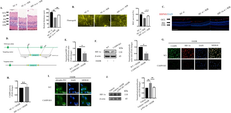
Methods: A murine retinal ischemia/ reperfusion (RIR) model and an in vitro oxygen and glucose deprivation/reoxygenation (OGDR) model were used to investigate the pathogenic mechanisms of acute glaucoma.

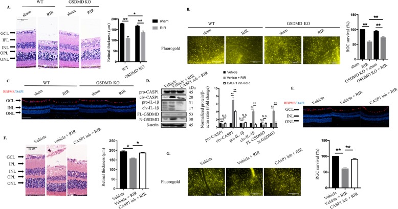
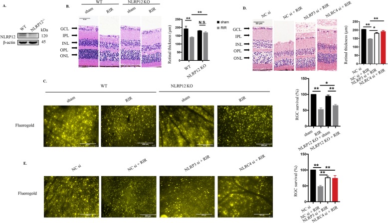
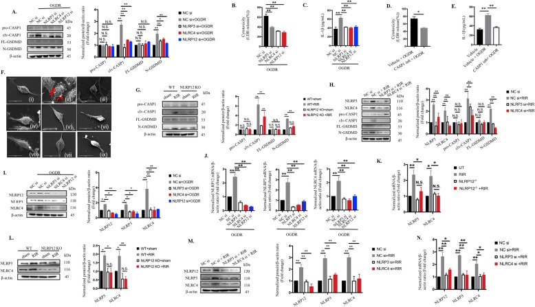
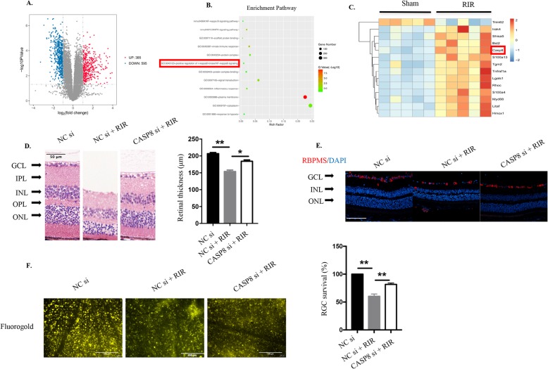
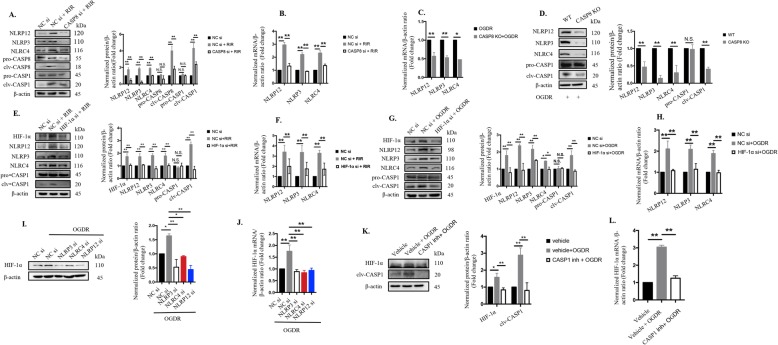
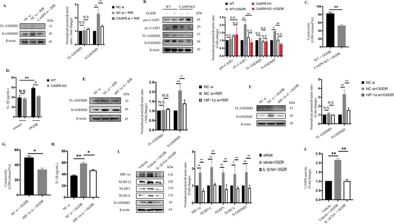
Results: Our findings reveal a novel mechanism of microglia-induced pyroptosis-mediated RGCs death associated with glaucomatous vision loss. Genetic deletion of gasdermin D (GSDMD), the effector of pyroptosis, markedly ameliorated the RGCs death and retinal tissue damage in acute glaucoma. Moreover, GSDMD cleavage of microglial cells was dependent on caspase-8 (CASP8)-hypoxia-inducible factor-1α (HIF-1α) signaling. Mechanistically, the newly identified nucleotide-binding leucine-rich repeat-containing receptor (NLR) family pyrin domain-containing 12 (NLRP12) collaborated with NLR family pyrin domain-containing 3 (NLRP3) and NLR family CARD domain-containing protein 4 (NLRC4) downstream of the CASP8-HIF-1α axis, to elicit pyroptotic processes and interleukin-1β (IL-1β) maturation through caspase-1 activation, facilitating pyroptosis and neuroinflammation in acute glaucoma. Interestingly, processing of IL-1β in turn magnified the CASP8-HIF-1α-NLRP12/NLRP3/NLRC4-pyroptosis circuit to accelerate inflammatory cascades.

Conclusions: These data not only indicate that the collaborative effects of NLRP12, NLRP3 and NLRC4 on pyroptosis are responsible for RGCs death, but also shed novel mechanistic insights into microglial pyroptosis, paving novel therapeutic avenues for the treatment of glaucoma-induced irreversible vision loss through simultaneously targeting of pyroptosis.

Keywords: Acute glaucoma; NOD-like receptor 12; caspase-8/ HIF-1α; pyroptosis.


Conflict of interest statement

The authors declare no competing financial interests.

Figures

Fig. 1.
Fig. 1.
CASP1-dependent pyroptosis plays a vital role in elevated IOP-induced retinal ischemic damage and RGCs loss. a HE staining and quantitative analysis of total retinal thickness in retina tissue harvested 7 days post RIR injury (n = 6). Scale bar: 50 μm. b Retrograde FG-labeled images and quantitative assay of RGCs survival in WT and GSDMD KO mice (n = 6). Scale bar: 200 μm. c Representative immunofluorescence images of RGCs in the retina. Anti-RBPMS was used to label RGCs (n = 6). Scale bar: 100 μm. d Western blot analysis of the indicated proteins (n = 6). The protein levels were normalized to β-actin levels. e Representative immunofluorescence images of RGCs in the retina from mice treated with or without CASP1 inhibitor. Anti-RBPMS was used to label RGCs (n = 6). Scale bar: 100 μm. f HE staining and quantitative analysis of total retinal thickness in retinal tissue (n = 6). Scale bar: 50 μm. g Retrograde FG-stained images and quantitative assay of RGCs amount from groups subjected to RIR and RIR concomitant with CASP1 suppression groups (200 μΜ, n = 6). Scale bar: 200 μm. WT: wide type; KO: knockout; RIR: retinal ischemia-reperfusion; GCL: ganglion cell layer; IPL: inner plexiform layer; INL: inner nuclear layer; OPL: outer plexiform layer; ONL: outer nuclear layer; CASP1 inh: caspase-1 inhibitor. All the experiments are representative of at least three independent experiments. Data are represented as the mean ± SD. *P < 0.05, **P < 0.01, one-way ANOVA and two-way ANOVA
Fig. 2
Fig. 2
Genetic deletion of NLRP12 and inhibition of NLRP3 or NLRC4 significantly attenuate retinal damage and improve RGC survival. a Western blot detection of NLRP12 in retinas from NLRP12-deficient mice (n = 6). b HE staining and quantitative measurement of total retinal thickness targeting retinal tissue morphology in WT and NLRP12 KO mice under high IOP (n = 6). Scale bar: 50 μm. c Retrograde FG labeling and RGC amount evaluation in WT and NLRP12 KO mice under high IOP (n = 6). Scale bar: 200 μm. d HE staining and quantitative evaluation of total retinal thickness in retinal tissue subjected to high IOP followed by NLRP3/NLRC4 knockdown (20 μΜ, n = 6). Scale bar: 50 μm. e Retrograde FG labeling and quantitative measurement of RGC survival in retinal tissue subjected to elevated IOP followed by NLRP3/NLRC4 knockdown (20 μΜ, n = 6). Scale bar: 200 μm. WT: wide type; KO: knockout; RIR: retinal ischemia-reperfusion; GCL: ganglion cell layer; IPL: inner plexiform layer; INL: inner nuclear layer; OPL: outer plexiform layer; ONL: outer nuclear layer. All of the data are representative of at least three independent experiments. The data are represented as the mean ± SD. *P < 0.05, **P < 0.01, one-way ANOVA and two-way ANOVA.
Fig. 3.
Fig. 3.
NLRP12 collaborates with NLRP3 and NLRC4 to elicit pyroptosis and promote IL-1β production in ischemic injury through CASP1-dependent GSDMD cleavage a-h NLRP12/NLRP3/NLRC4 promoted IL-1β release and induced pyroptosis by CASP1-dependent GSDMD cleavage: a Knockdown of NLRP12/NLRP3/NLRC4 reduced CASP1 and GSDMD cleavage in extracts from BV2 microglia after OGDR injury, as determined by western blot analysis (n = 6). The protein levels were normalized to β-actin levels. b LDH release (n = 6). c IL-1β secretion (n = 6). d-e LDH release (d) and IL-1β production (e) of BV2 microglia treated with the CASP1 inh, YVAD (200 µM) and subjected to OGDR injury (n = 6). Representative SEM images showed pyroptotic cell death and other morphological changes in BV2 microglia subjected to OGDR combined with or without different additional treatments (n = 5): (i): control; (ii): OGDR; (iii): OGDR plus CASP1 inh (YVAD, 200 µM); (iv): OGDR plus NLRP3 siRNA (si); (v): OGDR plus NLRP12 si; (vi): OGDR plus NLRC4 si; (vii): CASP8 CRISPR plus OGDR; (viii): OGDR plus HIF-1α si ; (ix): OGDR plus IL-1β neutralizing antibody. Scale bar: 20 µm. g-h Immunoblotting analysis for detection of pyroptotic proteins in the retinas of NLRP12-/- or WT mice with or without NLRP3/NLRC4 knockdown under RIR conditions (n = 6). The protein levels were normalized to β-actin levels. i-n Mutual regulatory relationships among NLRP12, NLRP3 and NLRC4: i-j Protein expression and mRNA levels of the indicated molecules, as determined by western blotting and qRT-PCR detection in BV2 microglia exposed to OGDR with or without NLRP3/NLRP12/NLRC4 knockdown (n = 6). The mRNA and protein levels were normalized to β-actin levels. k-n Western blotting and qRT-PCR analyses of NLRP3, NLRP12, and NLRC4 in retinas from WT mice and NLRP12 KO mice that were sacrificed 7 days after elevated IOP injury, combined with or without NLRP3/NLRC4 knockdown (n = 6). The mRNA and protein levels were normalized to β-actin levels. The data shown are representative of at least three independent experiments. The data are represented as the mean ± SD. *P < 0.05, **P < 0.01, one-way ANOVA, two-way ANOVA and two-tailed unpaired t-test.
Fig. 4
Fig. 4
CASP8 is a pivotal participant in RIR injury. a Volcano plot showing a total of 980 genes, of which 385 were upregulated and 595 were downregulated in retinal tissue in the RIR group compared with the sham group. A fold change > 2 or < 0.5 was considered statistically significant (n = 5 mice/group). b KEGG pathway enrichment analysis of differentially expressed genes between the ischemia/reperfusion and sham groups (n = 5 mice/group). The enriched pathways of hypoxia and inflammatory signaling are shown. The positive regulation of I-kappaB kinase/NF-kappaB signaling was chosen for further analysis. c Heat map showing the contents of the positive regulation of I-kappaB kinase/NF-kappaB signaling in the retinas of mice after the RIR process (n = 5 mice/group). RIR mice were sacrificed 7 days after reperfusion, and retinal tissue was collected for RNA sequence analysis. d HE staining of retinas from mice under RIR with or without CASP8 knockdown (20 μΜ). The retinal tissue was harvested on the seventh day after reperfusion (n = 6). Scale bar: 50 μm. The total retinal thicknesses were quantified for the three groups (n = 6). e Representative immunofluorescence images of RGCs in the retina from mice treated with or without CASP8 knockdown. Primary antibody against RBPMS was used to label RGCs (n = 6). Scale bar: 100 μm. f Retrograde FG staining of RGCs from RIR injury in the absence or presence of CASP8 interference (20 μΜ, n = 6). Scale bar: 200 μm. RGCs survival were analyzed comparing to the controls (n = 6). RIR: retinal ischemia-reperfusion; GCL: ganglion cell layer; IPL: inner plexiform layer; INL: inner nuclear layer; OPL: outer plexiform layer; ONL: outer nuclear layer; CASP8 si: caspase-8 siRNA. All of the data are representative of at least three independent experiments. Data are represented as the mean ± SD. *P < 0.05, **P < 0.01, one-way ANOVA
Fig. 5
Fig. 5
CASP8-mediated HIF-1α signaling is involved in retinal ischemic injury and RGCs loss. a HE staining and quantitative evaluation of total retinal thickness in retinal tissue subjected to high IOP followed by HIF-1α knockdown (20 μΜ, n = 6). Scale bar: 50 μm. b Retrograde FG labeling and quantitative measurement of RGCs survival from mice subjected to RIR injury in the absence or presence of HIF-1α interference (20 μΜ, n = 6). Scale bar: 200 μm. c Representative immunofluorescence images of RGCs in the retina from mice treated with or without HIF-1α blockage. Primary antibody against RBPMS was used to label RGCs (n = 6). Scale bar: 100 μm. d CRISPR-CAS9 design to knock out CASP8 in BV2 cell line. Targeted vector was designed based on the exon 3 to exon 5 in WT allele. e-f RNA level and protein levels of HIF-1α in WT and CASP8 KO BV2 cell line exposed to OGDR insult (n = 6, both). The protein and mRNA levels were normalized to β-actin levels. g BV2 microglia with the indicated genotypes were subjected to OGDR and stained with antibodies against cleaved-CASP8 and HIF-1α (n = 6). Scale bar: 100 μm. h CASP8 activity (n = 5). i Representative images of immunofluorescence staining targeting phospho-NF-kB P65 translocation in WT BV2 microglia and CASP8-specific KO cell line exposed to OGDR (n = 6). Scale bar: 20 μm. j The protein levels of HIF-1α were assayed by immunoblots in BV2 microglia treated with the NF-kB P65 inhibitor, JSH-23 (40 μM, n = 6). The protein expression was normalized to β-actin expression. RIR: retinal ischemia-reperfusion; GCL: ganglion cell layer; IPL: inner plexiform layer; INL: inner nuclear layer; OPL: outer plexiform layer; ONL: outer nuclear layer; HIF-1α si: HIF-1α siRNA. All of the data are representative of at least three independent experiments. Data are represented as the mean ± SD. *P < 0.05, **P < 0.01, one-way ANOVA and two-tailed unpaired t-test.
Fig. 6
Fig. 6
CASP8 promotes NLRP12/NLRP3/NLRC4 and CASP1 activation upon HIF-1α signaling. a-b The protein and mRNA levels of NLRP12/NLRP3/NLRC4 and CASP1 was detected in retinas from WT mice with or without CASP8 knockdown (20 μΜ) that were harvested at the seventh day after reperfusion (n = 6). The protein and mRNA levels were normalized to β-actin levels. c-d CASP8 elimination diminished the activation of NLRP12/NLRP3/NLRC4 and CASP1 in BV2 microglia exposed to OGDR (n = 6). The protein and mRNA levels were normalized to β-actin levels. e-h Immunoblot and qRT-PCR analyses of targeting NLRP12/NLRP3/NLRC4 and CASP1 in vivo and in vitro (n = 6) with or without HIF-1α knockdown. The protein and mRNA levels were normalized to β-actin levels. i-j Protein and mRNA levels of HIF-1α upon NLRP12/NLRP3/NLRC4 suppression in vitro (n = 6). The mRNA and protein levels were normalized to β-actin levels. k-l Knockdown of CASP1 suppressed the production of HIF-1α protein and mRNA in vitro (n = 6). The data shown are representative of at least three independent experiments. The data are represented as the mean ± SD. *P < 0.05, **P < 0.01, one-way ANOVA
Fig. 7
Fig. 7
Activation of the CASP8-HIF-1α pathway elicits pyroptosis and promotes IL-1β production which in turn magnifies inflammatory cascades via the CASP8-HIF-1α-NLR axis. a The protein levels of cleaved GSDMD were detected in retinas from WT mice with or without CASP8 knockdown (20 μΜ) that were harvested at the seventh day after reperfusion (n = 6). The protein levels were normalized to β-actin levels. b Western blot analysis of cleaved CASP1 and GSDMD in extracts from WT BV2 microglia and CASP8-specific KO cell line after OGDR injury (n = 6). The protein levels were normalized to β-actin levels. c-d Cytotoxicity c and IL-1β production d in BV2 microglia under OGDR injury (n = 6). e-f The protein levels of cleaved GSDMD were detected in vivo and in vitro with or without HIF-1α knockdown. The protein levels were normalized to β-actin levels. g-h Cytotoxicity g and IL-1β processing h were measured in the presence or absence of HIF-1α siRNA treatment in BV2 microglia (n = 6, both). i Western blotting detection of the indicated proteins in BV2 microglia subjected to OGDR and OGDR concomitant with IL-1β neutralizing antibody treatment (n = 6). The protein levels were normalized to β-actin levels. j CASP8 activity (n = 6). The data shown are representative of at least three independent experiments. Data are represented as the mean ± SD. *P < 0.05, **P < 0.01, experiments were assessed by one-way ANOVA, two-way ANOVA or two-tailed unpaired t-test
Fig. 8
Fig. 8
Diagram illustrating the pathway by which the CASP8-HIF-1α- NLRP12/NLRP3/NLRC4-IL1β-pyroptosis circuit contributes to the pathogenesis of acute glaucoma

References

    1. Tanner L, Gazzard G, Nolan WP, Foster PJ. Has the EAGLE landed for the use of clear lens extraction in angle-closure glaucoma? And how should primary angle-closure suspects be treated? Eye (London, England) 2020;34(1):40–50. doi: 10.1038/s41433-019-0634-5. - DOI - PMC - PubMed
    1. Tham YC, Li X, Wong TY, Quigley HA, Aung T, Cheng CY. Global prevalence of glaucoma and projections of glaucoma burden through 2040: a systematic review and meta-analysis. Ophthalmology. 2014;121(11):2081–2090. doi: 10.1016/j.ophtha.2014.05.013. - DOI - PubMed
    1. Zhu X, Zeng W, Wu S, Chen X, Zheng T, Ke M. Measurement of retinal changes in primary acute angle closure Glaucoma under different durations of symptoms. J Ophthalmol. 2019;2019:5409837. - PMC - PubMed
    1. Almasieh M, Wilson AM, Morquette B, Cueva Vargas JL, Di Polo A. The molecular basis of retinal ganglion cell death in glaucoma. Prog Retin Eye Res. 2012;31(2):152–181. doi: 10.1016/j.preteyeres.2011.11.002. - DOI - PubMed
    1. Minhas G, Sharma J, Khan N. Cellular stress response and immune signaling in retinal ischemia-reperfusion injury. Front Immunol. 2016;7:444. doi: 10.3389/fimmu.2016.00444. - DOI - PMC - PubMed

Publication types

MeSH terms

Substances