. 2020 May 7;9(5):bio051508.
doi: 10.1242/bio.051508.

A long lost key opens an ancient lock: Drosophila Myb causes a synthetic multivulval phenotype in nematodes

Affiliations

A long lost key opens an ancient lock: Drosophila Myb causes a synthetic multivulval phenotype in nematodes

Paul J Vorster et al. Biol Open. .

Abstract

The five-protein MuvB core complex is highly conserved in animals. This nuclear complex interacts with RB-family tumor suppressor proteins and E2F-DP transcription factors to form DREAM complexes that repress genes that regulate cell cycle progression and cell fate. The MuvB core complex also interacts with Myb family oncoproteins to form the Myb-MuvB complexes that activate many of the same genes. We show that animal-type Myb genes are present in Bilateria, Cnidaria and Placozoa, the latter including the simplest known animal species. However, bilaterian nematode worms lost their animal-type Myb genes hundreds of millions of years ago. Nevertheless, amino acids in the LIN9 and LIN52 proteins that directly interact with the MuvB-binding domains of human B-Myb and Drosophila Myb are conserved in Caenorhabditiselegans Here, we show that, despite greater than 500 million years since their last common ancestor, the Drosophila melanogaster Myb protein can bind to the nematode LIN9-LIN52 proteins in vitro and can cause a synthetic multivulval (synMuv) phenotype in vivo This phenotype is similar to that caused by loss-of-function mutations in C. elegans synMuvB-class genes including those that encode homologs of the MuvB core, RB, E2F and DP. Furthermore, amino acid substitutions in the MuvB-binding domain of Drosophila Myb that disrupt its functions in vitro and in vivo also disrupt these activities in C. elegans We speculate that nematodes and other animals may contain another protein that can bind to LIN9 and LIN52 in order to activate transcription of genes repressed by DREAM complexes.

Keywords: Development; Evolution; Myb; Oncogene; Tumor suppressor; synMuv.


Conflict of interest statement

Competing interestsThe authors declare no competing or financial interests.

Figures

Fig. 1.
Fig. 1.
Evolutionary conservation of Myb three-repeat (3R) DNA-binding domains, MuvB-binding domains of Myb proteins, and Myb-binding domains of LIN9 and LIN52. A partial phylogenetic tree of the current view of eukaryotic evolution (http://tolweb.org/tree/) shows the presence or absence of the indicated protein domains in representative species from diverse clades: human (Homo sapiens), lancelet (Branchiostoma belcheri), sea squirt (Ciona intestinalis), acorn worm (Saccoglossus kowalevskii), sea urchin (Strongylocentrotus purpuratus), fruit fly (D. melanogaster), nematode (C. elegans), penis worm (Priapulus caudatus), scallop (Mizuhopecten yessoensis), lamp shell (Lingula anatina), coral (Stylophora pistillata), trichoplax (Trichoplax adhaerens), fusarium (Fusarium sp. AF-4), dictyostelium (Dictyostelium discoideum), cacao (Theobroma cacao) and stentor (Stentor coeruleus). Displayed branch lengths are unscaled.
Fig. 2.
Fig. 2.
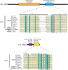
Sequence alignments of conserved animal-type Myb protein domains. A schematic diagram of the human B-Myb protein shows the relative positions and amino acid sequence numbers of the conserved domains that define animal-type Myb proteins. Local multiple protein sequence alignments were constructed using MACAW with the BLOSUM62 scoring matrix (Schuler et al., 1991). The alignment shading indicates the mean score at each position as shown in the color key. Horizontal bars above the DNA-binding domain alignment indicate three tandem Myb repeats (R1, R2, R3). Asterisks below the hinge domain alignments indicate known Cyclin A-CDK2 phosphorylation sites in the hinge region human B-Myb as described by Werwein et al. (2019). The central hinge domain alignment contains a binding site for the Plk1 polo-family protein kinase. Horizontal bars above the MuvB-binding domain alignment indicate α-helices in the human B-Myb crystal structure with human LIN9 and LIN52 (Guiley et al., 2018). Black dots below the MuvB-binding domain alignment indicate amino acids of human B-Myb that contact human LIN9 or LIN52 in the crystal structure. Arrows below the MuvB-binding domain indicate amino acids substituted by alanine in two Drosophila Myb mutants used in experiments in this study (Andrejka et al., 2011).
Fig. 3.
Fig. 3.
Sequence alignments of the Myb-binding domains of LIN9 and LIN52. Schematic diagrams of the human LIN9 and LIN52 proteins show the relative positions and amino acid sequence numbers of the conserved domains. Horizontal bars above the Myb-binding domain alignments indicate α-helices in the crystal structure of human LIN9 and LIN52 bound to human B-Myb (Guiley et al., 2018). Black dots below the Myb-binding domain alignments indicate amino acids that contact human B-Myb in the crystal structure. Alignments are not shown for the DIRP domain of LIN9 (pfam 06584) (White-Cooper et al., 2000), the function of which remains unknown, or for the pocket-binding domain of LIN52 that binds to the human RB-related p107 and p130 proteins but not to human RB itself (Guiley et al., 2015).
Fig. 4.
Fig. 4.
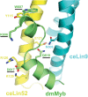
Structural modeling of the Drosophila Myb MuvB-binding domain bound to the Myb-binding domains of nematode LIN9 and LIN52. Protein sequences of Drosophila Myb (dmMyb; green), C. elegans LIN9 (ceLIN9; cyan) and C. elegans LIN52 (ceLIN52; yellow) were modeled into the human crystal structure (PDB ID: 6C48) using MODELLER (Webb and Sali, 2017). Amino acid numbering shown is for these Drosophila and C. elegans proteins. The four amino acids substituted by alanine in the two Drosophila Myb mutants used in experiments in this study are underlined.
Fig. 5.
Fig. 5.
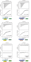
The Myb-binding domains of C. elegans LIN9 and LIN52 bind the Drosophila MuvB-binding domain of Drosophila Myb in vitro. Recombinant C. elegans LIN9-LIN52 heterodimeric Myb-binding domains produced in E. coli were purified and then assayed for binding to recombinant Drosophila Myb MuvB-binding domain, using isothermal titration calorimetry. Each panel displays a representative experiment using the proteins diagrammed below the panel. The raw data are presented above and the fitted binding curve is presented below. The mean calculated Kd values and standard deviations from three replicate experiments are shown below each panel.
Fig. 6.
Fig. 6.
Drosophila Myb causes a synthetic multivulval phenotype in C. elegans. Top panel: the indicated strains were heat-shocked as L2/L3 larvae, then scored as adults for the presence of a multivulval phenotype in a lin-15A mutant background. DIC images of two representative multivulval worms of the lin-15A; GFP::Myb genotype are shown (open arrowheads indicate the normal vulval opening, black arrows indicate ectopic vulvae). Middle panel: histograms show the incidence of multivulval worms in two different experiments using strains of the indicated genotypes. Statistical significance relative to the lin-15A; GFP control strain was determined using a two-tailed Z-test. One asterisk indicates a significance of 0.05 or less; two asterisks indicate a significance of 0.01 or less. Numbers within the bars indicate total number of worms scored for the indicated genotype. Bottom panel: schematic diagrams of the Drosophila Myb wild-type and mutant proteins expressed in transgenic worms. All of the Myb proteins contained a GFP tag fused at their amino termini (not shown).
Fig. 7.
Fig. 7.
Model for the mechanism of action of Drosophila Myb in C. elegans. The wild-type DREAM complex, which includes LIN9 and LIN52 and other synMuvB proteins, redundantly represses the ectopic expression of LIN3/EGF, resulting in a wild-type worm even in a lin-15A synMuvA mutant background. Loss-of-function mutants of synMuvB genes fail to repress ectopic expression of LIN3/EGF, resulting in a synthetic multivulal worm in a lin-15A mutant background. Ectopic expression of Drosophila Myb overrides repression by the wild-type DREAM complex, causing a synthetic multivulval worm in a lin-15A mutant background, presumably due to ectopic expression of LIN-3/EGF. It remains unknown whether nematodes and other animals have a second ‘key’ that can also open the highly conserved DREAM complex ‘lock’.

References

    1. Amatschek S., Koenig U., Auer H., Steinlein P., Pacher M., Gruenfelder A., Dekan G., Vogl S., Kubista E., Heider K.-H. et al. (2004). Tissue-wide expression profiling using cDNA subtraction and microarrays to identify tumor-specific genes. Cancer Res. 64, 844-856. 10.1158/0008-5472.CAN-03-2361 - DOI - PubMed
    1. Andrejka L., Wen H., Ashton J., Grant M., Iori K., Wang A., Manak J. R. and Lipsick J. S. (2011). Animal-specific C-terminal domain links myeloblastosis oncoprotein (Myb) to an ancient repressor complex. Proc. Natl. Acad. Sci. USA 108, 17438-17443. 10.1073/pnas.1111855108 - DOI - PMC - PubMed
    1. Ansieau S., Kowenz-Leutz E., Dechend R. and Leutz A. (1997). B-Myb, a repressed trans-activating protein. J. Mol. Med. 75, 815-819. 10.1007/s001090050170 - DOI - PubMed
    1. Beall E. L., Manak J. R., Zhou S., Bell M., Lipsick J. S. and Botchan M. R. (2002). Role for a Drosophila Myb-containing protein complex in site-specific DNA replication. Nature 420, 833-837. 10.1038/nature01228 - DOI - PubMed
    1. Beall E. L., Bell M., Georlette D. and Botchan M. R. (2004). Dm-myb mutant lethality in Drosophila is dependent upon mip130: positive and negative regulation of DNA replication. Genes Dev. 18, 1667-1680. 10.1101/gad.1206604 - DOI - PMC - PubMed

Publication types

MeSH terms