. 2020 Feb 14;5(1):13.
doi: 10.1038/s41392-020-0111-4.

Silencing MYH9 blocks HBx-induced GSK3β ubiquitination and degradation to inhibit tumor stemness in hepatocellular carcinoma

Affiliations

Silencing MYH9 blocks HBx-induced GSK3β ubiquitination and degradation to inhibit tumor stemness in hepatocellular carcinoma

Xian Lin et al. Signal Transduct Target Ther. .

Abstract

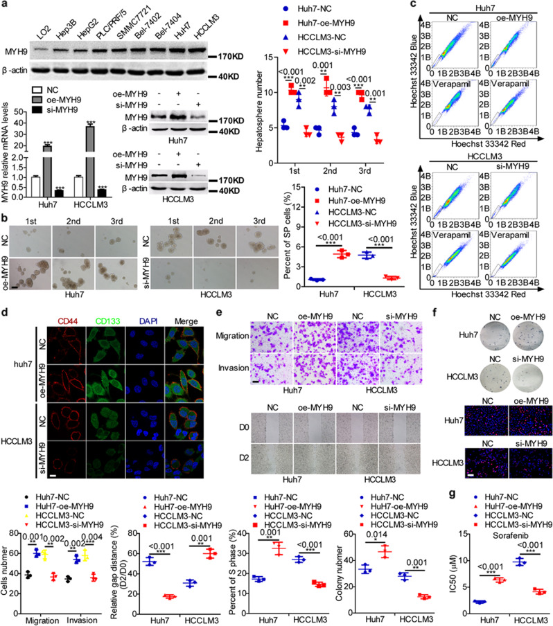
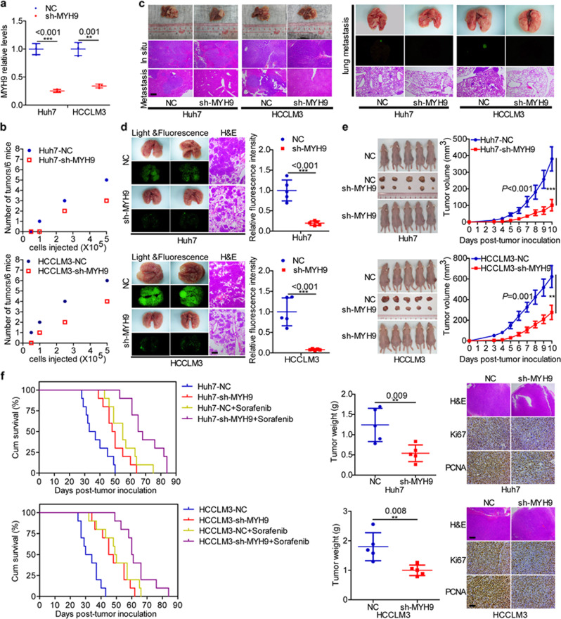
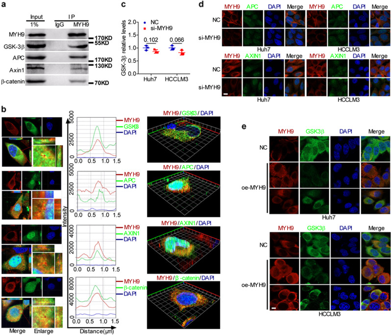
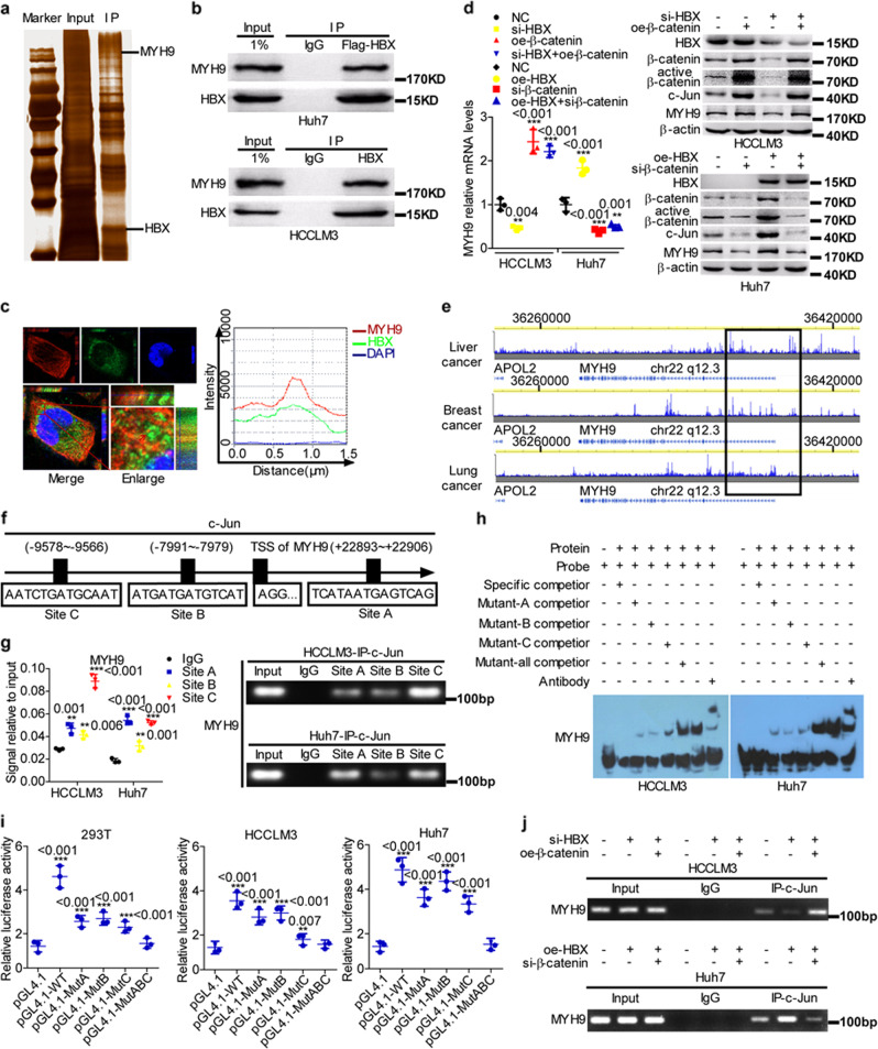
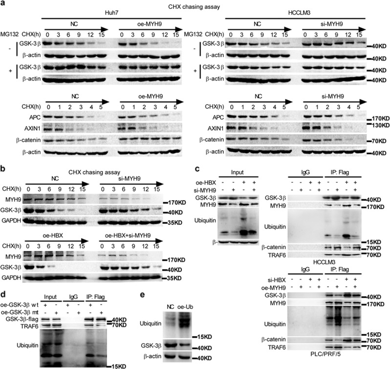
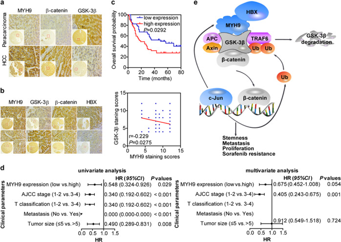
MYH9 has dual functions in tumors. However, its role in inducing tumor stemness in hepatocellular carcinoma (HCC) is not yet determined. Here, we found that MYH9 is an effective promoter of tumor stemness that facilitates hepatocellular carcinoma pathogenesis. Importantly, targeting MYH9 remarkably improved the survival of hepatocellular carcinoma-bearing mice and promoted sorafenib sensitivity of hepatocellular carcinoma cells in vivo. Mechanistic analysis suggested that MYH9 interacted with GSK3β and reduced its protein expression by ubiquitin-mediated degradation, which therefore dysregulated the β-catenin destruction complex and induced the downstream tumor stemness phenotype, epithelial-mesenchymal transition, and c-Jun signaling in HCC. C-Jun transcriptionally stimulated MYH9 expression and formed an MYH9/GSK3β/β-catenin/c-Jun feedback loop. X protein is a hepatitis B virus (HBV)-encoded key oncogenic protein that promotes HCC pathogenesis. Interestingly, we observed that HBV X protein (HBX) interacted with MYH9 and induced its expression by modulating GSK3β/β-catenin/c-Jun signaling. Targeting MYH9 blocked HBX-induced GSK3β ubiquitination to activate the β-catenin destruction complex and suppressed cancer stemness and EMT. Based on TCGA database analysis, MYH9 was found to be elevated and conferred poor prognosis for hepatocellular carcinoma patients. In clinical samples, high MYH9 expression levels predicted poor prognosis of hepatocellular carcinoma patients. These findings identify the suppression of MYH9 as an alternative approach for the effective eradication of CSC properties to inhibit cancer migration, invasion, growth, and sorafenib resistance in HCC patients. Our study demonstrated that MYH9 is a crucial therapeutic target in HCC.


Conflict of interest statement

The authors declare no competing interests.

Figures

Fig. 1
Fig. 1. MYH9 promotes stemness properties, migration, invasion, growth, and sorafenib resistance in HCC.
a Western blotting analyses of MYH9 expression in Hep3B, HepG2, PLC/PRF/5, SMMC-7721, Bel-7402, Bel-7404, Huh7, HCCLM3, and LO2 cells. Western blotting and QPCR analyses of MYH9 levels in MYH9-overexpressing HCC cells, MYH9-depleted HCC cells, and control cells. bf Hepatosphere formation assays (scale bar indicates 20 μm) b, flow cytometry analyses c, immunofluorescence analyses (scale bar indicates 5 μm) d, wound healing assays and transwell assays (scale bar indicates 10 μm) e, EdU incorporation assays (scale bar indicates 10 μm) and colony-formation assays f, and anticancer drug sensitivity tests g in MYH9-overexpressing Huh7 cells, MYH9-depleted HCCLM3 cells, and control cells.
Fig. 2
Fig. 2. MYH9 enhances the stemness properties, migration, invasion, growth, and sorafenib resistance of HCC cells in vivo.
a QPCR analyses of MYH9 levels in xenografts originating from Huh7 and HCCLM3 cells with stable silencing of MYH9 and corresponding control cells. b A subcutaneous xenograft mouse model was established to elucidate the impact of MYH9 on the tumor-initiating frequency (n = 6 per group). c The orthotopic tumor model was used to elucidate the effect of MYH9 on metastasis and proliferation (n = 7 per group). d A pulmonary metastasis model was generated to investigate the impact of MYH9 on metastasis (n = 5 per group). e A subcutaneous xenograft mouse model was used to elucidate the function of MYH9 on proliferation (general linear model, n = 5 per group). Xenografts were stained with H&E and subjected to immunohistochemistry for Ki67 and PCNA expression (n = 5 per group). f Survival analyses showing the overall survival of HCC-bearing mice in the indicated groups (log-rank test, n = 10 per group). Scale bars indicate 50 μm for H&E staining. Scale bars indicate 10 μm for immunohistochemistry.
Fig. 3
Fig. 3. MYH9 interacts with GSK3β to suppress its expression.
a Coimmunoprecipitation analyses of the interaction between MYH9 and the β-catenin destruction complex in HCCLM3 cells. b Immunofluorescence costaining of MYH9 and the β-catenin destruction complex to indicate the colocalization in HCCLM3 (left) and Huh7 (right) cells. The colocalization of MYH9 and the β-catenin destruction complex is shown by calculating the fluorescence intensities along the red arrow crossing the cytoplasm. c QPCR analyses of GSK3β mRNA levels in MYH9-depleted Huh7 and HCCLM3 cells and control cells. d Immunofluorescence staining of MYH9 and APC or AXIN1 expression and localization in MYH9-silenced Huh7 and HCCLM3 cells and control cells (scale bar indicates 5 μm). e MYH9 and GSK3β expression and localization were detected by immunofluorescence costaining in MYH9-overexpressing Huh7 and HCCLM3 cells and control cells (scale bar indicates 5 μm).
Fig. 4
Fig. 4. HBX protein interacts with MYH9 and induces its expression.
a Coimmunoprecipitation and silver staining analyses of HBX-associated proteins in HCC cells. b Coimmunoprecipitation analyses of the interaction between HBX and MYH9 in HBX-overexpressing Huh7 and HCCLM3 cells. c The colocalization of HBX and MYH9 was evaluated by immunofluorescence costaining. The colocalization of HBX and MYH9 was shown by calculating the fluorescence intensities along the red arrow crossing the cytoplasm. d QPCR and western blotting analyses were utilized to measure MYH9 expression levels in HCC cells. e ChIP-seq binding peaks searched using the Cistrome Data Browser. f Bioinformatics tools were adopted to identify c-Jun-binding sites inside the transcriptional regulatory region of MYH9. Chromatin immunoprecipitation analyses g, electrophoretic mobility shift assays h, and luciferase reporter assays i were conducted to elucidate c-Jun binding to the transcriptional regulatory sequences of MYH9. j Chromatin immunoprecipitation analyses were conducted to assess c-Jun binding to the transcriptional regulatory sequences of MYH9 in HCC cells.
Fig. 5
Fig. 5. MYH9 mediates HBX-induced GSK3β ubiquitination and degradation.
a Western blotting and quantification analyses of the effect of MYH9 depletion on GSK3β, β-catenin, APC, and AXIN1 stability in HCCLM3 cells incubated with cycloheximide or MG132 at the indicated time points. b Western blotting and quantification analyses of the impact of HBX and MYH9 on GSK3β stability in HCCLM3 cells incubated with cycloheximide at the indicated time points. c Coimmunoprecipitation analyses were conducted to identify the function of HBX and MYH9 on the interplay between MYH9, TRAF6, ubiquitin, β-catenin, and GSK3β in HCCLM3 and PLC/PRF/5 cells incubated with MG132. d Coimmunoprecipitation analyses of the interaction between TRAF6, ubiquitin, and wild-type GSK3β or mutant GSK3β in HCCLM3 cells treated with MG132. e Western blot analyses were used to assess the impact of ubiquitin on GSK3β expression levels in ubiquitin-overexpressing HCCLM3 cells and the corresponding control cells.
Fig. 6
Fig. 6. Immunohistochemical analyses of MYH9, GSK3β, β-catenin, and HBX expression in HCC.
a Comparisons of MYH9, GSK3β, and β-catenin expression between HCC and adjacent noncancerous tissues. b Correlations among HBX, MYH9, GSK3β, and β-catenin expression. c Kaplan–Meier survival analysis was performed for HCC patients according to MYH9 expression. d Univariate and multivariate survival analyses of clinicopathological characteristics of hepatocellular carcinoma patients. e Working model of the MYH9/GSK3β/β-catenin-c-Jun regulatory circuit activated by HBX via MYH9-modulated ubiquitination and degradation of GSK3β in Wnt signaling in HCC.

References

    1. Plaks V, Kong N, Werb Z. The cancer stem cell niche: how essential is the niche in regulating stemness of tumor cells? Cell Stem Cell. 2015;16:225–238. doi: 10.1016/j.stem.2015.02.015. - DOI - PMC - PubMed
    1. Sia D, Villanueva A, Friedman SL, Llovet JM. Liver cancer cell of origin, molecular class, and effects on patient prognosis. Gastroenterology. 2017;152:745–761. doi: 10.1053/j.gastro.2016.11.048. - DOI - PubMed
    1. Vicente-Manzanares M, Ma X, Adelstein RS, Horwitz AR. Non-muscle myosin II takes centre stage in cell adhesion and migration. Nat. Rev. Mol. Cell Biol. 2009;10:778–790. doi: 10.1038/nrm2786. - DOI - PMC - PubMed
    1. Li Y, et al. Chemical compound cinobufotalin potently induces FOXO1-stimulated cisplatin sensitivity by antagonizing its binding partner MYH9. Signal Transduct. Target Ther. 2019;4:48. doi: 10.1038/s41392-019-0084-3. - DOI - PMC - PubMed
    1. Liu D, et al. Clinicopathological significance of NMIIA overexpression in human gastric cancer. Int. J. Mol. Sci. 2012;13:15291–15304. doi: 10.3390/ijms131115291. - DOI - PMC - PubMed

Publication types

MeSH terms