. 2020 Apr;580(7803):396-401.
doi: 10.1038/s41586-020-2164-5. Epub 2020 Apr 1.

Germline Elongator mutations in Sonic Hedgehog medulloblastoma

Sebastian M Waszak #  1 Giles W Robinson #  2 Brian L Gudenas  3 Kyle S Smith  3 Antoine Forget  4 Marija Kojic  5 Jesus Garcia-Lopez  3 Jennifer Hadley  3 Kayla V Hamilton  6 Emilie Indersie  4 Ivo Buchhalter  7 Jules Kerssemakers  7 Natalie Jäger  8   9 Tanvi Sharma  8   9 Tobias Rausch  1 Marcel Kool  8   9   10 Dominik Sturm  8   11 David T W Jones  8   11 Aksana Vasilyeva  12 Ruth G Tatevossian  13 Geoffrey Neale  14 Bérangère Lombard  15 Damarys Loew  15 Joy Nakitandwe  13 Michael Rusch  16 Daniel C Bowers  17 Anne Bendel  18 Sonia Partap  19 Murali Chintagumpala  20 John Crawford  21   22 Nicholas G Gottardo  23 Amy Smith  24 Christelle Dufour  25 Stefan Rutkowski  26 Tone Eggen  27 Finn Wesenberg  28 Kristina Kjaerheim  28 Maria Feychting  29 Birgitta Lannering  30 Joachim Schüz  31 Christoffer Johansen  32   33 Tina V Andersen  34 Martin Röösli  35 Claudia E Kuehni  35   36 Michael Grotzer  37 Marc Remke  38 Stéphanie Puget  39 Kristian W Pajtler  8   9   40 Till Milde  8   40   41 Olaf Witt  8   40   41 Marina Ryzhova  42 Andrey Korshunov  43   44 Brent A Orr  13 David W Ellison  13 Laurence Brugieres  25 Peter Lichter  45 Kim E Nichols  6 Amar Gajjar  2 Brandon J Wainwright  5 Olivier Ayrault  4 Jan O Korbel  46 Paul A Northcott  47 Stefan M Pfister  48   49   50
Affiliations

Germline Elongator mutations in Sonic Hedgehog medulloblastoma

Sebastian M Waszak et al. Nature. 2020 Apr.

Abstract

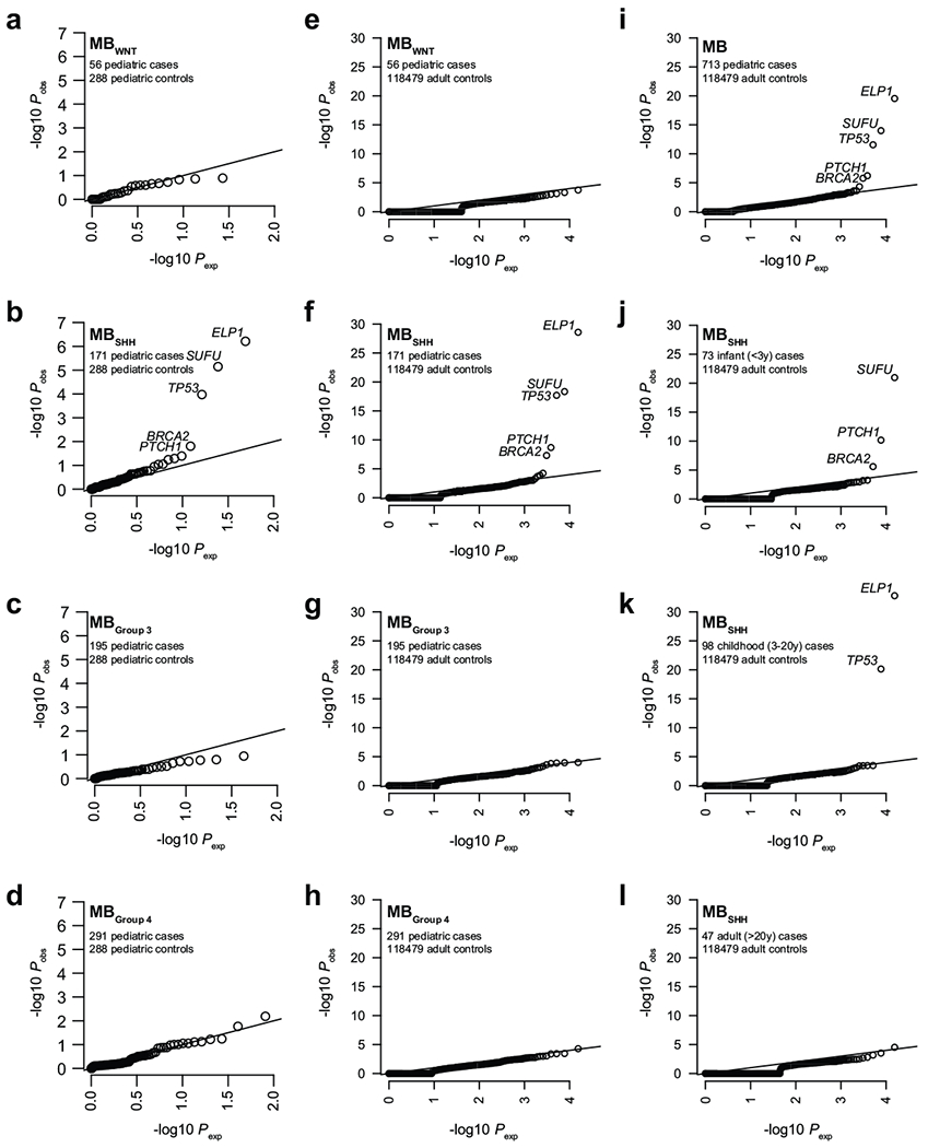
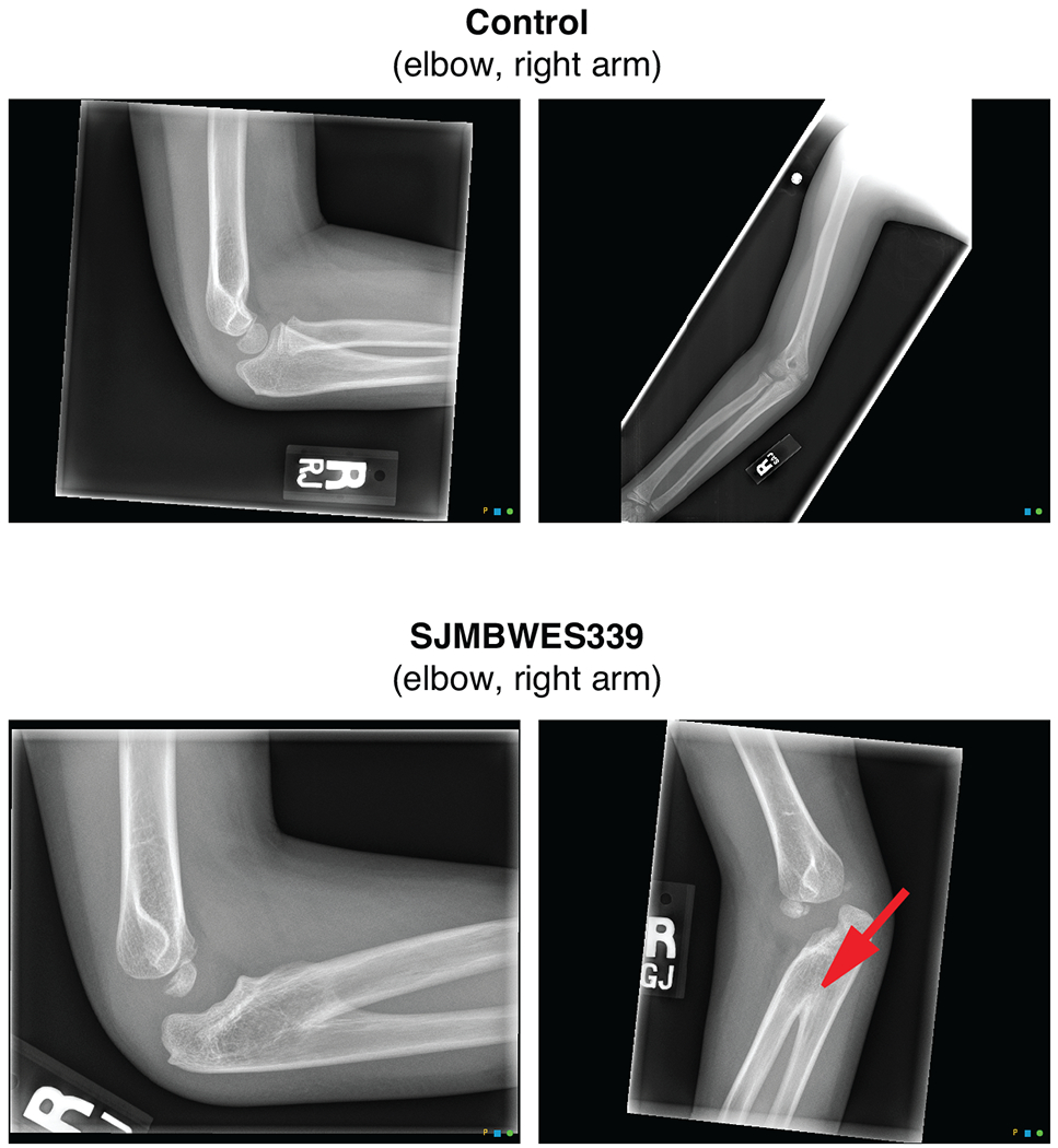
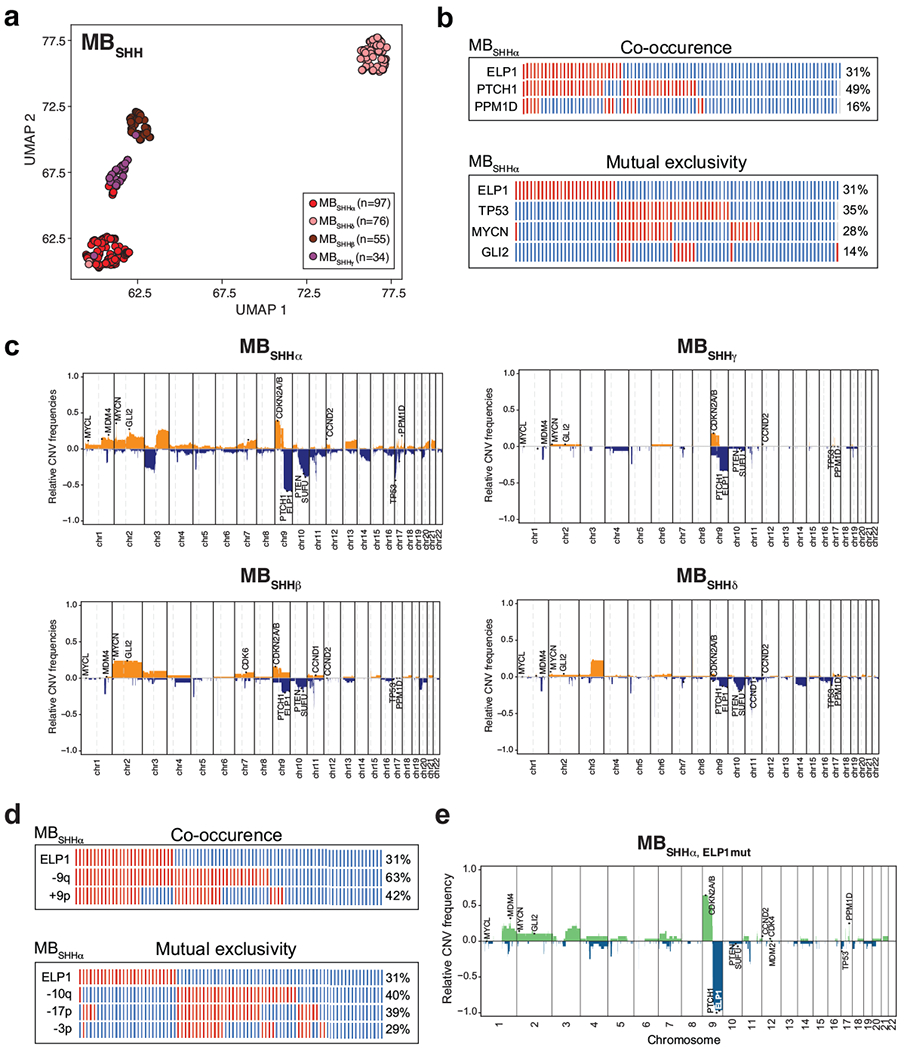
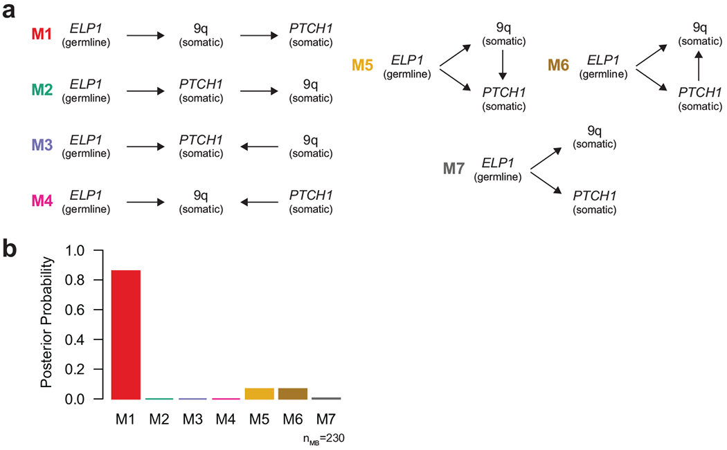
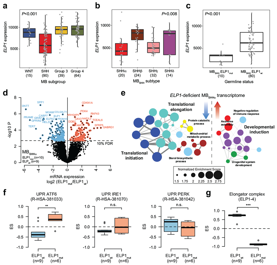
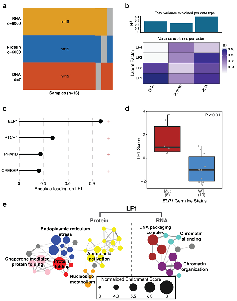
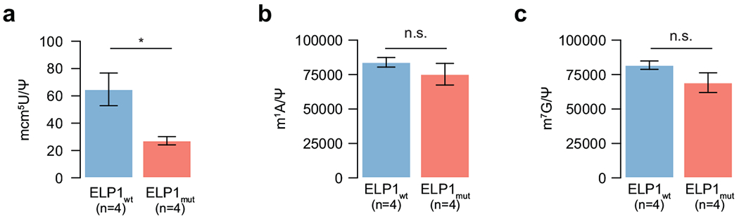
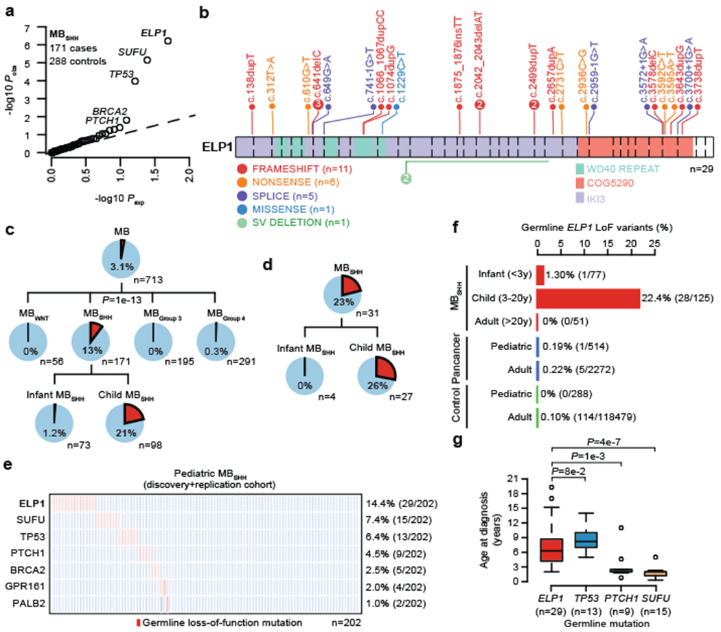
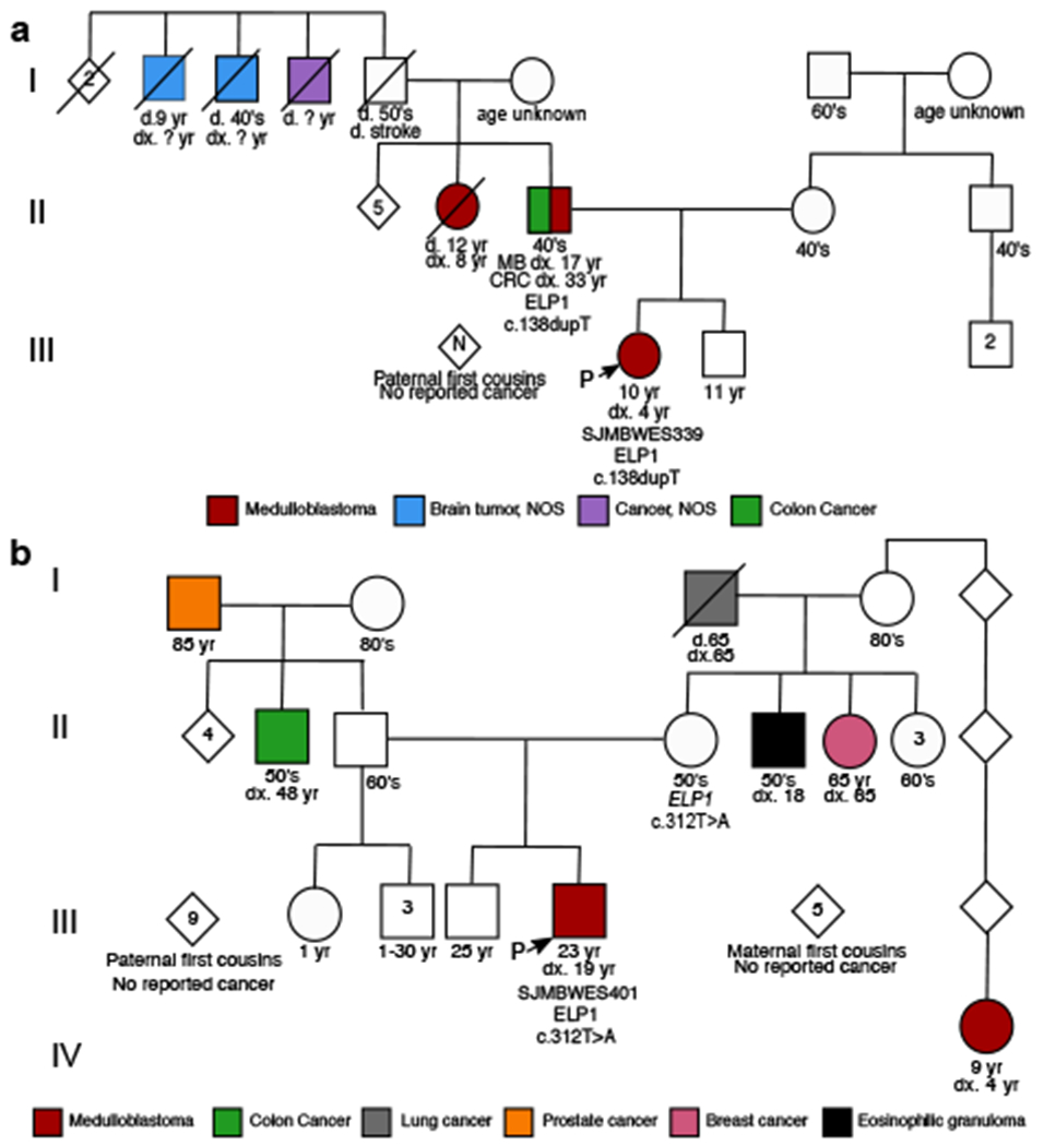
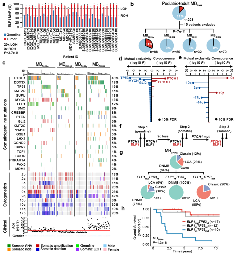
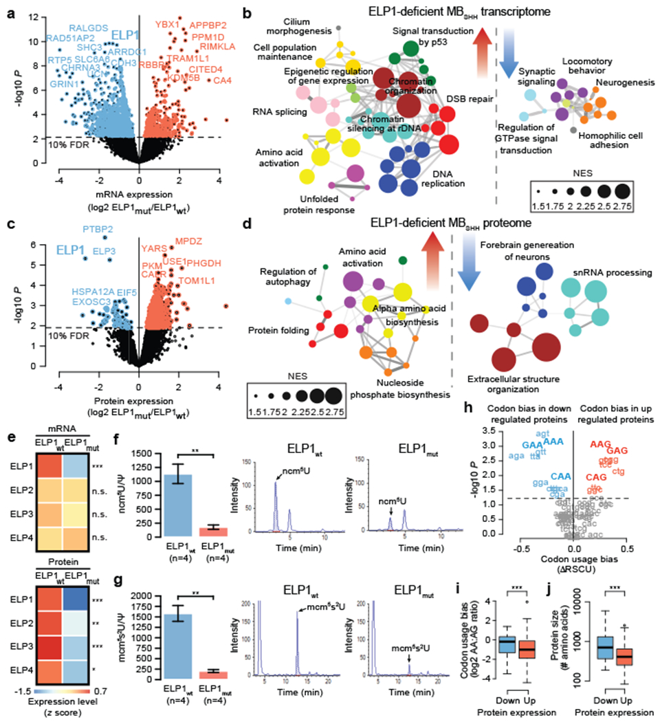
Cancer genomics has revealed many genes and core molecular processes that contribute to human malignancies, but the genetic and molecular bases of many rare cancers remains unclear. Genetic predisposition accounts for 5 to 10% of cancer diagnoses in children1,2, and genetic events that cooperate with known somatic driver events are poorly understood. Pathogenic germline variants in established cancer predisposition genes have been recently identified in 5% of patients with the malignant brain tumour medulloblastoma3. Here, by analysing all protein-coding genes, we identify and replicate rare germline loss-of-function variants across ELP1 in 14% of paediatric patients with the medulloblastoma subgroup Sonic Hedgehog (MBSHH). ELP1 was the most common medulloblastoma predisposition gene and increased the prevalence of genetic predisposition to 40% among paediatric patients with MBSHH. Parent-offspring and pedigree analyses identified two families with a history of paediatric medulloblastoma. ELP1-associated medulloblastomas were restricted to the molecular SHHα subtype4 and characterized by universal biallelic inactivation of ELP1 owing to somatic loss of chromosome arm 9q. Most ELP1-associated medulloblastomas also exhibited somatic alterations in PTCH1, which suggests that germline ELP1 loss-of-function variants predispose individuals to tumour development in combination with constitutive activation of SHH signalling. ELP1 is the largest subunit of the evolutionarily conserved Elongator complex, which catalyses translational elongation through tRNA modifications at the wobble (U34) position5,6. Tumours from patients with ELP1-associated MBSHH were characterized by a destabilized Elongator complex, loss of Elongator-dependent tRNA modifications, codon-dependent translational reprogramming, and induction of the unfolded protein response, consistent with loss of protein homeostasis due to Elongator deficiency in model systems7-9. Thus, genetic predisposition to proteome instability may be a determinant in the pathogenesis of paediatric brain cancers. These results support investigation of the role of protein homeostasis in other cancer types and potential for therapeutic interference.


Conflict of interest statement

Competing Interests

The authors declare no competing interests.

Figures

Extended Data Figure 1.
Extended Data Figure 1.. Case-control germline loss-of-function (LoF) variant burden analysis.
a-d. Case-control germline rare LoF variant association analysis in pediatric MB subgroups vs pediatric controls (CEFALO) using burden tests (implemented in SKAT). P values were corrected for multiple testing using Bonferroni correction. e-h. Case-control germline rare LoF variant association analysis in pediatric MB subgroups vs adult controls (gnomAD) using burden tests (two-sided Fisher’s Exact tests). P values were corrected for multiple testing using Bonferroni correction. i. Case-control germline LoF burden analysis in pediatric MB vs adult controls (gnomAD). j-l. Case-control germline LoF burden analysis in infant (j), childhood (k), and adult (1) MBSHH vs adult controls (gnomAD).
Extended Data Figure 2.
Extended Data Figure 2.. Congenital radioulnar synostosis
Right arm X-rays of (a) an unaffected child and (b) the ELP1-associated MBSHH patient SJMBWES339.
Extended Data Figure 3.
Extended Data Figure 3.. Molecular MBSHH subtypes
a. DNA methylation-based UMAP plot of MBSHH with inference of MBSHH subtypes (n = 262). b. Co-occurring and mutually exclusive somatic gene alterations in ELP1-associated medulloblastoma subtype SHHα. c. Co-occurring and mutually exclusive somatic chromosomal aberrations in ELP1-associated medulloblastoma subtype SHHα. d. Recurrent somatic copy-number alterations in ELP1-associated medulloblastoma subtype SHHα. e. Recurrent somatic copy-number alterations in ELP1-associated SHHα.
Extended Data Figure 4.
Extended Data Figure 4.. Inference of somatic evolution in ELP1-associated MBSHH.
a. Possible genetic models explaining the relationship between germline ELP1 mutation status and two somatic mutational events (PTCH1mut, and loss of chromosome 9q) in MBSHH. b. Posterior probabilities derived from Bayesian network analysis of all possible genetic models shown in a and data for 230 MBSHH patients.
Extended Data Figure 5.
Extended Data Figure 5.. Molecular features of ELP1-associated medulloblastoma
a. ELP1 expression stratified by consensus MB subgroup (n=208 patients). P value was calculated using likelihood ratio tests. Boxes show median, first and third quartile, and whiskers extend to 1.5× the interquartile range. b. ELP1 expression stratified by molecular MBSHH subtype (n=90 patients). P value was calculated using likelihood ratio tests. Boxes show median, first and third quartile, and whiskers extend to 1.5× the interquartile range. c. ELP1 expression stratified by germline ELP1 mutation status (n=90 patients). P value was calculated using likelihood ratio tests. Boxes show median, first and third quartile, and whiskers extend to 1.5× the interquartile range. d. Differential gene expression in ELP1mut (n=10) and ELP1wt SHHα (n=9). P values were derived from models that use negative binomal test statistics and were adjusted for multiple testing based on FDR correction. e. Functional gene enrichment in ELP1-associated SHHα. f. GSEA-based enrichment of UPR pathways in MBSHH proteomes. P values were calculated using a two-sided Mann-Whitney U-test. **P<0.01; ns P>0.05. Boxes show median, first and third quartile, and whiskers extend to 1.5× the interquartile range. g. GSEA-based enrichment of the Elongator complex in MBSHH proteomes. P value was calculated using a two-sided Mann-Whitney U-test. ***P<0.001; Boxes show median, first and third quartile, and whiskers extend to 1.5× the interquartile range.
Extended Data Figure 6.
Extended Data Figure 6.. Unsupervised multi-omics factor integration analysis of MBSHH
a. Overview of input samples and data types. b. Latent factors variance summary across data types. c. Somatic/germline gene alterations contributing to latent factor 1 (LF1). d. Association between germline ELP1 mutation status and LF1 score (n=16 patients). P value was calculated using a two-sided Mann-Whitney U-test. Boxes show median, first and third quartile, and whiskers extend to 1.5× the interquartile range. e. Functional enrichment of LF1-ranked proteins and mRNAs.
Extended Data Figure 7.
Extended Data Figure 7.. Quantification of tRNA modifications in ELP1-associated MBSHH
a. Quantification of mcm5U nucleosides in ELP1mut MBSHH PDX (n=4 biologically independent samples) and ELP1wt MBSHH PDX (n=4 biologically independent samples). Mean and standard errors are shown. P value was calculated using a two-sided Welch t-test. *P<0.05. b. Quantification of m1A nucleosides in ELP1mut MBSHH PDX (n=4 biologically independent samples) and ELP1wt MBSHH PDX (n=4 biologically independent samples). Mean and standard errors are shown. P value was calculated using a two-sided Welch i-test. *ns>0.05. c. Quantification of m7G nucleosides in ELP1mut MBSHH PDX (n=4 biologically independent samples) and ELP1wt MBSHH PDX (n=4 biologically independent samples). Mean and standard errors are shown. P value was calculated using a two-sided Welch t-test. ns P>0.05.
Extended Data Figure 8.
Extended Data Figure 8.. Spatio-temporal ELP1 expression in human and mouse.
a. ELP1 expression in adult human tissues (n=9-653 donors per tissue). Violin plots depict kernel density estimates and represent the density distribution b. ELP1 expression during human brain development (n=3-12 donors per tissue and time point). Boxes show median, first and third quartile, and whiskers extend to 1.5× the interquartile range. c. ELP1 expression during human organ development. Shaded areas define 90% confidence intervals (n=18-58 donors per tissue). d. Elp1 expression during mouse cerebellum development (n=27 animals). Center values and error bars define the mean expression of cells with non-zero ELP1 expression and standard error of the mean.
Figure 1.
Figure 1.. ELP1-associated medulloblastoma
a. QQ plot for LoF burden testing based on 171 pediatric MBSHH cases and 288 pediatric controls. P values were adjusted for multiple testing based on Bonferroni correction. b. Germline ELP1 mutation profile in MBSHH patients. c. Frequency of germline ELP1 LoF variants in the MB discovery cohort. d. Frequency of germline ELP1 LoF variants in a prospective pediatric MBSHH cohort. e. Frequency of known and recurrent genetic disorders in pediatric MBSHH. f. Frequency of germline ELP1 LoF variants in MBSHH, pediatric/adult pan-cancer, and control cohorts. g. Age at diagnosis for MBSHH patients stratified by known genetic disorders. P values were calculated using a two-sided Mann–Whitney U-test. Boxes show median, first and third quartile, and whiskers extend to 1.5× the interquartile range.
Figure 2.
Figure 2.. Familial transmission of ELP1-associated medulloblastoma
Pedigrees and family history for two ELP1-associated MBSHH patients. Circles, females; squares, males; diamond, sex unspecified; slash, deceased. Index patients are marked with an arrow. a. SJMBWES339. b. SJMBWES401.
Figure 3.
Figure 3.. Somatic mutation landscape of ELP1-associated medulloblastoma
a. Loss-of-heterozygosity (LOH) at the ELP1 locus for patients with germline ELP1 LoF variants (n=29 germline and n=29 MB samples). P values for LOH in MBs were calculated using a two-sided binomial test; *P<0.05. P value for the proportion of ELP1 LOH events was calculated using a two-sided binomial test. b. Germline ELP1 mutation status stratified by molecular MBSHH subtypes. c. Somatic mutation landscape of MBSHH subtypes. d. Association between germline ELP1 LoF variants and somatic gene alterations (n=238 MBSHH). P values were calculated using Bayesian logistic regression analysis, likelihood ratio tests, and adjusted for multiple testing based on FDR correction. e. Association between germline ELP1 LoF variants and somatic chromosomal alterations (n=238 MBSHH). P values were calculated using Bayesian logistic regression analysis, likelihood ratio tests, and adjusted for multiple testing based on FDR correction. Proposed three-step model of somatic evolution in ELP1-associated MBSHH. f. Histological classification within molecular SHHα subtypes. g. Overall survival within molecular SHHα subtypes based on Kaplan-Meier estimator and log-rank test.
Figure 4.
Figure 4.. Molecular features of ELP1-associated medulloblastoma
a. Differential gene expression between ELP1mut MBSHH (n=10) and ELP1wt, MBSHH (n=90). P values were derived from models that use negative binomal test statistics and were adjusted for multiple testing based on FDR correction. b. Functional gene enrichment in ELP1mut MBSHH. c. Differential protein expression between ELP1mut MBSHH (n=6) and ELP1wt, MBSHH (n=9). P values were derived from linear models that use empirical Bayes statistics and were adjusted for multiple testing based on FDR correction. d. Functional protein enrichment in ELP1mut MBSHH. e. Gene expression of Elongator subunits in ELP1mut MBSHH (n=10) and ELP1mut MBSHH (n=80) and protein expression of Elongator subunits in ELP1mut MBSHH (n=6) and ELP1mut MBSHH (n=9). P values were derived from models that use negative binomal test statistics and empirical Bayes statistics. *P<0.05, **P<0.01, ***P<0.001, n.s. P>0.10. f. Quantification of ncm5U nucleosides in ELP1mut MBSHH (n=4 biologically independent samples) and ELP1wt MBSHH (n=4 biologically independent samples). P values were calculated using two-sided Welch t-tests. Mean values and standard errors are shown. **P<0.01. g. Quantification of mcm5s2U nucleosides in ELP1mut MBSHH (n=4 biologically independent samples) and ELP1wt MBSHH (n=4 biologically independent samples). P values were calculated using two-sided Welch t-tests. Mean values and standard errors are shown. **P<0.01. h. Codon usage bias (ARSCU) in ELP1mut MBSHH (n=6) vs ELP1wt MBSHH (n=9). P values were calculated using two-sided Mann–Whitney U-tests and adjusted for multiple testing using FDR correction. i. Codon usage bias (AA- vs AG-ending codons) in ELP1mut MBSHH (n=6) vs ELP1wt MBSHH (n=9). P value was calculated using a two-sided Mann–Whitney U-test. ***P<0.001. Boxes show median, first and third quartile, and whiskers extend to 1.5× the interquartile range. j. Protein size distribution in ELP1mut MBSHH (n=6) vs ELP1wt MBSHH (n=9). P values were calculated using a two-sided Mann–Whitney U-test. ***P<0.001. Boxes show median, first and third quartile, and whiskers extend to 1.5× the interquartile range.

References

    1. Grobner SN, Worst BC, Weischenfeldt J, Buchhalter I, Kleinheinz K, Rudneva VA, Johann PD , Balasubramanian GP, Segura-Wang M, Brabetz S, Bender S, Hutter B, Sturm D, Pfaff E, Hubschmann D, Zipprich G, Heinold M, Eils J, Lawerenz C, Erkek S, Lambo S, Waszak S, Blattmann C, Borkhardt A, Kuhlen M, Eggert A, Fulda S, Gessler M, Wegert J, Kappler R, Baumhoer D, Burdach S, Kirschner-Schwabe R, Kontny U, Kulozik AE, Lohmann D, Hettmer S, Eckert C, Bielack S, Nathrath M, Niemeyer C, Richter GH, Schulte J, Siebert R, Westermann F, Molenaar JJ, Vassal G, Witt H, Project IP-S, Project IM-S, Burkhardt B, Kratz CP, Witt O, van Tilburg CM, Kramm CM, Fleischhack G, Dirksen U, Rutkowski S, Fruhwald M, von Hoff K, Wolf S, Klingebiel T, Koscielniak E, Landgraf P, Koster J, Resnick AC, Zhang J, Liu Y, Zhou X, Waanders AJ, Zwijnenburg DA, Raman P, Brors B, Weber UD, Northcott PA, Pajtler KW, Kool M, Piro RM, Korbel JO, Schlesner M, Eils R, Jones DTW, Lichter P, Chavez L, Zapatka M & Pfister SM The landscape of genomic alterations across childhood cancers. Nature 555, 321–327, doi:10.1038/nature25480 (2018). - DOI - PubMed
    1. Zhang J, Walsh MF, Wu G, Edmonson MN, Gruber TA, Easton J, Hedges D, Ma X, Zhou X, Yergeau DA, Wilkinson MR, Vadodaria B, Chen X, McGee RB, Hines-Dowell S, Nuccio R, Quinn E, Shurtleff SA, Rusch M, Patel A, Becksfort JB, Wang S, Weaver MS, Ding L, Mardis ER, Wilson RK, Gajjar A, Ellison DW, Pappo AS, Pui CH, Nichols KE & Downing JR Germline Mutations in Predisposition Genes in Pediatric Cancer. N Engl J Med 373, 2336–2346, doi:10.1056/NEJMoa1508054 (2015). - DOI - PMC - PubMed
    1. Waszak SM, Northcott PA, Buchhalter I, Robinson GW, Sutter C, Groebner S, Grand KB, Brugieres L, Jones DTW, Pajtler KW, Morrissy AS, Kool M, Sturm D, Chavez L, Ernst A, Brabetz S, Hain M, Zichner T, Segura-Wang M, Weischenfeldt J, Rausch T, Mardin BR, Zhou X, Baciu C, Lawerenz C, Chan JA, Varlet P, Guerrini-Rousseau L, Fults DW, Grajkowska W, Hauser P, Jabado N, Ra YS, Zitterbart K, Shringarpure SS, De La Vega FM, Bustamante CD, Ng HK, Perry A, MacDonald TJ, Hernaiz Driever P, Bendel AE, Bowers DC, McCowage G, Chintagumpala MM, Cohn R, Hassall T, Fleischhack G, Eggen T, Wesenberg F, Feychting M, Lannering B, Schuz J, Johansen C, Andersen TV, Roosli M, Kuehni CE, Grotzer M, Kjaerheim K , Monoranu CM, Archer TC, Duke E, Pomeroy SL, Shelagh R, Frank S, Sumerauer D, Scheurlen W, Ryzhova MV, Milde T, Kratz CP, Samuel D, Zhang J, Solomon DA, Marra M, Eils R, Bartram CR, von Hoff K, Rutkowski S, Ramaswamy V, Gilbertson RJ, Korshunov A, Taylor MD, Lichter P, Malkin D, Gajjar A, Korbel JO & Pfister SM Spectrum and prevalence of genetic predisposition in medulloblastoma: a retrospective genetic study and prospective validation in a clinical trial cohort. Lancet Oncol 19, 785–798, doi:10.1016/S1470-2045(18)30242-0 (2018). - DOI - PMC - PubMed
    1. Cavalli FMG, Remke M, Rampasek L, Peacock J, Shih DJH, Luu B, Garzia L, Torchia J, Nor C, Morrissy AS, Agnihotri S, Thompson YY, Kuzan-Fischer CM, Farooq H, Isaev K, Daniels C , Cho BK, Kim SK, Wang KC, Lee JY, Grajkowska WA, Perek-Polnik M, Vasiljevic A, Faure-Conter C, Jouvet A, Giannini C, Nageswara Rao AA, Li KKW, Ng HK, Eberhart CG, Pollack IF, Hamilton RL, Gillespie GY, Olson JM, Leary S, Weiss WA, Lach B, Chambless LB, Thompson RC, Cooper MK, Vibhakar R, Hauser P, van Veelen MC, Kros JM, French PJ , Ra YS, Kumabe T, Lopez-Aguilar E, Zitterbart K, Sterba J, Finocchiaro G, Massimino M, Van Meir EG, Osuka S, Shofuda T, Klekner A, Zollo M, Leonard JR, Rubin JB, Jabado N, Albrecht S, Mora J, Van Meter TE, Jung S, Moore AS, Hallahan AR, Chan JA, Tirapelli DPC, Carlotti CG, Fouladi M, Pimentel J, Faria CC, Saad AG, Massimi L, Liau LM, Wheeler H, Nakamura H, Elbabaa SK, Perezpena-Diazconti M, Chico Ponce de Leon F, Robinson S, Zapotocky M , Lassaletta A, Huang A, Hawkins CE, Tabori U, Bouffet E, Bartels U, Dirks PB., Rutka JT., Bader GD., Reimand J, Goldenberg A, Ramaswamy V & Taylor MD. Intertumoral Heterogeneity within Medulloblastoma Subgroups. Cancer Cell 31, 737–754 e736, doi:10.1016/j.ccell.2017.05.005 (2017). - DOI - PMC - PubMed
    1. Hawer H, Hammermeister A, Ravichandran KE, Glatt S, Schaffrath R & Klassen R Roles of Elongator Dependent tRNA Modification Pathways in Neurodegeneration and Cancer. Genes (Basel) 10, doi:10.3390/genes10010019 (2018). - DOI - PMC - PubMed