. 2020 Mar 31:11:158.
doi: 10.3389/fendo.2020.00158. eCollection 2020.

Expression Profiling, Downstream Signaling, and Inter-subunit Interactions of GPA2/GPB5 in the Adult Mosquito Aedes aegypti

Affiliations

Expression Profiling, Downstream Signaling, and Inter-subunit Interactions of GPA2/GPB5 in the Adult Mosquito Aedes aegypti

David A Rocco et al. Front Endocrinol (Lausanne). .

Abstract

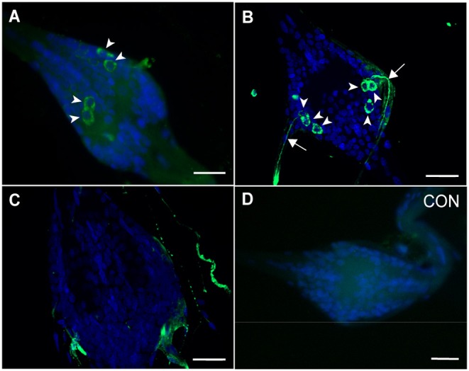
GPA2/GPB5 and its receptor constitute a glycoprotein hormone-signaling system native to the genomes of most vertebrate and invertebrate organisms. Unlike the well-studied gonadotropins and thyrotropin, the exact function of GPA2/GPB5 remains elusive, and whether it elicits its functions as heterodimers, homodimers or as independent monomers remains unclear. Here, the glycoprotein hormone signaling system was investigated in adult mosquitoes, where GPA2 and GPB5 subunit expression was mapped and modes of its signaling were characterized. In adult Aedes aegypti mosquitoes, GPA2 and GPB5 transcripts co-localized to bilateral pairs of neuroendocrine cells, positioned within the first five abdominal ganglia of the central nervous system. Unlike GPA2/GPB5 homologs in human and fly, GPA2/GPB5 subunits in A. aegypti lacked evidence of heterodimerization. Rather, cross-linking analysis to determine subunit interactions revealed A. aegypti GPA2 and GPB5 subunits may form homodimers, although treatments with independent subunits did not demonstrate receptor activity. Since mosquito GPA2/GPB5 heterodimers were not evident by heterologous expression, a tethered fusion construct was generated for expression of the subunits as a single polypeptide chain to mimic heterodimer formation. Our findings revealed A. aegypti LGR1 elicited constitutive activity with elevated levels of cAMP. However, upon treatment with recombinant tethered GPA2/GPB5, an inhibitory G protein (Gi/o) signaling cascade is initiated and forskolin-induced cAMP production is inhibited. These results further support the notion that heterodimerization is a requirement for glycoprotein hormone receptor activation and provide novel insight to how signaling is achieved for GPA2/GPB5, an evolutionary ancient neurohormone.

Keywords: GPA2/GPB5; glycoprotein hormone; heterodimer; homodimer; leucine-rich repeat-containing G protein coupled-receptor 1; mosquito; thyrostimulin.


Figures

Figure 1
Figure 1
GPA2/GPB5 subunit transcript expression and localization in adult A. aegypti. (A,B) RT-qPCR examining GPA2 (A) and GPB5 (B) transcript expression in the central nervous system of adult mosquitoes, with significant enrichment in the abdominal ganglia (AG). Subunit transcript abundance is shown relative to their expression in the thoracic ganglia (TG). Mean ± SEM of three biological replicates. Columns denoted with different letters are significantly different from one another. Multiple comparisons two-way ANOVA test with Tukey's multiple comparisons (P < 0.05) to determine sex- and tissue-specific differences. Reproductive tissues (RT), alimentary canal (Gut), carcass (Carc), brain (B). Fluorescence in situ hybridization anti-sense (C,D,G–J) and sense (E,F) probes to determine GPA2 and GPB5 transcript localization (GPA2 and/or GPB5 transcript, red; nuclei, blue) in the abdominal ganglia of adult mosquitoes. Unlike sense probe controls (E,F), two bilateral pairs of cells (arrowheads) were detected with GPA2 (C,G) and GPB5 (D,H) anti-sense probes in the first five abdominal ganglia of adult male and female A. aegypti. Co-localization of the GPA2 (G) and GPB5 (H) transcript was verified by treating abdominal ganglia dually with a combination of GPA2 and GPB5 anti-sense probes (I,J) that revealed two, intensely-stained bilateral pairs of cells. In (C–F,I,J), microscope settings were kept identical when acquiring images of control and experimental ganglia. The second abdominal ganglion is depicted in each image as a representative, given that no differences in the number nor staining intensity of cells were observed between ganglia of a given mosquito. Scale bars are 50 μm in (C–F,I,J) and 40 μm in (G,H). Experimental procedures were repeated three-four times with five mosquitoes (of each sex) per trial.
Figure 2
Figure 2
Immunolocalization of GPB5 subunit expression in the abdominal ganglia of adult A. aegypti. Experimental treatments demonstrated GPB5 immunoreactivity (green) in two (A), and in some mosquitoes, three (B) bilateral pairs of neuroendocrine cells (arrowheads) within the first five abdominal ganglia of the ventral nerve cord (DAPI, blue). Optical sections of ganglia revealed axonal projections emanate from GPB5-immunoreactive cells through the lateral nerves (arrows). No cells were detected in the sixth, terminal ganglion (C) and in control treatments (CON) where GPB5 antibody was preabsorbed with GPB5 synthetic antigen (D). In (A,D) or (B,C), microscope settings were kept identical when acquiring images of control and experimental ganglia. The second abdominal ganglion is depicted in (A,B,D) as a representative, given that no differences in the number nor staining intensity of cells were observed between ganglia of a given mosquito. Scale bars are 25 μm in (A–D) and 20 μm in (B,C). Experimental procedures were repeated three-four times with five mosquitoes (of each sex) per trial.
Figure 3
Figure 3
Western blot analyses to determine the effects of glycosylation on homo- and heterodimer formation on the glycoprotein hormone (GPA2/GPB5) subunits in the mosquito, A. aegypti. (A) In untreated conditions, western blot analysis of GPA2 subunit alone reveals two bands at 16 and 13 kDa. Whereas, following treatment with PNGase, the higher molecular weight band disappears and the 13 kDa band is intensified. A thick, additional band at ~32 kDa appears when GPA2 protein is cross-linked with DSS, and this band migrates slightly lower to ~30 kDa when GPA2 protein is treated with both DSS and PNGase. (B) A 24 kDa band is observed in lanes loaded with untreated GPB5 subunit alone. Upon PNGase treatment, the 24 kDa band is not affected; however, upon treatment with DSS, a second faint band appears at 48 kDa that is not affected by deglycosylation. (C) Western blot analyses of co-expressed GPA2 and GPB5 subunits shows three distinct band sizes at 24, 16, and 13 kDa, corresponding to the GPB5 subunit and two forms of GPA2 subunit protein. Similar to (A), after treatment with PNGase, the higher molecular weight form of GPA2 is eliminated and the 13 kDa band intensifies. When GPA2/GPB5 protein is cross-linked, two additional bands are detected at ~48 and ~32 kDa; however, following cross-linking and PNGase treatment, the ~32 kDa band is eliminated leaving only the 30 kDa band along with the unaffected ~48 kDa band. GPA2 (A) and GPA2/GPB5 (C) subunit protein was resolved on either a 10% (top) or 15% (bottom) polyacrylamide gel under denaturing conditions.
Figure 4
Figure 4
Elucidating heterodimerization of H. sapiens (human) (A) and A. aegypti (mosquito) (B) GPA2 and GPB5 subunits. Single promoter expression constructs for human and mosquito GPA2-FLAG and GPB5-His were used for transient expression in HEK 293T cells. Protein was harvested and subsequently concentrated, treated with DSS cross-linker, and probed with an anti-His or an anti-FLAG antibody after SDS-PAGE. (A,B) No bands were detected in lanes loaded with cross-linked GPA2-FLAG protein (Lane 1) or cross-linked GPB5-His protein (Lane 6), probed with an anti-His antibody or anti-FLAG antibody, respectively. (A) Bands corresponding to the monomeric form (18 kDa) and homodimer (36 kDa) of cross-linked human GPB5-His (Lane 3) and to the monomeric form (20 kDa) and homodimer (40 kDa) of cross-linked human GPA2-FLAG protein (Lane 4). A combination of the subunits with subsequent cross-linking of separately-produced human GPA2-FLAG and GPB5-His protein (Lane 2, 5) revealed a band size correlating to the human GPA2/GPB5 heterodimer (38 kDa), detected using an anti-His (Lane 2) or anti-FLAG (Lane 5) antibody. (B) Bands corresponding to the mosquito GPB5 monomer (24 kDa) (Lane 3), mosquito GPA2 glycosylated monomer (16 kDa) and homodimer pairs (30 and 32 kDa) (Lane 4). No detection of bands correlating to mosquito GPA2/GPB5 heterodimer (37–40 kDa) were observed, when probed with either anti-His (Lane 2) or anti-FLAG (Lane 5) antibodies.
Figure 5
Figure 5
cAMP-mediated bioluminescence assays to determine the effect of GPA2, GPB5, and GPA2/GPB5 on G-protein signaling of H. sapiens (human) TSHR (A), A. aegypti (mosquito) LGR1 (B,C), or cells expressing a red fluorescent protein, mCherry (D). Secreted protein fractions for each subunit were prepared separately from HEK 293T cells expressing human GPA2 (hA2), human GPB5 (hB5), mosquito GPA2 (A2), mosquito GPB5 (B5), mCherry, or co-expressing mosquito GPA2 and GPB5 in a dual promoter plasmid (A2/B5 coexp). Secreted protein fractions were then tested separately or combined (A2 + B5) and then incubated with cells co-expressing the cAMP biosensor along with either (A) human TSHR, (B,C) mosquito LGR1 or (D) mCherry, the latter of which was used as a negative control in the functional assay and also served to verify transfection efficiency of HEK 293T cells. (A–D) Luminescent values were recorded and normalized to luminescence response from treatments with 250 nM forskolin. (A) Unlike treatments with human GPA2 (hA2) or human GPB5 (hB5) applied singly, a significant increase in cAMP-mediated luminescence was observed when TSHR-expressing cells were incubated with culture media containing both human GPA2 and human GPB5 (hA2 + hB5), relative to incubations with mCherry controls. (B) No differences in luminescence were observed when LGR1-expressing cells were incubated with media containing mosquito GPA2/GPB5 subunits, compared to mCherry controls. (C,D) To test Gi/o signaling pathways, experiments were performed in the presence of 250 nM forskolin to determine the ability for each ligand to inhibit a forskolin-induced cAMP response. (C) The addition of GPB5 on LGR1-expressing cells significantly inhibited forskolin-induced luminescent response, compared to treatments with mCherry controls; (D) however, this inhibition was also observed when GPA2 and GPB5 proteins were incubated with HEK 293T cells in the absence of LGR1. Mean ± SEM of three (A,B,D) or six (C) biological replicates. Columns denoted with different letters are significantly different from one another. Multiple comparisons one-way ANOVA test with Tukey's multiple comparisons (P < 0.05).
Figure 6
Figure 6
Verification of A. aegypti GPA2/GPB5 tethered protein expressed in HEK 293T cells. (A) Western blot analysis of secreted or cell lysate protein fractions of HEK 293T expressing tethered GPA2/GPB5 (tA2/B5) or red fluorescent protein (mCherry) as a control. Tethered GPA2/GPB5 is represented as two bands at 32 and 37 kDa band in cell lysate fractions, and as two bands at 37 and 40 kDa in secreted fractions; however, no bands were detected in lanes loaded with proteins from mCherry transfected cells. (B) Upon treatment of tethered GPA2/GPB5 secreted protein fractions with PNGase, the 40 kDa band is eliminated and the 37 kDa band intensifies, indicating removal of N-linked oligosaccharides.
Figure 7
Figure 7
cAMP-mediated bioluminescence assay determining the effects of tethered GPA2/GPB5 on receptor activation and G protein signaling of LGR1. Secreted protein fractions (A,C) and cell lysates (B,D) derived from cells transiently expressing tethered GPA2/GPB5 (tA2/B5) or red fluorescent protein (mCherry) were tested for their ability to stimulate (Gs signaling) (A,B) or inhibit 1 μM forskolin-induced (Gi/o signaling) (C,D) cAMP-mediated luminescence. (A–D) Luminescence response was recorded and shown as normalized to the luminescence from sole treatments with 1 μM forskolin on LGR1-expressing cells. (C,D) In treatments involving mCherry proteins, the relative luminescence response was greater in LGR1-expressing cells (+ LGR1) compared to cells not expressing LGR1 (–LGR1). (A,B) Incubation of LGR1-expressing cells with tA2/B5 secreted (A) or cell lysate (B) proteins, failed to increase cAMP-mediated luminescence above background levels from incubations with mCherry proteins. (C,D) 1 μM forskolin along with either mCherry proteins or tA2/B5 proteins was added to cells in the presence or absence of LGR1 expression. The ability for each ligand treatment to reduce a forskolin-induced increase in cAMP luminescence was examined. (C) The tA2/B5 secreted protein samples incubated with LGR1-expressing cells did not significantly affect the forskolin-induced cAMP luminescence, compared to control treatments with secreted proteins from mCherry expressing cells. (D) Relative to incubations with mCherry cell lysate proteins, cell lysates containing tA2/B5 protein significantly inhibited forskolin-induced elevations of cAMP-mediated luminescence, and this inhibition was specific to LGR1-expressing cells. Mean ± SEM of three biological replicates. Columns denoted with different letters are significantly different from one another. Multiple comparisons two-way ANOVA test with Tukey's multiple comparisons (P < 0.05).

References

    1. Hartree AS, Renwick AGC. Molecular structures of glycoprotein hormones and functions of their carbohydrate components. Biochem J. (1992) 287(Pt 3):665–79. 10.1042/bj2870665 - DOI - PMC - PubMed
    1. Pierce JG, Parsons TF. Glycoprotein hormones: structure and function. Annu Rev Biochem. (1981) 50:465–95. 10.1146/annurev.bi.50.070181.002341 - DOI - PubMed
    1. Hsu SY, Nakabayashi K, Bhalla A. Evolution of glycoprotein hormone subunit genes in bilateral metazoa: identification of two novel human glycoprotein hormone subunit family genes, GPA2 and GPB5. Mol Endocrinol. (2002) 16:1538–51. 10.1210/mend.16.7.0871 - DOI - PubMed
    1. Nakabayashi K, Matsumi H, Bhalla A, Bae J, Mosselman S, Hsu SY, et al. . Thyrostimulin, a heterodimer of two new human glycoprotein hormone subunits, activates the thyroid-stimulating hormone receptor. J Clin Investig. (2002) 109:1445–52. 10.1172/JCI0214340 - DOI - PMC - PubMed
    1. Dos Santos S, Mazan S, Venkatesh B, Cohen-Tannoudji J, Quérat B. Emergence and evolution of the glycoprotein hormone and neurotrophin gene families in vertebrates. BMC Evol Biol. (2011) 11:332. 10.1186/1471-2148-11-332 - DOI - PMC - PubMed

Publication types