. 2020 Mar 31:8:230.
doi: 10.3389/fbioe.2020.00230. eCollection 2020.

A Developmental Engineering-Based Approach to Bone Repair: Endochondral Priming Enhances Vascularization and New Bone Formation in a Critical Size Defect

Affiliations

A Developmental Engineering-Based Approach to Bone Repair: Endochondral Priming Enhances Vascularization and New Bone Formation in a Critical Size Defect

Fiona E Freeman et al. Front Bioeng Biotechnol. .

Abstract

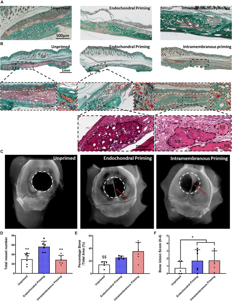
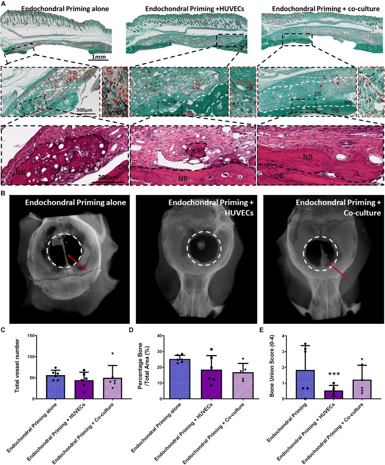
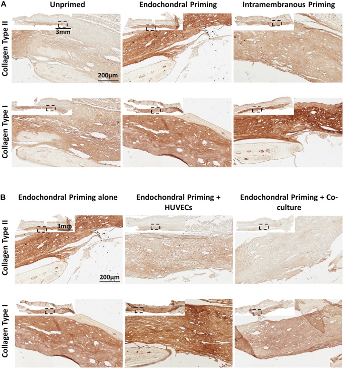
There is a distinct clinical need for new therapies that provide an effective treatment for large bone defect repair. Herein we describe a developmental approach, whereby constructs are primed to mimic certain aspects of bone formation that occur during embryogenesis. Specifically, we directly compared the bone healing potential of unprimed, intramembranous, and endochondral primed MSC-laden polycaprolactone (PCL) scaffolds. To generate intramembranous constructs, MSC-seeded PCL scaffolds were exposed to osteogenic growth factors, while endochondral constructs were exposed to chondrogenic growth factors to generate a cartilage template. Eight weeks after implantation into a cranial critical sized defect in mice, there were significantly more vessels present throughout defects treated with endochondral constructs compared to intramembranous constructs. Furthermore, 33 and 50% of the animals treated with the intramembranous and endochondral constructs respectively, had full bone union along the sagittal suture line, with significantly higher levels of bone healing than the unprimed group. Having demonstrated the potential of endochondral priming but recognizing that only 50% of animals completely healed after 8 weeks, we next sought to examine if we could further accelerate the bone healing capacity of the constructs by pre-vascularizing them in vitro prior to implantation. The addition of endothelial cells alone significantly reduced the healing capacity of the constructs. The addition of a co-culture of endothelial cells and MSCs had no benefit to either the vascularization or mineralization potential of the scaffolds. Together, these results demonstrate that endochondral priming alone is enough to induce vascularization and subsequent mineralization in a critical-size defect.

Keywords: bone tissue engineering; endochondral ossification; intramembranous ossification; mesenchymal stem cells; pre-vascularization.


Figures

FIGURE 1
FIGURE 1
(A) Morphology of human bone marrow MSCs attached to scaffolds 1.5 h after cell seeding as observed by SEM. Black scale bars represent 300 and 100 μm on the left and right, respectively. (B) Confocal microscopy of MSCs on scaffolds 24 h and 4 days after seeding cells. Actin cytoskeleton arrangement is shown in green by fluorescent staining with rhodamine phalloidin and nuclei are depicted in blue by DAPI staining. White scale bars represent 50 μm. (C) MSCs infiltration into scaffolds after 21 days post-culture as shown by fluorescent DAPI staining of cell nuclei in scaffold cryo-sections and H&E staining. Collagen matrix formation is observed in pink by pico-sirus red staining. Scale bars represents 250 μm.
FIGURE 2
FIGURE 2
(A) Masson’s Trichrome stained sections of all groups after 8 weeks in vivo, showing the osteoconductive nature of the scaffolds. (B) Masson’s Trichrome and H&E stained sections of all groups taken in the middle of the defect after 8 weeks in vivo. All Images taken at 20X. White dashed lines denoting periphery and center, OB denoted original bone, and NB denotes new bone. Red arrow heads denote vessels. (C) Representative X-ray images of the three experimental groups 8 weeks after implantation. Quantification of the amount of panel (D) total number of vessels, (E) percentage new bone, and (F) bone union score for all three experimental groups 8 weeks post implantation. Error bars denote standard deviation, **p < 0.01 vs. Endochondral Priming group, $$p < 0.01 vs. Intramembranous Priming group, n = 6 animals.
FIGURE 3
FIGURE 3
(A) Masson’s Trichrome and H&E stained sections of all groups taken in the middle of the defect after 8 weeks in vivo. All Images taken at 20X. White dashed lines denoting periphery and centre, OB denoted original bone, and NB denotes new bone. Red arrow heads denote vessels. (B) Representative X-ray images of the three experimental groups 8 weeks after implantation. Quantification of the amount of panel (C) total number of vessels, (D) percentage new bone, and (E) bone union score for all three experimental groups 8 weeks post implantation. Error bars denote standard deviation, ***p < 0.01 vs. Endochondral Priming alone group, n = 6 animals.
FIGURE 4
FIGURE 4
(A) Immunohistochemistry (Collage Type I and Collagen Type II) stained sections of Unprimed, Endochondral and Intramembranous primed groups taken in the middle of the defect after 8 weeks in vivo. All Images taken at 20X. (B) Immunohistochemistry (Collage Type I and Collagen Type II) stained sections of Endochondral priming alone, Endochondral priming + HUVECs and Endochondral priming + Co-culture groups taken in the middle of the defect after 8 weeks in vivo. All Images taken at 20X.

Similar articles

Cited by

References

    1. Abdal-hay A., Sheikh F. A., Lim J. K. (2013). Air jet spinning of hydroxyapatite/poly(lactic acid) hybrid nanocomposite membrane mats for bone tissue engineering. Colloids Surf. B Biointerfaces 102 635–643. 10.1016/j.colsurfb.2012.09.017 - DOI - PubMed
    1. Ahlmann E., Patzakis M., Roidis N., Shepherd L., Holtom P. (2002). Comparison of anterior and posterior iliac crest bone grafts in terms of harvest-site morbidity and functional outcomes. J. Bone Joint Surg. 84 716–720. 10.2106/00004623-200205000-00003 - DOI - PubMed
    1. Amini A. R., Laurencin C. T., Nukavarapu S. P. (2012). Bone tissue engineering: recent advances and challenges. Crit. Rev. Biomed. Eng. 40 363–408. 10.1615/critrevbiomedeng.v40.i5.10 - DOI - PMC - PubMed
    1. Brennan M. A., Renaud A., Amiaud J., Rojewski M. T., Schrezenmeier H., Heymann D., et al. (2014). Pre-clinical studies of bone regeneration with human bone marrow stromal cells and biphasic calcium phosphate. Stem Cell Res. Ther. 5:114. 10.1186/scrt504 - DOI - PMC - PubMed
    1. Brennan M. A., Renaud A., Gamblin A. L., D’Arros C., Nedellec S., Trichet V., et al. (2015). 3D cell culture and osteogenic differentiation of human bone marrow stromal cells plated onto jet-sprayed or electrospun micro-fiber scaffolds. Biomed. Mater. 10:045019. 10.1088/1748-6041/10/4/045019 - DOI - PubMed

LinkOut - more resources