Review
. 2020 Mar 31:8:203.
doi: 10.3389/fcell.2020.00203. eCollection 2020.

Multidimensional Imaging of Mammary Gland Development: A Window Into Breast Form and Function

Affiliations
Review

Multidimensional Imaging of Mammary Gland Development: A Window Into Breast Form and Function

Bethan Lloyd-Lewis. Front Cell Dev Biol. .

Abstract

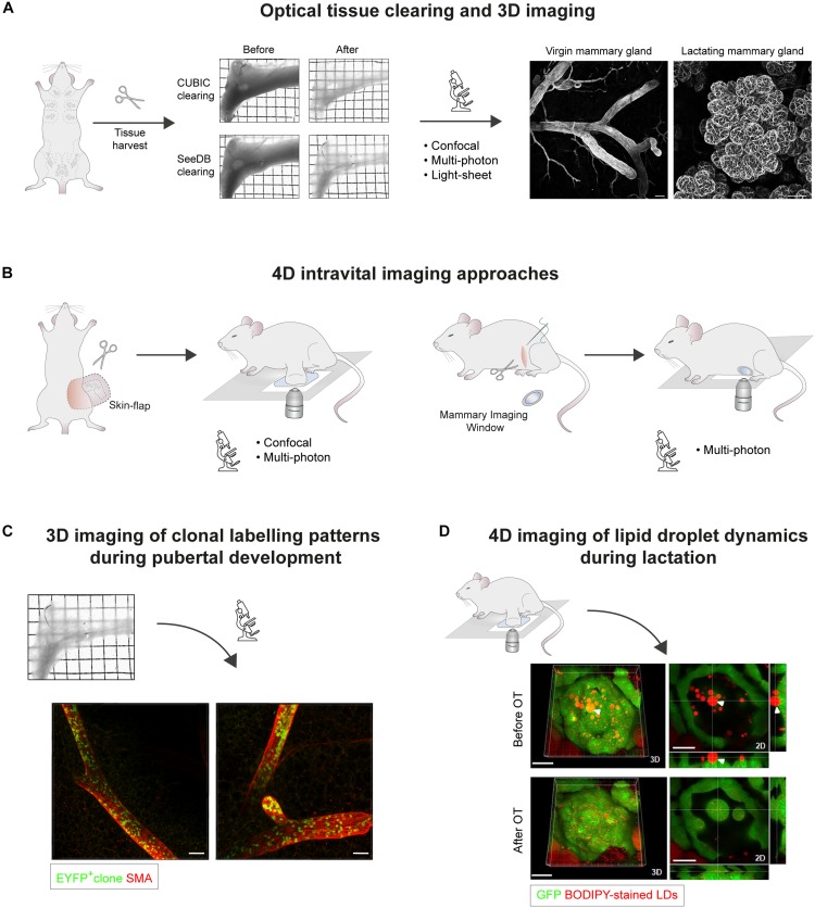
An in-depth appreciation of organ form and function relies on the ability to image intact tissues across multiple scales. Difficulties associated with imaging deep within organs, however, can preclude high-resolution multidimensional imaging of live and fixed tissues. This is particularly challenging in the mammary gland, where the epithelium lies deeply encased within a stromal matrix. Recent advances in deep-tissue and live imaging methodologies are increasingly facilitating the visualization of complex cellular structures within their native environment. Alongside, refinements in optical tissue clearing and immunostaining methods are enabling 3D fluorescence imaging of whole organs at unprecedented resolutions. Collectively, these methods are illuminating the dynamic biological processes underlying tissue morphogenesis, homeostasis, and disease. This review provides a snapshot of the current and state-of-the-art multidimensional imaging techniques applied to the postnatal mammary gland, illustrating how these approaches have revealed important new insights into mammary gland ductal development and lactation. Continual evolution of multidimensional image acquisition and analysis methods will undoubtedly offer further insights into mammary gland biology that promises to shed new light on the perturbations leading to breast cancer.

Keywords: 3D imaging; 4D imaging; breast cancer; intravital microscopy; lactation; mammary gland development; mammary stem cells.


Figures

FIGURE 1
FIGURE 1
Microscopic 3D and 4D imaging of mammary gland ductal development and lactation. (A) Optical tissue clearing and 3D imaging of fixed mammary tissues. Transmission images of harvested abdominal mammary glands before and after tissue clearing using CUBIC or SeeDB protocols. Grid width: 2 mm. 3D confocal imaging of mammary epithelial structures immunostained for Smooth Muscle Actin (SMA) in cleared virgin and lactating mammary tissues. Scale bars, 100 μm. (B) 4D intravital imaging approaches. Intravital microscopy can be performed either by surgically exposing the tissue via a skin-flap incision for multiple hours (non-recovery imaging, <40 h), or by implanting optical imaging windows for longitudinal imaging spanning multiple days to weeks. While confocal microscopy is suitable for imaging superficial tissue regions, multiphoton excitation is required for deep-tissue imaging, particularly through mammary imaging windows. (C) Clonal patterns arising from the genetic labeling of a single EYFP+ epithelial cell in the mammary gland of a ∼7 week old R26[CA]30EYFP mouse. SeeDB tissue clearing and immunostaining were performed prior to 3D imaging by confocal microscopy. Labeled progeny span multiple ducts and branches, and exhibit a sporadic, interspersed labeling pattern, emphasizing the importance of performing whole-gland and/or deep tissue 3D imaging for accurate clonal analysis. These patterns likely arise from the proliferation and intermixing of both labeled and unlabeled terminal end bud (TEB)-resident precursors, which have equipotent potential to contribute to ductal elongation. Scale bars, 100 μm. (D) Confocal intravital imaging of fluorescent BODIPY-stained lipid droplets (LDs) in surgically-exposed lactating mammary glands. Release of LDs from the apical surface is mediated by oxytocin (OT)-induced myoepithelial cell contractions. 3D images and 2D sections of the same alveolus before and after OT exposure are shown. White arrowhead points to an LD that was embedded in the cytoplasm prior to alveolus contraction. Scale bars, 30 μm. Images in (A,C) adapted from Davis et al. (2016) Nature Communications, under https://creativecommons.org/licenses/by/4.0/. Images in (D) adapted from Masedunskas et al. (2017) Mol Biol Cell, under https://creativecommons.org/licenses/by-nc-sa/3.0/.

References

    1. Alieva M., Ritsma L., Giedt R. J., Weissleder R., van Rheenen J. (2014). Imaging windows for long-term intravital imaging: general overview and technical insights. Intravital 3:e29917. 10.4161/intv.29917 - DOI - PMC - PubMed
    1. Andresen V., Alexander S., Heupel W. M., Hirschberg M., Hoffman R. M., Friedl P. (2009). Infrared multiphoton microscopy: subcellular-resolved deep tissue imaging. Curr. Opin. Biotechnol. 20 54–62. 10.1016/j.copbio.2009.02.008 - DOI - PubMed
    1. Banerjee M. R., Wagner J. E. (1972). Gene amplification in mammary gland at differentiation. Biochem. Biophys. Res. Commun. 49 480–487. 10.1016/0006-291X(72)90436-6 - DOI - PubMed
    1. Banerjee M. R., Wagner J. E., Kinder D. L. (1971). DNA synthesis in the absence of cell reproduction during functional differentiation of mouse mammary gland. Life Sci. 10 867–877. 10.1016/0024-3205(71)90199-8 - DOI - PubMed
    1. Benninger R. K. P., Piston D. W. (2013). Two-photon excitation microscopy for unit 4.11 the study of living cells and tissues. Curr. Protoc. Cell Biol. Chapter 4 Unit 4.11.1–24. 10.1002/0471143030.cb0411s59. - DOI - PMC - PubMed

LinkOut - more resources