Review
. 2020 Feb 24;3(2):221-236.
doi: 10.1021/acsptsci.0c00006. eCollection 2020 Apr 10.

Emerging Role of Compartmentalized G Protein-Coupled Receptor Signaling in the Cardiovascular Field

Affiliations
Review

Emerging Role of Compartmentalized G Protein-Coupled Receptor Signaling in the Cardiovascular Field

Bianca Plouffe et al. ACS Pharmacol Transl Sci. .

Abstract

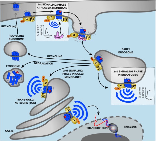
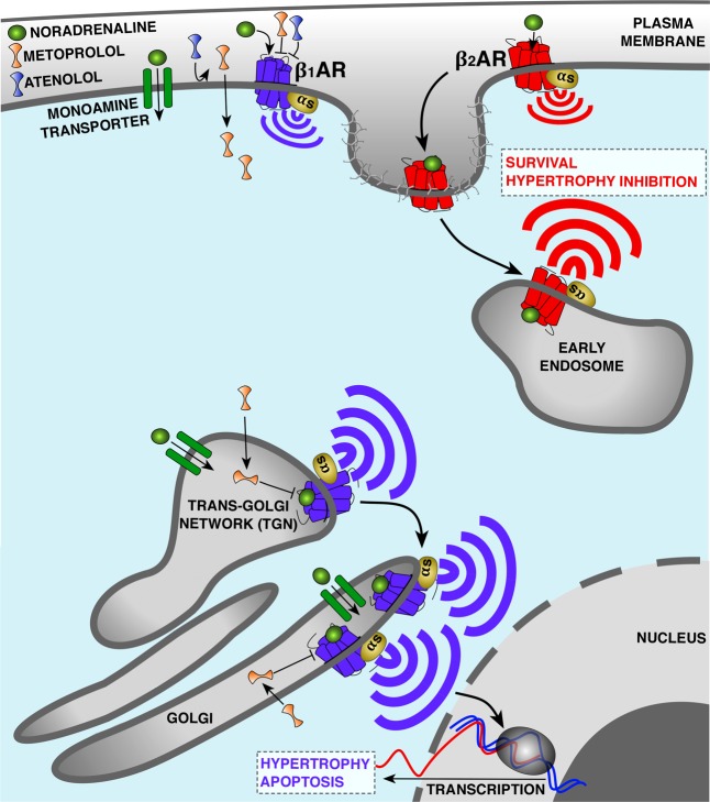
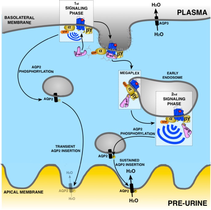
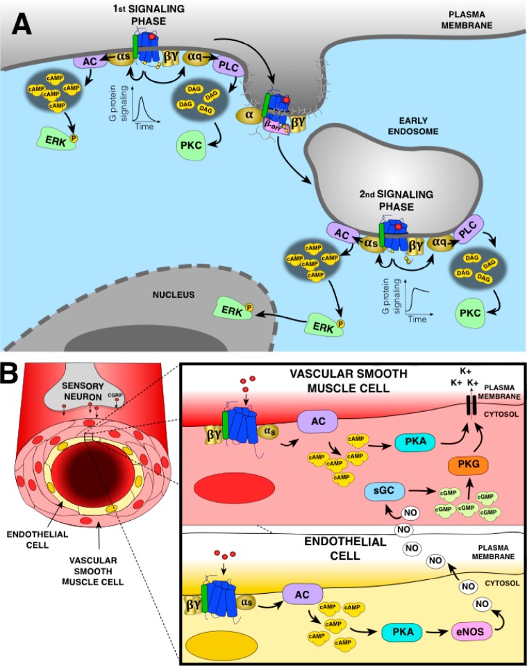
G protein-coupled receptors (GPCRs) are cell surface receptors that for many years have been considered to function exclusively at the plasma membrane, where they bind to extracellular ligands and activate G protein signaling cascades. According to the conventional model, these signaling events are rapidly terminated by β-arrestin (β-arr) recruitment to the activated GPCR resulting in signal desensitization and receptor internalization. However, during the past decade, emerging evidence suggest that many GPCRs can continue to activate G proteins from intracellular compartments after they have been internalized. G protein signaling from intracellular compartments is in general more sustained compared to G protein signaling at the plasma membrane. Notably, the particular location closer to the nucleus is beneficial for selective cellular functions such as regulation of gene transcription. Here, we review key GPCRs that undergo compartmentalized G protein signaling and discuss molecular considerations and requirements for this signaling to occur. Our main focus will be on receptors involved in the regulation of important physiological and pathological cardiovascular functions. We also discuss how sustained G protein activation from intracellular compartments may be involved in cellular functions that are distinct from functions regulated by plasma membrane G protein signaling, and the corresponding significance in cardiovascular physiology.


Conflict of interest statement

The authors declare no competing financial interest.

Figures

Figure 1
Figure 1
Updated model of G protein signaling. Agonist binding to GPCRs is detected by the Gα subunit of heterotrimeric G proteins. This induces exchange of GDP for GTP causing dissociation of the GTP-bound Gα from Gβγ which both activate membrane-localized effectors. This signaling is followed by the phosphorylation of the receptor by GRKs leading to recruitment of β-arr to the phosphorylated receptor. This event causes G protein uncoupling from the receptor and signaling desensitization. Recruitment of β-arr also promotes GPCR internalization into CCPs and receptor trafficking to early endosomes where desensitized receptors dissociate from β-arr and recycle back to the plasma membrane, are directed to lysosomes for degradation, or can undergo another round of G protein activation from early endosomes or Golgi membranes. In contrast to G protein signaling at the plasma membrane which is rapidly dampened by β-arr, this second activation upon GPCR internalization is generally more sustained. The particular duration and location of G protein signaling is critical for many cellular processes.
Figure 2
Figure 2
Compartmentalized Gαs signaling by β-adrenerigic receptors (βAR) in cardiomyocytes. β1AR and β2AR are coupled to Gαs and stimulate cAMP production but elicit different effects on cardiac functions. In the case of heart failure (HF), β1AR promotes cardiomyocyte hypertrophy and apoptosis, whereas β2AR prevents these events. One hypothesis explaining this difference is the distinct subcellular localization of β1AR and β2AR. Both subtypes bind noradrenaline and activate Gαs at the plasma membrane. However, upon receptor internalization β2AR preferentially activates Gαs from early endosomes, while the biosynthetic pool of β1AR activates Gαs from the Golgi membranes. Proximity of active Golgi-localized β1AR with the nucleus allows transcription of genes mediating hypertrophy. Activation of Golgi-localized β1AR depends on the uptake 2 monoamine transporter, facilitating the transport of noradrenaline to the pool of receptors at the Golgi. β1AR antagonists are among the most widely used drugs to treat HF. Metoprolol, a hydrophobic and membrane-permeable β1AR selective antagonist, blocks both the plasma membrane and the Golgi pool of β1AR, whereas atenolol, a hydrophilic and membrane-impermeable antagonist, only inhibits β1AR signaling at the plasma membrane. Correlated with the differential abilities of these drugs to access internal sites of signaling, metoprolol is more efficient than atenolol at reducing cAMP response and heart rate as well as contraction/relaxation responses.
Figure 3
Figure 3
Regulation of water transport in epithelial cells of the kidney by V2R. Vasopressin binding to V2R inactivates Gαs at the plasma membrane. This promotes phosphorylation of aquaporin-2 (AQP2) favoring its insertion into the apical membrane and entry of water. This is followed by a rapid recruitment of β-arr binding to V2R in core conformation leading to uncoupling from G proteins and internalization of the V2R-β-arr complex into early endosomes promoting the removal of AQP2 from the apical membrane. However, the change of β-arr from core to tail conformation in the endosome allows the core region of V2R to be available to interact with Gαs and activate this G protein in early endosomes. As β-arr in tail conformation does not uncouple V2R from G proteins, Gαs activation can occur causing a sustained AQP2 phosphorylation and insertion into apical membrane for a long-lasting entry of water. Aquaporin-3 (AQP3) allows water transport from epithelial cells to plasma to increase arterial volume and pressure and to decrease plasma sodium concentration.
Figure 4
Figure 4
(A) CGRP signaling at the plasma membrane and endosomes in sensory neurons. At the plasma membrane, CGRP receptor activates Gαs and Gαq. Gαs activation stimulates adenylyl cyclase (AC) to generate cAMP leading to phosphorylation of cytosolic ERK. Gαq activation stimulates phospholipase Cβ (PLC), production of second messengers, and activation of cytosolic protein kinase C (PKC). Recruitment of β-arr to the CGRP receptor induces receptor uncoupling from G proteins and internalization in early endosomes where the receptor can activate again G proteins. Endosomal Gαs activation induces sustained ERK phosphorylation and its translocation to the nucleus, while endosomal Gαq activation activate PKC in the cytosol. (B) Action of CGRP on peripheral vasculature. In the vascular smooth muscle cells (VSMC), activation of CGRP receptor induces Gαs activation, AC-mediated production of cAMP leading to activation of protein kinase A (PKA) opening the potassium channels to induce relaxation. In the endothelial cells, Gαs activation by CGRP receptor also lead to PKA activation. Although in these cells PKA activates the endothelial nitric oxide synthase (eNOS) responsible of nitric oxide (NO) production, NO diffuses into VSMC to mediate vasorelaxation by activating the soluble guanylate cyclase (sGC) producing cGMP-activating protein kinase G (PKG) also opening the potassium channels.

References

    1. Pierce K. L.; Premont R. T.; Lefkowitz R. J. (2002) Seven-transmembrane receptors. Nat. Rev. Mol. Cell Biol. 3, 639–650. 10.1038/nrm908. - DOI - PubMed
    1. Hauser A. S.; Attwood M. M.; Rask-Andersen M.; Schioth H. B.; Gloriam D. E. (2017) Trends in GPCR drug discovery: new agents, targets and indications. Nat. Rev. Drug Discovery 16, 829–842. 10.1038/nrd.2017.178. - DOI - PMC - PubMed
    1. Wettschureck N.; Offermanns S. (2005) Mammalian G proteins and their cell type specific functions. Physiol. Rev. 85, 1159–1204. 10.1152/physrev.00003.2005. - DOI - PubMed
    1. Lefkowitz R. J. (2004) Historical review: a brief history and personal retrospective of seven-transmembrane receptors. Trends Pharmacol. Sci. 25, 413–422. 10.1016/j.tips.2004.06.006. - DOI - PubMed
    1. Pitcher J. A.; Freedman N. J.; Lefkowitz R. J. (1998) G protein-coupled receptor kinases. Annu. Rev. Biochem. 67, 653–692. 10.1146/annurev.biochem.67.1.653. - DOI - PubMed