. 2020 Aug 17;22(8):1150-1161.
doi: 10.1093/neuonc/noaa093.

In vitro modeling of glioblastoma initiation using PDGF-AA and p53-null neural progenitors

Affiliations

In vitro modeling of glioblastoma initiation using PDGF-AA and p53-null neural progenitors

Alexandra K Bohm et al. Neuro Oncol. .

Abstract

Background: Imagining ways to prevent or treat glioblastoma (GBM) has been hindered by a lack of understanding of its pathogenesis. Although overexpression of platelet derived growth factor with two A-chains (PDGF-AA) may be an early event, critical details of the core biology of GBM are lacking. For example, existing PDGF-driven models replicate its microscopic appearance, but not its genomic architecture. Here we report a model that overcomes this barrier to authenticity.

Methods: Using a method developed to establish neural stem cell cultures, we investigated the effects of PDGF-AA on subventricular zone (SVZ) cells, one of the putative cells of origin of GBM. We microdissected SVZ tissue from p53-null and wild-type adult mice, cultured cells in media supplemented with PDGF-AA, and assessed cell viability, proliferation, genome stability, and tumorigenicity.

Results: Counterintuitive to its canonical role as a growth factor, we observed abrupt and massive cell death in PDGF-AA: wild-type cells did not survive, whereas a small fraction of null cells evaded apoptosis. Surviving null cells displayed attenuated proliferation accompanied by whole chromosome gains and losses. After approximately 100 days in PDGF-AA, cells suddenly proliferated rapidly, acquired growth factor independence, and became tumorigenic in immune-competent mice. Transformed cells had an oligodendrocyte precursor-like lineage marker profile, were resistant to platelet derived growth factor receptor alpha inhibition, and harbored highly abnormal karyotypes similar to human GBM.

Conclusion: This model associates genome instability in neural progenitor cells with chronic exposure to PDGF-AA and is the first to approximate the genomic landscape of human GBM and the first in which the earliest phases of the disease can be studied directly.

Keywords: GBM; PDGF-AA; genome instability; glioblastoma; model; p53.


Figures

Fig. 1
Fig. 1
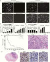
P53-null SVZ cell cultures acquire growth factor independence and become tumorigenic when cultured in PDGF-AA. (A) P53-null cells in media supplemented with EGF/FGF proliferate rapidly and form large spheres in early and late passage cultures but cease proliferating when EGF/FGF is removed (scale bar = 200 µM). (B) Early passage null cells display attenuated proliferation and sphere formation in PDGF-AA and remain PDGF-AA dependent. At late passages, cells rapidly form spheres in the presence or absence of PDGF-AA (scale bar = 200 µM). (C) Annexin V staining reveals that a high percentage of null SVZ cells (>70%) in early passage PDGF-AA cultures are dead or apoptotic; this proportion decreases over time. (D) EdU-labeling of null cultures supplemented with PDGF-AA shows an increase in the percentage of cells synthesizing DNA over time, increasing further when PDGF-AA independent proliferation is achieved. (E) Annexin-V/PI staining reveals that the proportion of dead, apoptotic and live null cells remains constant in EGF/FGF over time. (F) EdU-labeling shows that a high percentage EGF/FGF cultured null SVZ cells are replicating DNA and remain dependent on EGF/FGF supplementation. (G‒I) PDGF-AA independent transformed p53-null cells form infiltrating, hemorrhagic GBMs in wild-type mice. Tumor bearing mice (n = 32) had a median survival of 73 days (range, 50–420). Early passage p53-null SVZ cells cultured in PDGF-AA, or long-term EGF/FGF cultures did not form tumors. H&E staining reveals typical GBM features, including high cellularity, mitoses, and necrosis (scale bar = 50 μm). (J) Immunohistochemistry reveals presence of GBM markers, including GFAP, Nestin, and Olig2 positive cells (scale bar = 50 μm). (NS = P> 0.05; *P < 0.05; **P < 0.01; ***P < 0.001; ****P < 0.0001)
Fig. 2
Fig. 2
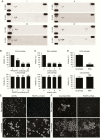
Transformed cells have variable RTK phosphorylation and are unaffected by PDGFR-α inactivation. (A) In PDGF-AA independent transformed lines and intracerebral xenografts, multiple patterns of RTK phosphorylation were observed including: PDGFR-α alone (I); PDGFR-α with EGFR (II); PDGFR-α with EGFR and HGFR (III); PDGFR-α with HGFR and AXLR [1 = PDGFR-α, 2 = EGFR, 3 = HFGR, 4 = AXLR] (B, C, D). Antibody-mediated inhibition of PDGFR-α (B), treatment with a PDGFR-α Imatinib (C), and knockdown of PDGFR-α (D) decrease cell viability in early but not late passage transformed cultures. (E, F) Blockade or knockdown of PDGFR-α leads to a decrease in cell proliferation and sphere formation in early but not late passage transformed p53-null SVZ cultures. (NS = P > 0.05; *P < 0.05; **P < 0.01; ***P< 0.001; ****P < 0.0001)
Fig. 3
Fig. 3
Transformed p53-null SVZ cells have an OPC lineage profile, proliferate in the presence of DNA damage, and evade apoptosis. (A, B) Assessment of stem and progenitor markers in primary versus transformed null cell cultures reveals that the percentage of markers consistent with a neural stem/progenitor identity increases significantly in transformed cultures, while markers of mature neural cells remain relatively constant and low. (C) Stem and cancer related surface marker analysis reveal heterogeneous expression of CD15, CD33, CD44, and CD133 in 4 independent transformed lines (I-IV). (D) Null cultures established in EGF/FGF retain the capacity to transform when transferred to PDGF-AA, but are not viable in the absence of growth factors. (E) γH2AX staining reveals an increase in DNA damage foci in PDGF-AA cultured cells compared with EGF/FGF. (F) γH2AX foci in null SVZ cells cultured in EGF/FGF or PDGF-AA increase with time in culture and are more abundant in PDGF-AA. (G) P53 wild-type and null cultures stop synthesizing DNA 6 days after being transferred from EGF/FGF to PDGF-AA. (H) P53 Wild-type and null cells undergo increased apoptosis when transferred from EGF/FGF to PDGF-AA. (I) Trypan blue exclusion demonstrates that wild-type and null cells undergo decreased proliferation in PDGF-AA; however, after 10 days null cells resume proliferating. (NS = P> 0.05; *P < 0.05; **P< 0.01; ***P< 0.001; ****P< 0.0001)
Fig. 4
Fig. 4
P53-null SVZ cells transformed in PDGF-AA have chromosomal instability. (A) G-band karyotyping of early passage p53-null cells cultured in PDGF-AA reveal heterozygous loss and gains of chromosomes (arrows). (B) G-band karyotypes of early (P4) to late (P18) passage p53-null cultures are shown. Transformed cells display chromosomal alterations that appear early and become increasing complex. (C) G-band karyotyping of p53-null EGF/FGF cultures reveals relatively normal chromosomal number and structure. (D) P53-null EGF/FGF cultures have relatively stable karyotypes over time. (E) aCGH of an early passage p53-null cells in EGF/FGF versus 3 pairs of early passage PDGF-AA dependent null cells and matched cell lines transformed in PDGF-AA. Cells in EGF/FGF have few alterations, whereas null cells in PDGF-AA have many large chromosomal alterations that increase over time in culture.

References

    1. Brennan CW, Verhaak RG, McKenna A, et al. ; TCGA Research Network The somatic genomic landscape of glioblastoma. Cell. 2013;155(2):462–477. - PMC - PubMed
    1. Ozawa T, Riester M, Cheng YK, et al. . Most human non-GCIMP glioblastoma subtypes evolve from a common proneural-like precursor glioma. Cancer Cell. 2014;26(2):288–300. - PMC - PubMed
    1. Korber V, Yang J, Barah P, et al. . Evolutionary trajectories of IDH(wt) glioblastomas reveal a common path of early tumorigenesis instigated years ahead of initial diagnosis. Cancer Cell. 2019; 35(4):692–704 e612. - PubMed
    1. Koga T, Benitez JA, Chaim IA, et al. . Glioblastomas derived from genetically modified pluripotent stem cells recapitulate pathobiology. BioRxiv. 2019. doi: 10.1101/576009. - DOI
    1. Jun HJ, Appleman VA, Wu HJ, et al. . A PDGFRα-driven mouse model of glioblastoma reveals a stathmin1-mediated mechanism of sensitivity to vinblastine. Nat Commun. 2018;9(1):3116. - PMC - PubMed

Publication types

MeSH terms

Substances