

Radiomics for liver tumours
- PMID: 32296901
- PMCID: PMC7498486
- DOI: 10.1007/s00066-020-01615-x
Radiomics for liver tumours
Abstract
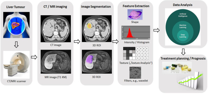
Current research, especially in oncology, increasingly focuses on the integration of quantitative, multiparametric and functional imaging data. In this fast-growing field of research, radiomics may allow for a more sophisticated analysis of imaging data, far beyond the qualitative evaluation of visible tissue changes. Through use of quantitative imaging data, more tailored and tumour-specific diagnostic work-up and individualized treatment concepts may be applied for oncologic patients in the future. This is of special importance in cross-sectional disciplines such as radiology and radiation oncology, with already high and still further increasing use of imaging data in daily clinical practice. Liver targets are generally treated with stereotactic body radiotherapy (SBRT), allowing for local dose escalation while preserving surrounding normal tissue. With the introduction of online target surveillance with implanted markers, 3D-ultrasound on conventional linacs and hybrid magnetic resonance imaging (MRI)-linear accelerators, individualized adaptive radiotherapy is heading towards realization. The use of big data such as radiomics and the integration of artificial intelligence techniques have the potential to further improve image-based treatment planning and structured follow-up, with outcome/toxicity prediction and immediate detection of (oligo)progression. The scope of current research in this innovative field is to identify and critically discuss possible application forms of radiomics, which is why this review tries to summarize current knowledge about interdisciplinary integration of radiomics in oncologic patients, with a focus on investigations of radiotherapy in patients with liver cancer or oligometastases including multiparametric, quantitative data into (radio)-oncologic workflow from disease diagnosis, treatment planning, delivery and patient follow-up.
Keywords: Artificial intelligence; Big data; Computed tomography; Magnetic resonance imaging; Stereotactic body radiation therapy.
Conflict of interest statement
C. Dreher, P. Linde, J. Boda-Heggemann and B. Baessler declare that they have no competing interests.
Figures
Similar articles
-
Applications of radiomics and machine learning for radiotherapy of malignant brain tumors.Strahlenther Onkol. 2020 Oct;196(10):856-867. doi: 10.1007/s00066-020-01626-8. Epub 2020 May 11. Strahlenther Onkol. 2020. PMID: 32394100 Free PMC article. Review.
-
Radiomics in radiation oncology-basics, methods, and limitations.Strahlenther Onkol. 2020 Oct;196(10):848-855. doi: 10.1007/s00066-020-01663-3. Epub 2020 Jul 9. Strahlenther Onkol. 2020. PMID: 32647917 Free PMC article. Review.
-
MR-guided adaptive versus ITV-based stereotactic body radiotherapy for hepatic metastases (MAESTRO): a randomized controlled phase II trial.Radiat Oncol. 2022 Mar 27;17(1):59. doi: 10.1186/s13014-022-02033-2. Radiat Oncol. 2022. PMID: 35346270 Free PMC article. Clinical Trial.
-
Radiomics and deep learning in lung cancer.Strahlenther Onkol. 2020 Oct;196(10):879-887. doi: 10.1007/s00066-020-01625-9. Epub 2020 May 4. Strahlenther Onkol. 2020. PMID: 32367456 Review.
-
Reinventing radiation therapy with machine learning and imaging bio-markers (radiomics): State-of-the-art, challenges and perspectives.Methods. 2021 Apr;188:44-60. doi: 10.1016/j.ymeth.2020.07.003. Epub 2020 Jul 19. Methods. 2021. PMID: 32697964 Review.
Cited by
-
Gd-EOB-DTPA enhanced MRI based radiomics combined with clinical variables in stratifying hepatic functional reserve in HBV infected patients.Abdom Radiol (NY). 2024 Apr;49(4):1051-1062. doi: 10.1007/s00261-023-04176-6. Epub 2024 Jan 31. Abdom Radiol (NY). 2024. PMID: 38294541
-
An overview of artificial intelligence in oncology.Future Sci OA. 2022 Feb 10;8(4):FSO787. doi: 10.2144/fsoa-2021-0074. eCollection 2022 Apr. Future Sci OA. 2022. PMID: 35369274 Free PMC article. Review.
-
Imaging of pediatric liver tumors: A COG Diagnostic Imaging Committee/SPR Oncology Committee White Paper.Pediatr Blood Cancer. 2023 Jun;70 Suppl 4(Suppl 4):e29965. doi: 10.1002/pbc.29965. Epub 2022 Sep 14. Pediatr Blood Cancer. 2023. PMID: 36102690 Free PMC article.
-
Machine Learning and AI in Cancer Prognosis, Prediction, and Treatment Selection: A Critical Approach.J Multidiscip Healthc. 2023 Jun 26;16:1779-1791. doi: 10.2147/JMDH.S410301. eCollection 2023. J Multidiscip Healthc. 2023. PMID: 37398894 Free PMC article. Review.
-
Deep Learning Algorithm for Differentiating Patients with a Healthy Liver from Patients with Liver Lesions Based on MR Images.Cancers (Basel). 2023 Jun 11;15(12):3142. doi: 10.3390/cancers15123142. Cancers (Basel). 2023. PMID: 37370752 Free PMC article.
References
Publication types
MeSH terms
LinkOut - more resources
Full Text Sources
Medical
