

The role of imaging in 2019 novel coronavirus pneumonia (COVID-19)
- PMID: 32296940
- PMCID: PMC7156903
- DOI: 10.1007/s00330-020-06827-4
The role of imaging in 2019 novel coronavirus pneumonia (COVID-19)
Abstract
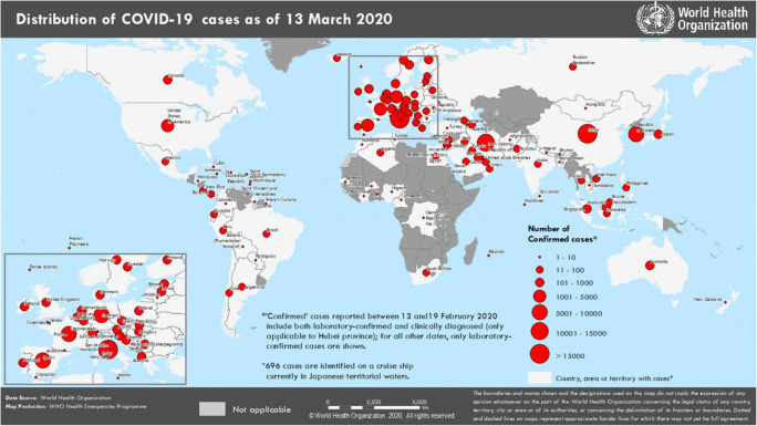
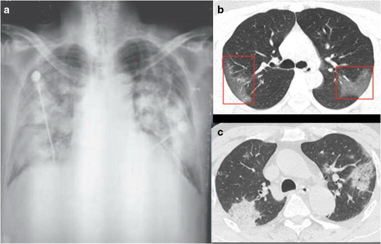
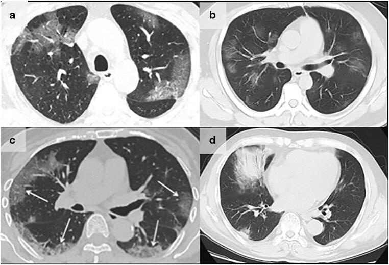
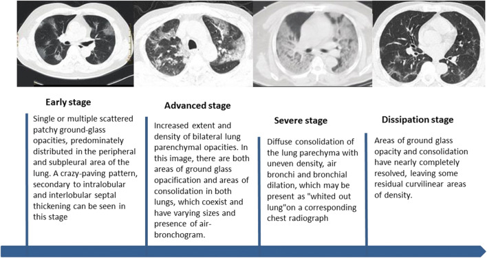
Almost the entire world, not only China, is currently experiencing the outbreak of a novel coronavirus that causes respiratory disease, severe pneumonia, and even death. The outbreak began in Wuhan, China, in December of 2019 and is currently still ongoing. This novel coronavirus is highly contagious and has resulted in a continuously increasing number of infections and deaths that have already surpassed the SARS-CoV outbreak that occurred in China between 2002 and 2003. It is now officially a pandemic, announced by WHO on the 11th of March. Currently, the 2019 novel coronavirus (SARS-CoV-2) can be identified by virus isolation or viral nucleic acid detection; however, false negatives associated with the nucleic acid detection provide a clinical challenge and thus make the imaging examination crucial. Imaging exams have been a main clinical diagnostic criteria for the 2019 novel coronavirus disease (COVID-19) in China. Imaging features of multiple patchy areas of ground glass opacity and consolidation predominately in the periphery of the lungs are characteristic manifestations on chest CT and extremely helpful in the early detection and diagnosis of this disease, which aids prompt diagnosis and the eventual control of this emerging global health emergency. Key Points • In December 2019, China, an outbreak of pneumonia caused by a novel, highly contagious coronavirus raised grave concerns and posed a huge threat to global public health. • Among the infected patients, characteristic findings on CT imaging include multiple, patchy, ground-glass opacity, crazy-paving pattern, and consolidation shadows, mainly distributed in the peripheral and subpleural areas of both lungs, which are very helpful for the frontline clinicians. • Imaging examination has become the indispensable means not only in the early detection and diagnosis but also in monitoring the clinical course, evaluating the disease severity, and may be presented as an important warning signal preceding the negative RT-PCR test results.
Keywords: Coronavirus; Diagnostic imaging; Mass chest X-ray; Multidetector computed tomography; Pneumonia.
Conflict of interest statement
The authors have no conflict of interest to disclose.
Figures
References
-
- Mo Q, Qin W, Fei QH, et al. Correctly understand the influencing factors of nucleic acid detection of novel coronavirus. Chin J Lab Med. 2020;43(00):E002–E002.
Publication types
MeSH terms
Grants and funding
- 2019PT310025/Construction of Key Laboratory (Cultivation) of Chinese Academy of Medical Sciences
- 81971588 and 81771811/National Natural Science Foundation of China
- Z191100006619021/Capital Clinically Characteristic Applied Research Fund
- G20190001630/National Foreign Expert Talent Project
- 10023201900204/Education Reform Project of Peking Union Medical College
LinkOut - more resources
Full Text Sources
Miscellaneous
