Review
. 2021 Apr;44(2):1083-1091.
doi: 10.1007/s10143-020-01293-4. Epub 2020 Apr 15.

Surgical management of primary and secondary pilocytic astrocytoma of the cerebellopontine angle (in adults and children) and review of the literature

Affiliations
Review

Surgical management of primary and secondary pilocytic astrocytoma of the cerebellopontine angle (in adults and children) and review of the literature

Sasan Darius Adib et al. Neurosurg Rev. 2021 Apr.

Abstract

Glial tumors in the cerebellopontine angle (CPA) are uncommon and comprise less than 1% of CPA tumors. We present four cases of pilocytic astrocytoma of the CPA (PA-CPA) that were treated in our department. Patients who received surgical treatment for PA-CPA from January 2004 to December 2019 were identified by a computer search of their files from the Department of Neurosurgery, Tübingen. Patients were evaluated for initial symptoms, pre- and postoperative facial nerve function and cochlear function, complications, and recurrence rate by reviewing surgical reports, patient documents, neuroradiological data, and follow-up data. We identified four patients with PA-CPA out of about 1500 CPA lesions (~ 0.2%), which were surgically treated in our department in the last 16 years. Of the four patients, three were male, and one was a female patient. Two were adults, and two were children (mean age 35 years). A gross total resection was achieved in three cases, and a subtotal resection was attained in one case. Two patients experienced a moderate facial palsy immediately after surgery (House-Brackmann grade III). In all cases, the facial function was intact or good (House-Brackmann grades I-II) at the long-term follow-up (mean follow-up 4.5 years). No mortality occurred during follow-up. Three of the patients had no recurrence at the latest follow-up (mean latest follow-up 4.5 years), while one patient had a slight recurrence. PA-CPA can be safely removed, and most complications immediately after surgery resolve in the long-term follow-up.

Keywords: Brainstem; CPA; Cerebellopontine angle; Facial nerve; Glioma; Pilocytic astrocytoma.


Conflict of interest statement

The authors declare that they have no conflicts of interest.

Figures
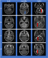
Fig. 1
Fig. 1
a Left (case 1), b left (case 2), c left (case 3), d left (case 4): Preoperative axial contrast-enhanced T1-weighted magnetic resonance imaging scans of each patient with PA-CPA (A+B: pediatric PA-CPA presented typically with a cystic cerebellar mass with enhancing mural nodule; C+D: adult PA-CPA presented atypically with only solid tumor components). a Middle, b middle, c middle, d middle: Postoperative axial contrast-enhanced T1-weighted magnetic resonance imaging scans; a gross total resection (GTR) of the PA-CPA was achieved in three cases (3/4). In one case, only a subtotal removal of the tumor was performed (blue block arrow). a Right, b right, c right, d right: Postoperative axial T2-weighted magnetic resonance images; anatomical facial nerve preservation was achieved during surgery in all cases (red block arrows)
Fig. 2
Fig. 2
Different steps of tumor resection strategy: in case of large cystic component, a decompression of the cyst should be performed (a) and, furthermore, if possible, a removal of the cyst wall (b). After removal of the cyst, the surface of the solid tumor portion was incised (c) and a debulking was performed (d)

Similar articles

Cited by

References

    1. Ahn MS, Jackler RK. Exophytic brain tumors mimicking primary lesions of the cerebellopontine angle. Laryngoscope. 1997;107:466–471. doi: 10.1097/00005537-199704000-00008. - DOI - PubMed
    1. Arnautovic KI, Husain MM, Linskey ME. Cranial nerve root entry zone primary cerebellopontine angle gliomas: a rare and poorly recognized subset of extraparenchymal tumors. J Neuro-Oncol. 2000;49:205–212. doi: 10.1023/a:1006488905526. - DOI - PubMed
    1. Beutler AS, Hsiang JK, Moorhouse DF, Hansen LA, Alksne JF. Pilocytic astrocytoma presenting as an extra-axial tumor in the cerebellopontine angle: case report. Neurosurgery. 1995;37:125–128. doi: 10.1227/00006123-199507000-00019. - DOI - PubMed
    1. Bonneville F, Savatovsky J, Chiras J. Imaging of cerebellopontine angle lesions: an update. Part 1: enhancing extra-axial lesions. Eur Radiol. 2007;17:2472–2482. doi: 10.1007/s00330-007-0679-x. - DOI - PubMed
    1. Bonneville F, Savatovsky J, Chiras J. Imaging of cerebellopontine angle lesions: an update. Part 2: intra-axial lesions, skull base lesions that may invade the CPA region, and non-enhancing extra-axial lesions. Eur Radiol. 2007;17:2908–2920. doi: 10.1007/s00330-007-0680-4. - DOI - PubMed

MeSH terms