. 2020 Jun;23(2):217-221.
doi: 10.1007/s40477-020-00458-7. Epub 2020 Apr 16.

Thoracic ultrasound and SARS-COVID-19: a pictorial essay

Affiliations

Thoracic ultrasound and SARS-COVID-19: a pictorial essay

Soccorsa Sofia et al. J Ultrasound. 2020 Jun.

Abstract

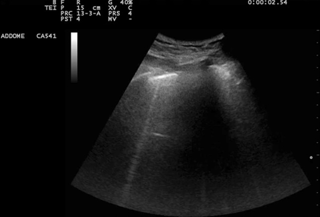
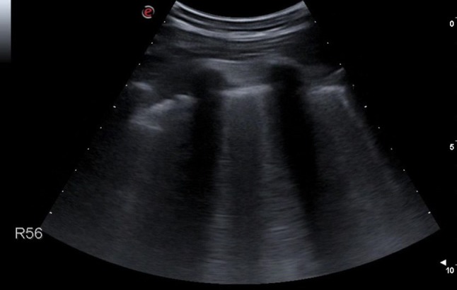
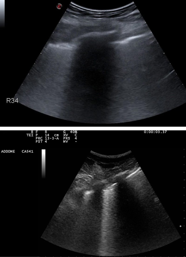
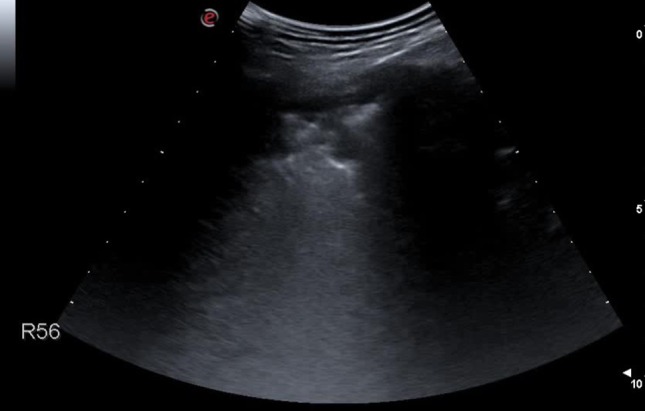
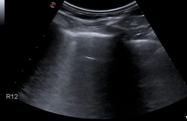
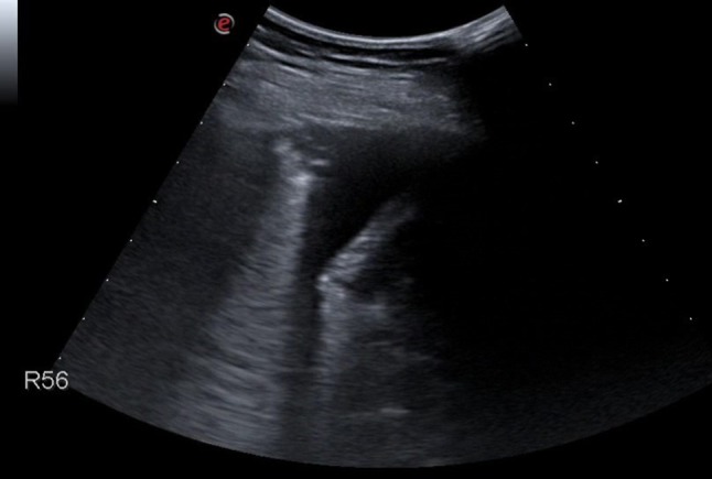
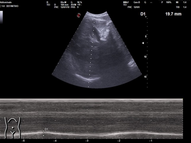
Thoracic ultrasound seems to adapt to the screening for lung involvement of patients with suspected or ascertained SARS-COVID-19 infection due to its characteristics of easy applicability. It can be also a relevant method in monitoring patients. B lines are early finding of COVID-19, even in mild-symptomatic subjects; in the most serious cases such as pre-ARDS or ARDS, the B lines end up filling the ultrasound image almost completely, until it merges, so as to create a single hyperechoic image named as "white lung", with distortion and irregularity of the pleural line. In advanced stage, lung consolidations are present, representing pulmonary pathological areas that are no longer normally ventilated.

Keywords: COVID-19; Lung; Ultrasound; Virus.


Conflict of interest statement

The authors declare no conflicts of interest and no funding sources.

Figures

Fig. 1
Fig. 1
Lateral chest scan. Single vertical hyperechoic artifact originating from the pleural line, called “B line”
Fig. 2
Fig. 2
Posterior chest area scan. Vertical artifacts (“B lines”), isolated and non-confluent
Fig. 3–4
Fig. 3–4
Lateral chest scans. Vertical artifacts (“B lines”), numerous and coalescent at a stretch of pleural line of increased thickness
Fig. 5
Fig. 5
Postero-basal chest scan. Lung field is occupied by numerous B lines that come together to form a single hyperechoic image. This aspect is named “white lung,” referring to the ground-glass opacities found in the computed chest tomography
Fig. 6
Fig. 6
Longitudinal posterior chest scan. Coarse subpleural thickening with very irregular margins
Fig. 7
Fig. 7
Lateral chest scan. Coarse supleural thickening with thin aerial bronchograms
Fig. 8
Fig. 8
Anterior chest longitudinal scan. Minute subpleural parenchymal thickening
Fig. 9
Fig. 9
Posterior chest longitudinal scan. Small basal pleural effusion
Fig. 10
Fig. 10
Ultrasound evaluation of diaphragm excursion. An oblique subcostal scan should be performed by convex probe (scan for the gallbladder), by adjusting the depth of the image so as to completely highlight the liver margins, with the diaphragm represented as a hyperechoic line surrounding the liver. The M-mode option is activated by centering the line on the bed of the gallbladder and evaluating the degree of excursion of the diaphragm

References

    1. Zhu N. A novel coronavirus from patients with pneumonia in China, 2019. N Engl J Med. 2020;382(8):727–733. doi: 10.1056/NEJMoa2001017. - DOI - PMC - PubMed
    1. Huang C. Clinical features of patients infected with 2019 novel coronavirus in Wuhan. China Lancet. 2020;395(10223):497–506. doi: 10.1016/S0140-6736(20)30183-5. - DOI - PMC - PubMed
    1. National Health Commission of People’s Republic of China (2020) Pneumonia diagnosis and treatment of 2019-nCoV infection from Chinese NHC and CDC 2020. https://www.nhc.gov.cn/xcs/zhengcwj/202001/4294563ed35b43209b31739bd0785.... Accessed 1 Feb 2020
    1. Weiss P, Murdoch DR. Clinical course and mortality risk of severe COVID-19. Lancet. 2020 doi: 10.1016/S0140-6736(20)30633-4. - DOI - PMC - PubMed
    1. Shi H, Han X, Jiang N, Cao Y, Alwalid O, Gu J, et al. Radiological findings from 81 patients with COVID-19 pneumonia in Wuhan, China: a descriptive study. Lancet Infect Dis. 2020 doi: 10.1016/S1473-3099(20)30086-4. - DOI - PMC - PubMed

LinkOut - more resources