. 2020 Jul 1;130(7):3684-3698.
doi: 10.1172/JCI136908.

IL-1β suppression of VE-cadherin transcription underlies sepsis-induced inflammatory lung injury

Affiliations

IL-1β suppression of VE-cadherin transcription underlies sepsis-induced inflammatory lung injury

Shiqin Xiong et al. J Clin Invest. .

Erratum in

Abstract

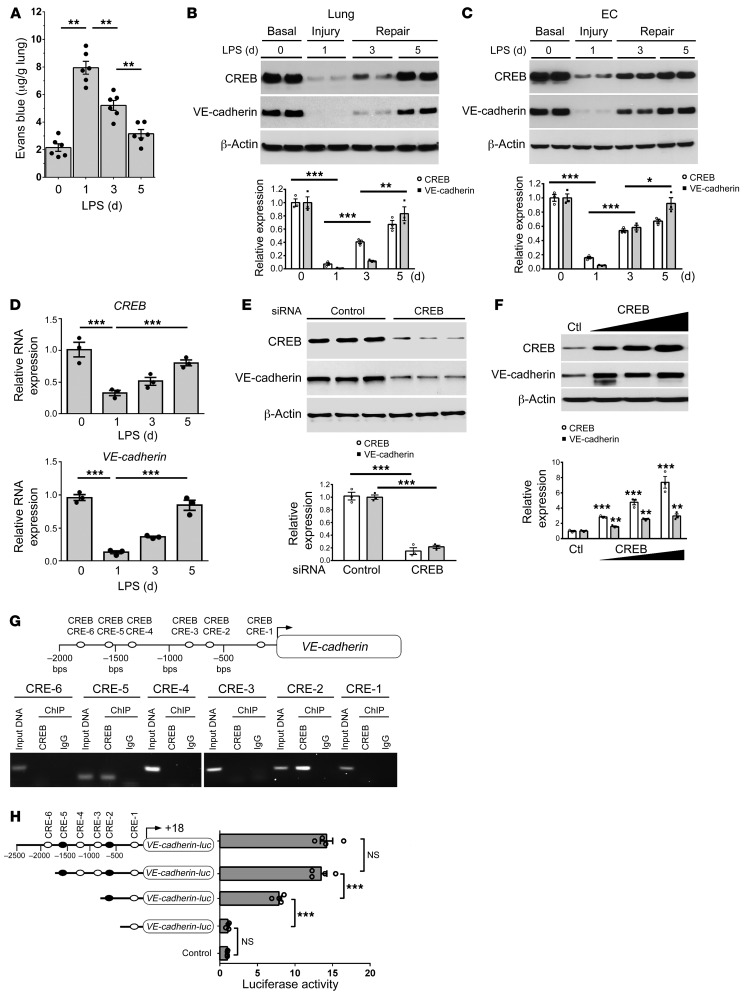
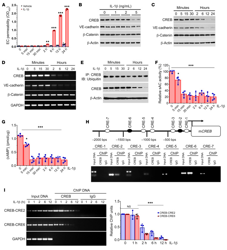
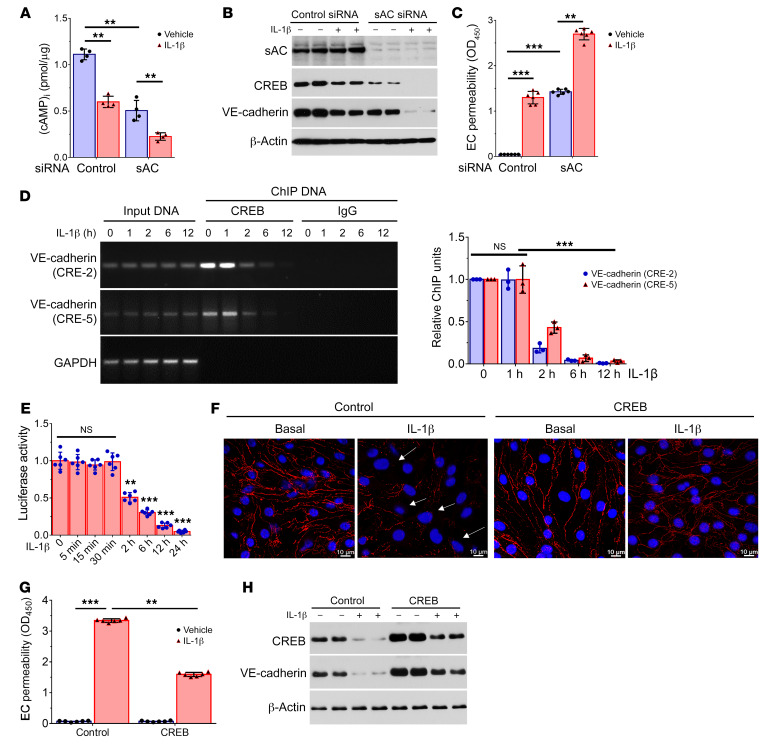
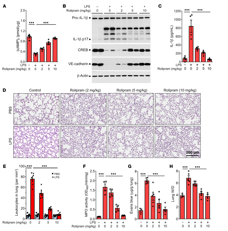
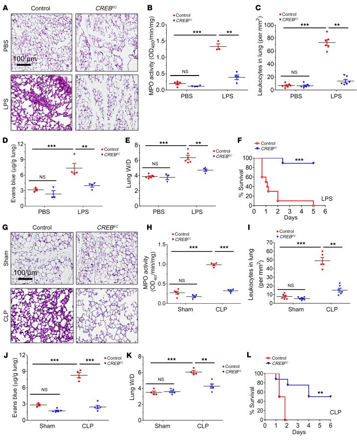
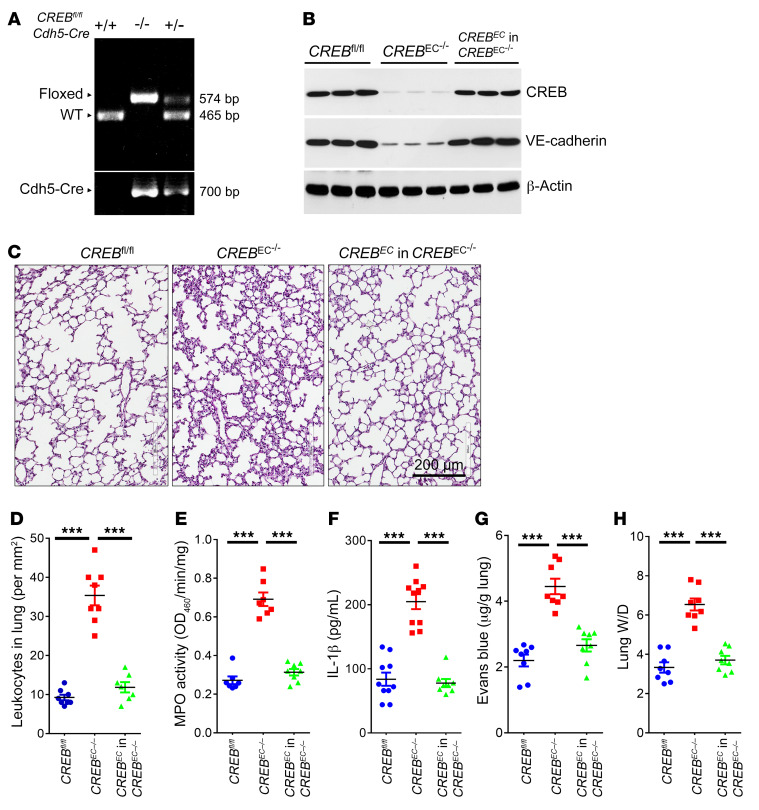
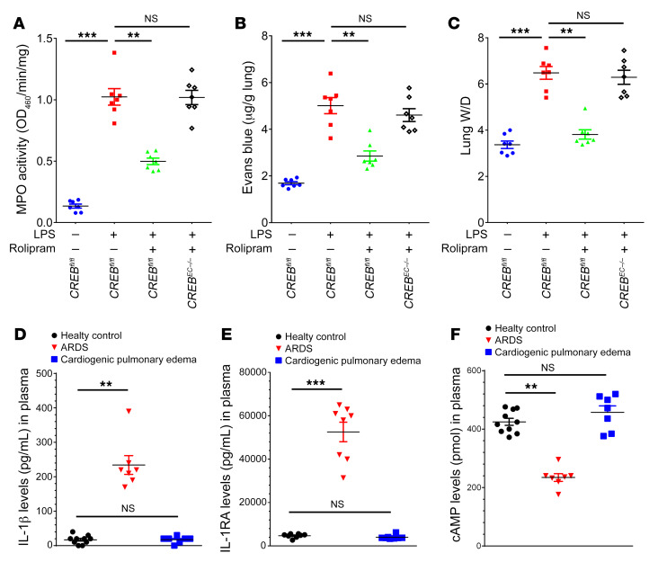
Unchecked inflammation is a hallmark of inflammatory tissue injury in diseases such as acute respiratory distress syndrome (ARDS). Yet the mechanisms of inflammatory lung injury remain largely unknown. Here we showed that bacterial endotoxin lipopolysaccharide (LPS) and cecal ligation and puncture-induced (CLP-induced) polymicrobial sepsis decreased the expression of transcription factor cAMP response element binding (CREB) in lung endothelial cells. We demonstrated that endothelial CREB was crucial for VE-cadherin transcription and the formation of the normal restrictive endothelial adherens junctions. The inflammatory cytokine IL-1β reduced cAMP generation and CREB-mediated transcription of VE-cadherin. Furthermore, endothelial cell-specific deletion of CREB induced lung vascular injury whereas ectopic expression of CREB in the endothelium prevented the injury. We also observed that rolipram, which inhibits type 4 cyclic nucleotide phosphodiesterase-mediated (PDE4-mediated) hydrolysis of cAMP, prevented endotoxemia-induced lung vascular injury since it preserved CREB-mediated VE-cadherin expression. These data demonstrate the fundamental role of the endothelial cAMP-CREB axis in promoting lung vascular integrity and suppressing inflammatory injury. Therefore, strategies aimed at enhancing endothelial CREB-mediated VE-cadherin transcription are potentially useful in preventing sepsis-induced lung vascular injury in ARDS.

Keywords: Inflammation; Innate immunity; Vascular Biology; endothelial cells.


Conflict of interest statement

Conflict of interest: The authors have declared that no conflict of interest exists.

Figures

Figure 1
Figure 1. Endotoxin impairs CREB-mediated VE-cadherin expression and promotes lung vascular injury.
(AD) C57BL/6J mice (n = 6) were injected intraperitoneally with LPS (12 mg/kg) for 1, 3, and 5 days. Lung vascular permeability was analyzed (A). Western blot detection of protein expression in whole lung lysates (B) and in fresh isolated endothelial cells (C) from LPS-challenged mice. (D) Real-time PCR detection of CREB and VE-cadherin mRNA levels in freshly isolated endothelial cells (n = 3). (E) CREB depletion by siRNA decreases VE-cadherin expression in mLMVECs (n = 3). (F) Overexpression of CREB dose-dependently increases VE-cadherin levels in mLMVECs (n = 3). (G) CREB binds to the mouse VE-cadherin promoter in mLMVECs by ChIP assays. Soluble chromatin was coimmunoprecipitated with anti-CREB or an equal amount of rabbit IgG. DNA purified from starting (1% input) and immunoprecipitated samples was subjected to PCR. The amplified region surrounds each proposed CRE binding element within the mouse VE-cadherin promoter regions. (H) Identification of central CREB CREs enabling VE-cadherin transcription by reporter assay (n = 4). mLMVECs were transiently transfected with the indicated sequential deletion luciferase constructs or pGL3.0 basic control vector together with CREB expression vector, and the internal control p-RL-TK vector. Representative immunoblots and the bar graph quantification are shown. *P < 0.05; **P < 0.01; ***P < 0.001.
Figure 2
Figure 2. IL-1β represses cAMP-CREB signaling.
(A) mLMVECs were treated with IL-1β (5 ng/mL) or vehicle for the indicated time. Transwell permeability to HRP was measured (n = 6). (B and C) mLMVECs were stimulated with increasing doses of IL-1β for 24 hours (B) or stimulated with IL-1β (5 ng/mL) over time (C). Protein expression was detected by Western blot. (DG) mLMVECs were stimulated with IL-1β (5 ng/mL) over time. RNA expression levels were analyzed by RT-PCR (D). Ubiquitination status of CREB was analyzed by immunoprecipitation with CREB antibody and blotting with an anti-ubiquitin antibody (E). sAC activity was detected (F) (n = 4). Intracellular cAMP levels were measured (G) (n = 4). (H) CREB itself binds to the putative CREs within the CREB promoter in mLMVECs by ChIP assays. The amplified region surrounds each proposed CRE binding element within the mouse CREB promoter regions. (I) mLMVECs were stimulated with IL-1β (5 ng/mL) for the indicated time. Effect of IL-1β treatment on CREB binding to the promoter regions (CRE2 and CRE6) in mLMVECs was investigated by ChIP assays (n = 3). *P < 0.05; **P < 0.01; ***P < 0.001; NS, no significant difference. Statistics obtained from ANOVA.
Figure 3
Figure 3. IL-1β transcriptionally downregulates CREB-mediated VE-cadherin expression to increase endothelial permeability.
(AC) mLMVECs were transfected with siRNA of sAC or control scrambled siRNA for 3 days. Cells were then stimulated with IL-1β (5 ng/mL) for 12 hours. Intracellular cAMP levels were measured (A) (n = 4). Protein expression by Western blot (B) and permeability to HRP (C) were determined (n = 6). (D) mLMVECs were stimulated with IL-1β (5 ng/mL) for the indicated time. Effect of IL-1β treatment on CREB binding to the mouse VE-cadherin promoter regions (CRE2 and CRE5) in mLMVECs was investigated by ChIP assays (n = 3). (E) mLMVECs were transiently transfected with the 2.5-kb VE-cadherin promoter luciferase construct, CREB expression vector, and the internal control p-RL-TK vector by lipofectamine 3000 reagent for 2 days. Cells were then treated with IL-1β (5 ng/mL) over time. CREB-regulated VE-cadherin transcription activity was measured using the dual luciferase reporter assay system (n = 6). (FH) mLMVECs were transfected with CREB plasmids or control for 2 days, and stimulated with IL-1β (5 ng/mL) for 1 day. Immunofluorescence staining of VE-cadherin (F); arrows denote disruption of endothelial barrier integrity. Permeability to HRP (n = 6) (G) and protein expression by Western blot (H) were determined. **P < 0.01; ***P < 0.001; NS, no significant difference. Statistics obtained from ANOVA. Scale bars: 10 μm.
Figure 4
Figure 4. Augmentation of cAMP signaling by rolipram suppresses inflammatory lung injury.
(A) C57BL/6J mice (n = 4) were administrated with rolipram (2, 5, and 10 mg/kg) for 1 hour, and injected intraperitoneally with LPS (12 mg/kg) for 1 day. Intracellular cAMP levels of the lung lysates were measured by ELISA. (B) Lung lysates were prepared for expression detection of IL-1β maturation, CREB, and VE-cadherin by Western blot. (C) Measurement of IL-1β levels in mouse serum, as determined by ELISA (n = 4). (D) H&E staining of the lung is shown (scale bar: 200 μm; n = 3). (E) Quantitative analysis for leukocyte infiltration in lungs (n = 6). (F) Quantitative analysis of neutrophil infiltration by measurement of lung tissue MPO activity (n = 4). (G) Lung vascular permeability is detected by EBD leakage in the lungs (n = 3–5). Extracted dye contents in the formamide extracts are quantified by measuring at 620 nm. (H) The ratios of the wet lung to dry lung weight were assessed (n = 3–5). Results are shown as mean ± SEM. ***P < 0.001. Statistics obtained from ANOVA.
Figure 5
Figure 5. Ectopic expression of CREB in endothelial cells prevents inflammatory lung injury.
(AE) The 2.5-kb VE-cadherin promoter–directed CREB expression constructs (CREBEC) and control plasmids were delivered into C57BL/6J mice by liposome-mediated retroorbital injection for 7 days. Mice were then injected intraperitoneally with LPS (12 mg/kg) for 1 day. (A) H&E-stained cross-section of the lung from control and CREBEC gene–delivered mice shows inflammation and lung injury in control, but not in mice delivered by CREBEC (scale bar: 100 μm; n = 3). Quantitative analyses for lung tissue MPO activity (B) and leukocyte accumulation in lungs (C) were performed (n = 3–6). (D) Lung vascular permeability was detected by EBD leakage in the lungs isolated from control and CREBEC gene–delivered mice (n = 3–4). Extracted dye contents in the formamide extracts are quantified by measuring at 620 nm. (E) The ratio of the wet lung to dry lung weight was assessed (n = 4–6). (F) Mice (n = 10) were delivered by control and CREBEC for 7 days. Survival of mice subjected to a lethal dose of LPS (20 mg/kg i.p.) for the indicated days was monitored and is presented as a Kaplan-Meier plot. (GK) Mice were delivered by control and CREBEC for 7 days, and then subjected to CLP surgery for 1 day. (G) Representative H&E staining of lung sections from control and CREBEC gene–delivered mice is shown (scale bar: 100 μm; n = 3). (H) Quantitative analyses for lung tissue MPO activity (n = 4–5) and (I) leukocyte accumulation in lungs were measured (n = 5–6). (J) Lung vascular permeability was detected by EBD leakage (n = 3–5). (K) The ratio of the wet lung to dry lung weight was determined (n = 4–5). (L) Survival of mice after CLP surgery for the indicated days was monitored and is presented as a Kaplan-Meier plot (n = 10). **P < 0.01; ***P < 0.001; NS, no significant difference. Statistics obtained from ANOVA.
Figure 6
Figure 6. Endothelial CREB expression is required and sufficient for inflammatory lung injury in mice.
(A) EC-specific CREB knockout mice were generated by backcrossing CREBfl/fl mice with Cdh5-Cre-ERT mice. (B) The 2.5-kb VE-cadherin promoter–directed CREB expression constructs were delivered into CREBEC–/– mice (n = 3) by liposome-mediated retroorbital injection for 7 days. Endothelial cells were isolated from mice using anti-CD31 beads. EC-specific CREB restoration was confirmed by Western blot. (C) Representative H&E staining of lung sections from CREBfl/fl, CREBEC–/–, and the EC-CREB–restored CREBEC–/– mice (scale bar: 200 μm; n = 3). (D) Quantitative analysis for leukocyte infiltration in lungs (n = 7). (E) Measurement of lung tissue MPO activity (n = 7). (F) Inflammatory cytokine levels of IL-1β in mice serum were measured by ELISA (n = 7–10). (G) Lung vascular permeability is detected in lungs from CREBfl/fl, CREBEC–/–, and the EC-CREB–restored CREBEC–/– mice (n = 8). (H) The ratio of the wet lung to dry lung weight was determined (n = 8). ***P < 0.001. Statistics obtained from 1-way ANOVA.
Figure 7
Figure 7. CREB–VE-cadherin axis is required for ameliorative effects of rolipram on LPS-induced lung vascular injury.
(AC) CREBfl/fl and CREBEC–/– mice (n = 7) were injected i.p. with LPS (8 mg/kg) for 1 day, and administrated with rolipram (5 mg/kg) or control vehicle for another day. (A) Measurement of lung tissue MPO activity. (B) Changes in lung vascular permeability under different conditions. (C) The ratio of wet lung to dry lung weight measurements to assess pulmonary edema. (DF) Circulating concentrations of IL-1β (D), IL-1 receptor antagonist (IL-1RA) (E), and cAMP (F) in plasma of healthy volunteers (control) and ALI/ARDS and CPE patients (n = 7–10) were measured by ELISA. **P < 0.01; ***P < 0.001; NS, no significant difference by 1-way ANOVA.

References

    1. Matthay MA, et al. Acute respiratory distress syndrome. Nat Rev Dis Primers. 2019;5(1):18. doi: 10.1038/s41572-019-0069-0. - DOI - PMC - PubMed
    1. Polat G, Ugan RA, Cadirci E, Halici Z. Sepsis and septic shock: current treatment strategies and new approaches. Eurasian J Med. 2017;49(1):53–58. doi: 10.5152/eurasianjmed.2017.17062. - DOI - PMC - PubMed
    1. Lee WL, Slutsky AS. Sepsis and endothelial permeability. N Engl J Med. 2010;363(7):689–691. doi: 10.1056/NEJMcibr1007320. - DOI - PubMed
    1. Matthay MA, Ware LB, Zimmerman GA. The acute respiratory distress syndrome. J Clin Invest. 2012;122(8):2731–2740. doi: 10.1172/JCI60331. - DOI - PMC - PubMed
    1. Hoyer FF, et al. Tissue-specific macrophage responses to remote injury impact the outcome of subsequent local immune challenge. Immunity. 2019;51(5):899–914.e7. doi: 10.1016/j.immuni.2019.10.010. - DOI - PMC - PubMed

Publication types

MeSH terms