Review
. 2020 Apr 16;14(4):e0008005.
doi: 10.1371/journal.pntd.0008005. eCollection 2020 Apr.

Host immune responses during Taenia solium Neurocysticercosis infection and treatment

Affiliations
Review

Host immune responses during Taenia solium Neurocysticercosis infection and treatment

Ulrich Fabien Prodjinotho et al. PLoS Negl Trop Dis. .

Abstract

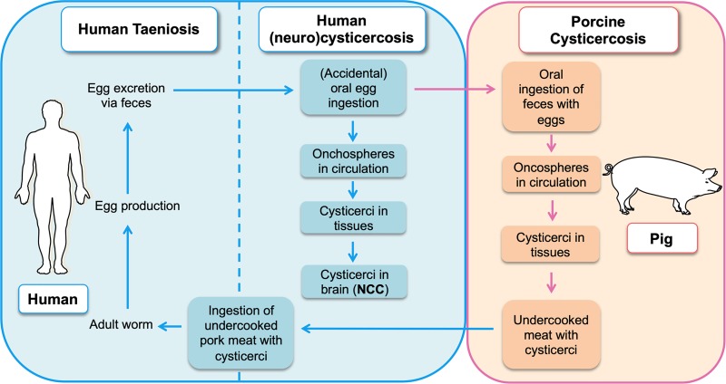
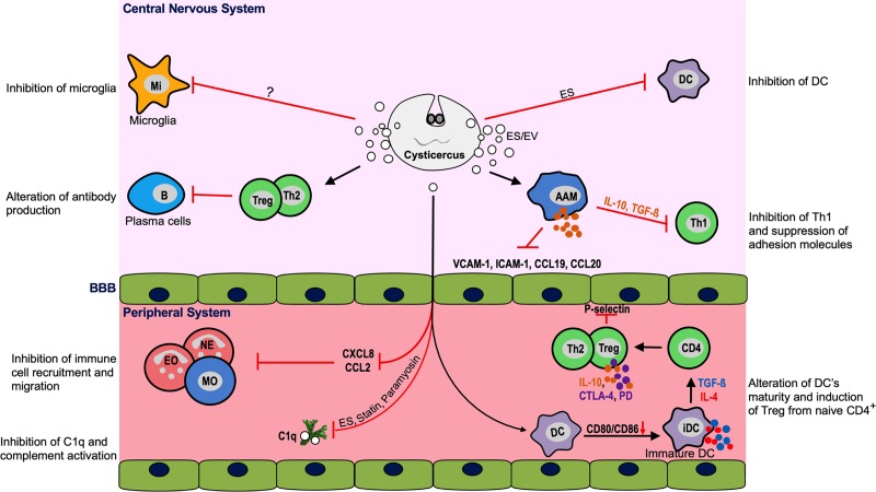
Taenia solium cysticercosis and taeniasis (TSCT), caused by the tapeworm T. solium, is a foodborne and zoonotic disease classified since 2010 by WHO as a neglected tropical isease. It causes considerable impact on health and economy and is one of the leading causes of acquired epilepsy in most endemic countries of Latin America, Sub-Saharan Africa, and Asia. There is some evidence that the prevalence of TSCT in high-income countries has recently increased, mainly due to immigration from endemic areas. In regions endemic for TSCT, human cysticercosis can manifest clinically as neurocysticercosis (NCC), resulting in epileptic seizures and severe progressive headaches, amongst other neurological signs and/or symptoms. The development of these symptoms results from a complex interplay between anatomical cyst localization, environmental factors, parasite's infective potential, host genetics, and, especially, host immune responses. Treatment of individuals with active NCC (presence of viable cerebral cysts) with anthelmintic drugs together with steroids is usually effective and, in the majority, reduces the number and/or size of cerebral lesions as well as the neurological symptoms. However, in some cases, treatment may profoundly enhance anthelmintic inflammatory responses with ensuing symptoms, which, otherwise, would have remained silent as long as the cysts are viable. This intriguing silencing process is not yet fully understood but may involve active modulation of host responses by cyst-derived immunomodulatory components released directly into the surrounding brain tissue or by the induction of regulatory networks including regulatory T cells (Treg) or regulatory B cells (Breg). These processes might be disturbed once the cysts undergo treatment-induced apoptosis and necrosis or in a coinfection setting such as HIV. Herein, we review the current literature regarding the immunology and pathogenesis of NCC with a highlight on the mobilization of immune cells during human NCC and their interaction with viable and degenerating cysticerci. Moreover, the immunological parameters associated with NCC in people living with HIV/AIDS and treatments are discussed. Eventually, we propose open questions to understand the role of the immune system and its impact in this intriguing host-parasite crosstalk.


Conflict of interest statement

The authors have declared that no competing interests exist.

Figures

Fig 1
Fig 1. Schematic diagram of T. solium infection showing the different steps of human and porcine cysticercosis including human NCC. NCC, neurocysticercosis.
Fig 2
Fig 2. Cyst involution in brain parenchyma and associated immunopathogenesis.
The presence of viable vesicular cysts (A) is usually asymptomatic and associated with suppression of the host immune responses through induction of anti-inflammatory cytokines. Upon successful attack of the host immune system, the cyst passes through a series of involuting stages (B–D). The BBB is disrupted, and granuloma, mainly composed of macrophages, granulocytes, and lymphocytes, encompasses the cyst that contracts (colloidal stage) (B) and becomes nodular and the scolex granulated (granular-nodular stage) (C); this is followed by the formation of collagenous and fibrotic structures and calcification of the cyst (calcified stage) (D). Each involuting stage of the cyst releases material (ES and Ag) and elicits an inflammatory reaction and release of mediators in the brain that lead to the development of pathology and symptoms (pale orange box). Ag, cyst antigen; B, lymphocyte B; BBB, blood–brain barrier; EO, eosinophil; ES, excretory–secretory; EV, extracellular vesicle; IFN, interferon; IL, interleukin; M, macrophage; Mi, microglia/dendritic cell; NE, neutrophil; TGF-ß, transforming growth factor beta; Th, T helper cell; TNF, tumor necrosis factor.
Fig 3
Fig 3. Host immune regulation during parenchymal NCC.
Viable cyst releases ES products and EVs that interact with resident brain cells (microglia and DCs) and induce AAMs, which suppress adhesion molecules and local Th1 response via TGF-ß and IL-10. Excreted products reach peripheral system where they drive the expansion of suppressive Tregs from CD4+ cells through the alteration of the maturation of DCs. Furthermore, cyst products inhibit complement C1q activity and prevent the infiltration and migration of neutrophils, eosinophils and MO from the peripheral system into the brain via production of immunomodulatory cytokines and blocking of chemokines and adhesion molecules. AAM, alternatively activated macrophages; B, ; BBB, blood–brain barrier; CCL, chemokine C-C ligand; CD4, cluster of differentiation 4; C1q, complement first component; CTLA-4, cytotoxic T-lymphocyte associated protein 4; CXCL, chemokine C-X-C ligand; DC, dendritic cell; EO, eosinophil; ES, excretory–secretory; EV, extracellular vesicle; ICAM-1, intercellular adhesion molecule 1; iDC, immature DC; IL, interleukin; Mi, microglia; MO, monocytes; NCC, neurocysticercosis; TGF-ß, transforming growth factor beta; Th, T helper; Treg, regulatory T cell; VCAM-1, vascular cell adhesion protein 1.

References

    1. Braae UC, Devleesschauwer B, Gabriel S, Dorny P, Speybroeck N, Magnussen P, et al. CystiSim—An Agent-Based Model for Taenia solium Transmission and Control. PLoS Negl Trop Dis. 2016;10(12):e0005184 10.1371/journal.pntd.0005184 - DOI - PMC - PubMed
    1. Winkler AS. Neurocysticercosis in sub-Saharan Africa: a review of prevalence, clinical characteristics, diagnosis, and management. Pathog Glob Health. 2012;106(5):261–74. 10.1179/2047773212Y.0000000047 - DOI - PMC - PubMed
    1. Ronsholt FF, Ostrowski SR, Katzenstein TL, Ullum H, Gerstoft J. T cell subsets in HIV infected patients after successful combination antiretroviral therapy: impact on survival after 12 years. PLoS One. 2012;7(7):e39356 10.1371/journal.pone.0039356 - DOI - PMC - PubMed
    1. Serpa JA, Moran A, Goodman JC, Giordano TP, White AC Jr., Neurocysticercosis in the HIV era: a case report and review of the literature. Am J Trop Med Hyg. 2007;77(1):113–7. . - PubMed
    1. Garcia HH, Gonzalez AE, Evans CA, Gilman RH, Cysticercosis Working Group in P. Taenia solium cysticercosis. Lancet. 2003;362(9383):547–56. 10.1016/S0140-6736(03)14117-7 - DOI - PMC - PubMed

Publication types

MeSH terms