Accuracy of a deep convolutional neural network in the detection of myopic macular diseases using swept-source optical coherence tomography
- PMID: 32298265
- PMCID: PMC7161961
- DOI: 10.1371/journal.pone.0227240
Accuracy of a deep convolutional neural network in the detection of myopic macular diseases using swept-source optical coherence tomography
Abstract
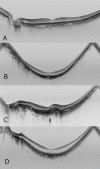
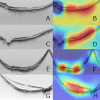
This study examined and compared outcomes of deep learning (DL) in identifying swept-source optical coherence tomography (OCT) images without myopic macular lesions [i.e., no high myopia (nHM) vs. high myopia (HM)], and OCT images with myopic macular lesions [e.g., myopic choroidal neovascularization (mCNV) and retinoschisis (RS)]. A total of 910 SS-OCT images were included in the study as follows and analyzed by k-fold cross-validation (k = 5) using DL's renowned model, Visual Geometry Group-16: nHM, 146 images; HM, 531 images; mCNV, 122 images; and RS, 111 images (n = 910). The binary classification of OCT images with or without myopic macular lesions; the binary classification of HM images and images with myopic macular lesions (i.e., mCNV and RS images); and the ternary classification of HM, mCNV, and RS images were examined. Additionally, sensitivity, specificity, and the area under the curve (AUC) for the binary classifications as well as the correct answer rate for ternary classification were examined. The classification results of OCT images with or without myopic macular lesions were as follows: AUC, 0.970; sensitivity, 90.6%; specificity, 94.2%. The classification results of HM images and images with myopic macular lesions were as follows: AUC, 1.000; sensitivity, 100.0%; specificity, 100.0%. The correct answer rate in the ternary classification of HM images, mCNV images, and RS images were as follows: HM images, 96.5%; mCNV images, 77.9%; and RS, 67.6% with mean, 88.9%.Using noninvasive, easy-to-obtain swept-source OCT images, the DL model was able to classify OCT images without myopic macular lesions and OCT images with myopic macular lesions such as mCNV and RS with high accuracy. The study results suggest the possibility of conducting highly accurate screening of ocular diseases using artificial intelligence, which may improve the prevention of blindness and reduce workloads for ophthalmologists.
Conflict of interest statement
The authors have declared that no competing interests exist.
Figures


References
-
- McBrien NA, Adams DW. A longitudinal investigation of adult-onset and adult-progression of myopia in an occupational group. Refractive and biometric findings. Invest Ophthalmol Vis Sci. 1997;38: 321–333. - PubMed
Publication types
MeSH terms
LinkOut - more resources
Full Text Sources

