. 2020 Apr 9;61(4):14.
doi: 10.1167/iovs.61.4.14.

Six-Year Changes in Myopic Macular Degeneration in Adults of the Singapore Epidemiology of Eye Diseases Study

Affiliations

Six-Year Changes in Myopic Macular Degeneration in Adults of the Singapore Epidemiology of Eye Diseases Study

Yee-Ling Wong et al. Invest Ophthalmol Vis Sci. .

Abstract

Purpose: To examine the 6-year incidence, progression, associated risk factors, and impact of myopic macular degeneration (MMD) in a myopic population in Singapore.

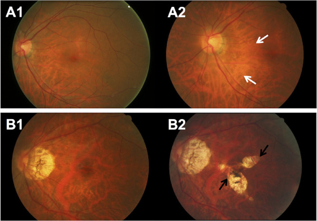
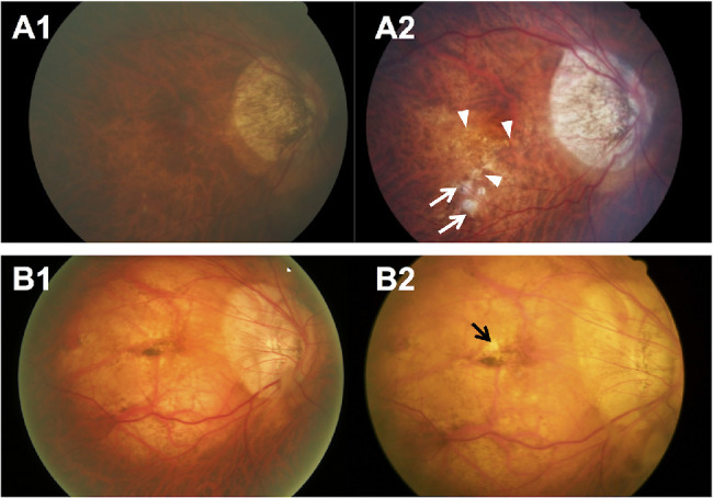
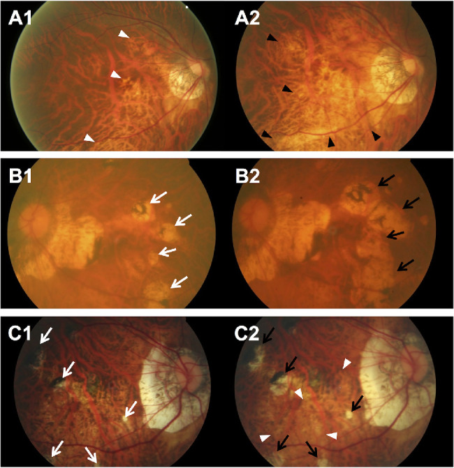
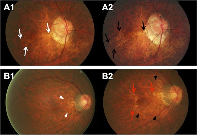
Methods: We examined myopic (spherical equivalent ≤-0.5 diopters) adults (N = 2157 persons and 3661 eyes) who were phakic at baseline and participated in both baseline and 6-year follow-up visits of the Singapore Epidemiology of Eye Diseases study. Eye examinations, including visual acuity, subjective refraction and axial length (AL) measurements, were performed. MMD was graded from fundus photographs following the META-PM classification. Vision-related quality of life was assessed with Rasch-transformed scores from reading, mobility, and emotional domains of the Impact of Vision Impairment questionnaire.

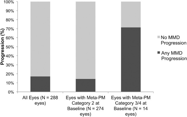
Results: The 6-year eye-specific incidence of MMD among myopic eyes was 1.2% (95% CI, 0.9%-1.6%). Older age, worse spherical equivalent, and longer AL at baseline were associated with MMD incidence (all P < 0.001). The 6-year eye-specific progression of MMD in 288 eyes with baseline MMD was 17.0% (95% CI, 12.6%-21.4%). More severe MMD at baseline, worse spherical equivalent, and longer AL (all P < 0.05) were associated with MMD progression. Patients with Meta-PM categories 3 and 4 had worse best-corrected visual acuity and poorer vision-related quality of life outcomes than those without MMD (all P < 0.05).

Conclusions: Over a 6-year period, one in 80 myopic eyes developed MMD and one in six with existing MMD had MMD progression. Myopia severity and AL were strong predictors of MMD development and progression. Eyes with severe MMD were at higher risk of MMD progression than those with less severe MMD, and were associated with poorer visual acuity and vision-related quality of life.


Conflict of interest statement

Disclosure: Y.-L. Wong, Essilor International (E); C. Sabanayagam, None; C.-W. Wong, None; Y.-B. Cheung, None; R.E.K. Man, None; A. Chwee-Hong Yeo, Essilor International (E); G. Cheung, None; A. Chia, None; A. Kuo, None; M. Ang, None; K. Ohno-Matsui, None; T.-Y. Wong, None; J.J. Wang, None; C.-Y. Cheng, None; Q.V. Hoang, None; E. Lamoureux, None; S.-M. Saw, None

Figures

Figure 1.
Figure 1.
Fundus photographs of eyes with development of new MMD over a 6-year period. (A) Fundus photographs from a 48-year-old Malay woman showing development of incident MMD (development of diffuse chorioretinal atrophy) over a follow-up of 6 years. (A1) Image from the left eye obtained at baseline visit (SE of –6.9 D and AL of 27.7 mm) showing fundus tessellation only. (A2) Six years later, diffuse chorioretinal atrophy (white arrows) developed. (B) Fundus photographs from a 64-year-old Chinese woman showing development of incident MMD (development of patchy chorioretinal atrophy) over a follow-up of 6 years. (B1) Image from the left eye obtained at baseline visit (SE of –10.3 D and AL of 27.8 mm) showing fundus tessellation only. (B2) Six years later, several well-defined patchy chorioretinal atrophies (black arrows) in the macular region and choroidal neovascularization (CNV) developed at the fovea.
Figure 2.
Figure 2.
Fundus photographs of eyes showing MMD progression (from Meta-PM category 2 at baseline to Meta-PM category 3 at follow-up) with development of new patchy chorioretinal atrophy over a 6-year period. (A) Fundus photographs from a 62-year-old Chinese woman showing MMD progression with development of new patchy chorioretinal atrophy over a follow-up of 6 years. (A1) Image from the right eye obtained at baseline visit (SE of –2.8 D and AL of 28.4 mm) showing diffuse chorioretinal atrophy on the inferior–temporal region of the macula. (A2) Six years later, several spotted patchy chorioretinal atrophies (white arrows) and lacquer cracks (white arrowheads) developed, and the preexisting diffuse chorioretinal atrophy expanded. Enlargement of PPA was observed as well. (B) Fundus photographs from a 50-year-old Malay man showing MMD progression with development of new patchy chorioretinal atrophy over a follow-up of 6 years. (B1) Image from the right eye obtained at baseline visit (SE of –12.3 D) showing diffuse chorioretinal atrophy on the entire macular region and Fuchs’ spot at the fovea. (B2) Six years later, a small new patchy chorioretinal atrophy (black arrow) developed in the macular region near the Fuchs’ spot. Enlargement of the PPA was seen.
Figure 3.
Figure 3.
Fundus photographs of eyes showing MMD progression with enlargement of existing chorioretinal atrophy (no change in Meta-PM category) over a 6-year period. (A. Fundus photographs from a 60-year-old Malay woman showing progression of MMD (enlargement of diffuse chorioretinal atrophy) over a follow-up of 6 years. (A1) Image from the right eye obtained at baseline visit (SE of –5.6 D and AL of 28.8 mm) showing peripapillary diffuse chorioretinal atrophy (white arrowheads). (A2) Six years later, enlargement of diffuse chorioretinal atrophy (black arrowheads) across the macular region was seen, and enlargement of the PPA was observed. (B) Fundus photographs from a 73-year-old Malay woman showing progression of MMD (enlargement of patchy chorioretinal atrophy) over a follow-up of 6 years. (B1) Image from the left eye obtained at baseline visit (SE of –14.4 D and AL of 28.0 mm) showing several patchy chorioretinal atrophies temporal to macular (white arrows). (B2) Six years later, the fundus showed an enlargement of the patchy chorioretinal atrophy coalescing with other previous patchy chorioretinal atrophies (black arrows) toward the macula, but not involving macula. Enlargement of the PPA was seen as well. (C) Fundus photographs from a 45-year-old Chinese man showing progression of MMD (enlargement of both diffuse and patchy chorioretinal atrophy) over a follow-up of 6 years. (C1) Image from the right eye obtained at baseline visit (SE of –15.6 D and AL of 33.1 mm) showing multiple small pigmented patchy chorioretinal atrophies (white arrows) around the macula. (C2) Six years later, the fundus showed slight enlargement of the patchy chorioretinal atrophies (black arrows) around the macula and enlargement of the diffuse chorioretinal atrophy inferior to the macula (white arrowheads). Enlargement of the PPA was observed.
Figure 4.
Figure 4.
Fundus photographs of eyes showing MMD progression with changes in plus lesions over a 6-year period. (A) Fundus photographs from a 50-year-old Chinese man showing progression of MMD (widening of existing lacquer cracks) over a follow-up of 6 years. (A1) Image from the right eye obtained at baseline visit (SE of –14.0 D and AL of 30.0 mm) showing linear lacquer cracks across the macula (white arrows). (A2) Six years later, widening of existing lacquer cracks temporal to the macula was seen (black arrows). (B) Fundus photographs from a 47-year-old woman showing progression of MMD (enlargement of diffuse chorioretinal atrophy and new lacquer crack) over a follow-up of 6 years. (B1) Image from the right eye obtained at baseline visit (SE of –9.0 D and AL of 29.1 mm) showing slight peripapillary diffuse chorioretinal atrophy (white arrowheads). (B2) Six years later, enlargement of diffuse chorioretinal atrophy (black arrowheads) toward the macula and development of lacquer cracks (red arrows) superior and temporal to the macula were observed.
Figure 5.
Figure 5.
Progression of MMD lesions over a 6-Year period in myopic eyes with preexisting MMD in the SEED Study (n = 202 patients and 288 eyes) stratified by meta-PM categories at baseline examination.
Figure 6.
Figure 6.
Fundus photographs of eyes with previous cataract surgery showing MMD progression over a 6-year period. (A) Fundus photographs from a 58-year-old Chinese man showing progression of MMD with development of new macular atrophy involving the fovea and new patchy chorioretinal atrophy over a follow-up of 6 years. (A1) Image from the right eye obtained at baseline visit (AL of 27.9 mm and with cataract surgery) showing diffuse chorioretinal atrophy in the macular region near the fovea. (A2) Six years later, macular atrophy (black arrow), several surrounding spotted patchy chorioretinal atrophies (white arrows), and choroidal neovascularization (black arrowhead) developed. (B) Fundus photographs from a 73-year-old Malay woman showing MMD progression (enlargement of both macular atrophy and patchy chorioretinal atrophy) over a follow-up of 6 years. (B1) Image from the left eye obtained at baseline visit (with cataract surgery) showing several patchy chorioretinal atrophies temporal to macular (white arrows). (B2) Six years later, the fundus showed an enlargement of the coalescence of macular atrophy and patchy chorioretinal atrophy (black arrows) from the macula with increased pigmentation.

References

    1. Saw S. How blinding is pathological myopia? Br J Ophthalmol. 2006; 90: 525–526. - PMC - PubMed
    1. Saw SM, Gazzard G, Shih-Yen EC, Chua WH. Myopia and associated pathological complications. Ophthalmic Physiol Opt. 2005; 25: 381–391. - PubMed
    1. Ohno-Matsui K. What is the fundamental nature of pathologic myopia? Retina. 2017; 37: 1043–1048. - PubMed
    1. Wong TY, Ferreira A, Hughes R, Carter G, Mitchell P. Epidemiology and disease burden of pathologic myopia and myopic choroidal neovascularization: an evidence-based systematic review. Am J Ophthalmol. 2014; 157: 9–25 e12. - PubMed
    1. Jonas JB, Nangia V, Gupta R, Bhojwani K, Nangia P, Panda‐Jonas S. Prevalence of myopic retinopathy in rural Central India. Acta Ophthalmologica. 2016; 95: e399–e404. - PubMed

Publication types

MeSH terms