. 2020 Apr 9;61(4):15.
doi: 10.1167/iovs.61.4.15.

PPARα-Dependent Effects of Palmitoylethanolamide Against Retinal Neovascularization and Fibrosis

Affiliations

PPARα-Dependent Effects of Palmitoylethanolamide Against Retinal Neovascularization and Fibrosis

Sihao Ye et al. Invest Ophthalmol Vis Sci. .

Abstract

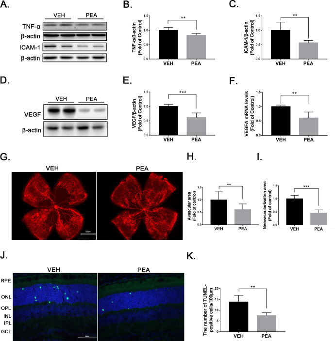
Purpose: Pathological neovascularization and fibrosis are common pathological changes of many retinal diseases, such as proliferative retinopathy (PR) and age-related macular degeneration (AMD). Treatment modalities for these pathological changes are limited. The purpose of the present study was to test the effects of palmitoylethanolamide (PEA), an endocannabinoid mimetic amide, on retinal neovascularization and fibrosis and to determine its molecular mechanism of action.

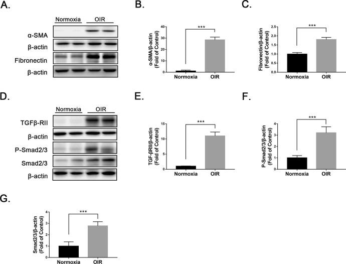
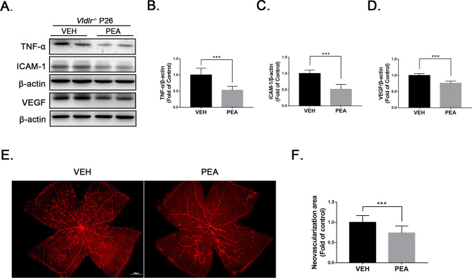
Methods: A rat Müller cell line (rMC-1), a mouse model of oxygen-induced retinopathy (OIR), and the very-low-density lipoprotein receptor (VLDLR) knockout mouse model were used. PEA was intraperitoneally injected or orally administrated in animal models. Inflammation and profibrotic changes were evaluated by western blot analysis. Glial fibrillary acidic protein (GFAP) and peroxisome proliferator-activated receptor alpha (PPARα) were measured by RT-PCR and western blot analysis.

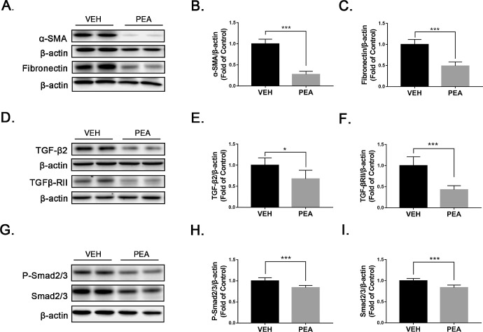
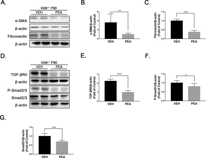
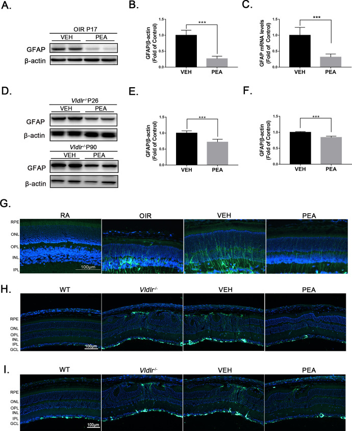
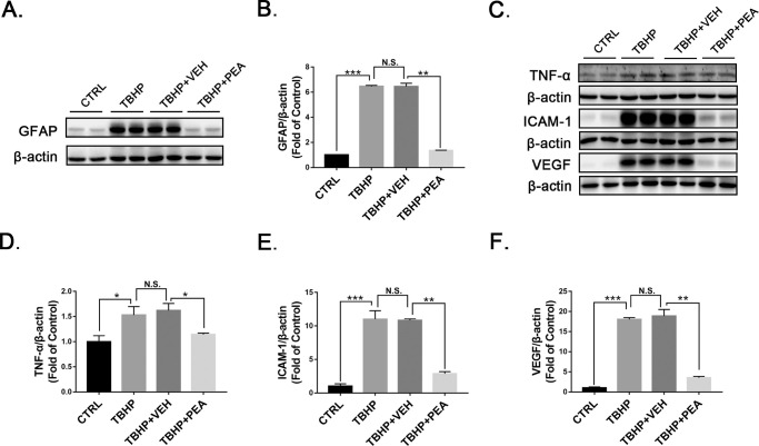
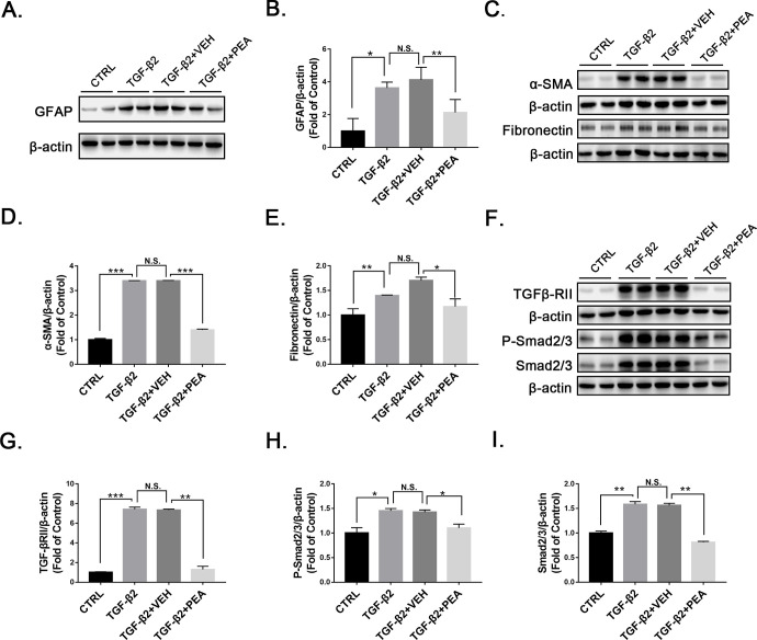
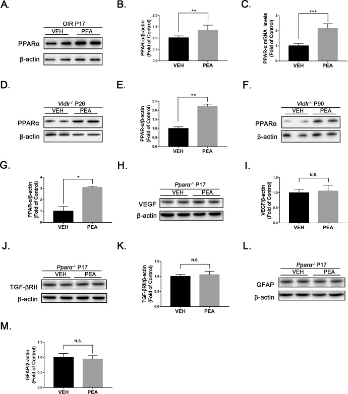
Results: Profibrotic changes were present in OIR and Vldlr-/- retinas. PEA significantly alleviated inflammation and inhibited neovascularization in OIR and Vldlr-/- retinas and suppressed profibrotic changes in OIR and Vldlr-/- retinas. Moreover, PEA potently suppressed Müller gliosis in these retinas. In rMC-1 cells, PEA suppressed Müller gliosis, reduced inflammatory cytokines, and attenuated profibrotic changes. Further, both mRNA and protein levels of PPARα were elevated in the retina under PEA treatment, and the effects of PEA were abolished in Pparα-/- OIR mice.

Conclusions: PEA reduced retinal neovascularization and fibrotic changes and suppressed Müller gliosis in experimental PR and neovascular AMD by activating PPARα. PEA may be a potential treatment for retinopathies with pathological neovascularization and fibrosis.


Conflict of interest statement

Disclosure: S. Ye, None; Q. Chen, None; N. Jiang, None; X. Liang, None; J. Li, None; R. Zong, None; C. Huang, None; Y. Qiu, None; J.-X. Ma, None; Z. Liu, None

Figures

Figure 1.
Figure 1.
Anti-inflammatory and anti-angiogenic effects of PEA in OIR. (A) Representative images of western blotting for TNF-α and ICAM1 in OIR retinas treated with VEH or PEA. Protein levels of (B) TNF-α and (C) ICAM-1 were quantified by densitometry and normalized to β-actin levels (n = 6). (D) Representative images of western blotting for VEGF in OIR retinas treated with VEH or PEA. (E) Protein level of VEGF was quantified by densitometry and normalized to β-actin levels (n = 6). (F) Quantitative RT-PCR of Vegfa mRNA expression in VEH-OIR and PEA-OIR groups (n = 5). (G) Retinas from VEH- and PEA-treated OIR mice were flat-mounted and stained with isolectin GS-IB4 (red). Areas of (H) retinal vaso-obliteration and (I) neovascularization were quantified in VEH- and PEA-treated OIR retinas (n = 8). (J) Retinal cryosections from VEH- and PEA-treated OIR mice were stained with TUNEL (green) and 4′,6-diamidino-2-phenylindole (DAPI) (blue). Representative images are shown. (K) Apoptotic cells were quantified and compared in VEH- and PEA-treated OIR mice. Data are presented as mean ± SEM; **P < 0.01, ***P < 0.001.
Figure 2.
Figure 2.
Presence of ECM remodeling and profibrotic changes in OIR retinas. (AC) Protein levels of (A, B) α-SMA and (A, C) fibronectin were measured by western blot analysis and quantified by densitometry in wild-type (WT) and OIR retinas at P17 (n = 6). (DG) Similarly, protein levels of (D, E) TGFβ-RII, (D, F) P-Smad2/3, and (D, G) Smad2/3 were measured by western blot analysis and quantified by densitometry (n = 6). Data are presented as mean ± SEM; ***P < 0.001.
Figure 3.
Figure 3.
Antifibrotic effects of PEA in OIR retinas. (AC) Protein levels of (A, B) α-SMA and (A, C) fibronectin were measured by western blot analysis and quantified by densitometry in VEH- and PEA-treated OIR retinas (n = 6). (DI) Similarly, protein levels of (D, E) TGF-β2, (D, F) TGFβ-RII, (G, H) P-Smad2/3, and (G, I) Smad2/3 were measured by western blot analysis and quantified by densitometry (n = 6). Data are presented as mean ± SEM; *P < 0.05, ***P < 0.001.
Figure 4.
Figure 4.
Anti-angiogenic effects of PEA in Vldlr−/− retinas. (A) Protein levels of TNF-α, ICAM-1 and VEGF were measured by western blot analysis in the eyecups of Vldlr−/− mice treated with VEH or PEA. (B) TNF-α, (C) ICAM-1, and (D) VEGF levels were quantified by densitometry (n = 6). (E) Retinas from VEH- and PEA-treated Vldlr−/− mice were flat-mounted and stained with isolectin GS-IB4 (red). (F) Areas of neovascularization were quantified (n = 8). Data are presented as mean ± SEM; *P < 0.05, **P < 0.01, ***P < 0.001.
Figure 5.
Figure 5.
Antifibrotic effects of PEA in Vldlr−/− retinas. (A) Protein levels of α-SMA and fibronectin were measured by western blot analysis in the eyecups of Vldlr−/− mice treated with VEH or PEA. (B) α-SMA and (C) fibronectin were quantified by densitometry (n = 6). (D) Similarly, protein levels of TGF-βRII, P-Smad2/3, and Smad2/3 were measured by western blot analysis, and (E) TGF-βRII, (F) P-Smad2/3, and (G) Smad2/3 were quantified by densitometry (n = 6). Data are presented as mean ± SEM; *P < 0.05, **P < 0.01, ***P < 0.001.
Figure 6.
Figure 6.
PEA inhibited gliosis in OIR and Vldlr−/− retinas. (A) Protein levels of GFAP in the retinas of OIR mice treated with VEH or PEA were measured by western blot analysis. (B) Protein levels of GFAP were quantified by densitometry and normalized to β-actin levels (n = 6). (C) Quantitative RT-PCR of Gfap gene expression in the retina of VEH- and PEA-treated OIR mice (n = 5). (D) Vldlr−/− mice at P14 were intraperitoneally injected with VEH or PEA for 2 weeks. Protein levels of GFAP were measured in Vldlr−/− retinas at P26. In addition, Vldlr−/− mice at P60 were orally administrated PEA or VEH for 30 days. Protein levels of GFAP were then measured in the retina of these Vldlr−/− mice at P90. (E) Levels of GFAP in Vldlr−/− retinas at P26 were quantified by densitometry (n = 6). (F) Levels of GFAP in Vldlr−/− retinas at P90 were quantified by densitometry (n = 6). (G) Representative images of immunostaining for GFAP in the retinal sections of OIR mice treated with VEH or PEA. (H) Representative images of immunostaining for GFAP in the retinal sections of Vldlr−/− mice at P26 treated with VEH or PEA. (I) Representative images of immunostaining for GFAP in the retinal sections of Vldlr−/− mice at P90 treated with VEH or PEA. Data are presented as mean ± SEM; ***P < 0.001.
Figure 7.
Figure 7.
Effects of PEA in TBHP-treated rMC-1 cells. Representative images of western blotting for (A) GFAP and (C) TNF-α, ICAM-1, and VEGF in Müller cells treated with VEH or PEA in the presence of TBHP. Protein levels of (B) GFAP, (D) TNF-α, (E) ICAM-1, and (F) VEGF were quantified by densitometry and normalized to β-actin levels. Data are presented as mean ± SEM; *P < 0.05, **P < 0.01, ***P < 0.001. TBHP, tertiary-butyl hydrogen peroxide.
Figure 8.
Figure 8.
Effects of PEA in TGF-β2-treated rMC-1 cells. (A) Representative images of western blotting for GFAP in rMC-1 cells treated with VEH or PEA in the presence of TGF-β2. (B) Protein levels of GFAP were quantified by densitometry and normalized by β-actin levels. Similarly, protein levels of (C, D) α-SMA and (C, E) fibronectin were measured and quantified. Further, components of TGF-β/Smad2/3 signaling, (F, G) TGF-βRII, (F, H) P-Smad2/3, and (F, I) Smad2/3 were measured and quantified (n = 6). Data are presented as mean ± SEM; *P < 0.05, **P < 0.01, ***P < 0.001.
Figure 9.
Figure 9.
The effects of PEA were PPARα dependent. (A) Representative images of western blotting for PPARα in the retinas of OIR mice treated with VEH or PEA. (B) Protein levels of PPARα were quantified by densitometry and normalized to β-actin levels (n = 8). (C) Quantitative RT-PCR of Pparα mRNA expression in PEA- and VEH-treated OIR retinas (n = 5). (DG) Western blot analysis of PPARα was performed in Vldlr−/− mice treated with PEA or VEH at (D, E) P26 and (F, G) P90 (n = 6). (HM) Further, OIR was induced in Pparα−/− mice. At P17, protein levels of (H) VEGF, (J) TGF-βRII, and (L) GFAP were measured by western blot analysis in Pparα−/− OIR retinas under VEH or PEA treatment. Protein levels of (I) VEGF, (K) TGF-βRII, and (M) GFAP were quantified by densitometry and normalized to β-actin levels (n = 6). Data are presented as mean ± SEM; *P < 0.05, **P < 0.01, ***P < 0.001.

References

    1. Antonetti DA, Klein R, Gardner TW. Diabetic retinopathy. N Engl J Med. 2012; 366: 1227–1239. - PubMed
    1. Roy S, Amin S, Roy S. Retinal fibrosis in diabetic retinopathy. Exp Eye Res. 2016; 142: 71–75. - PMC - PubMed
    1. Keppel Hesselink JM, Costagliola C, Fakhry J, Kopsky DJ. Palmitoylethanolamide, a natural retinoprotectant: its putative relevance for the treatment of glaucoma and diabetic retinopathy. J Ophthalmol. 2015; 2015: 430596. - PMC - PubMed
    1. Di Zazzo A, Roberti G, Mashaghi A, Abud TB, Pavese D, Bonini S. Use of topical cannabinomimetic palmitoylethanolamide in ocular surface disease associated with antiglaucoma medications. J Ocul Pharmacol Ther. 2017; 33: 670–677. - PubMed
    1. Paterniti I, Di Paola R, Campolo M, et al. .. Palmitoylethanolamide treatment reduces retinal inflammation in streptozotocin-induced diabetic rats. Eur J Pharmacol. 2015; 769: 313–323. - PubMed

Publication types

MeSH terms