. 2020 May 14;181(4):848-864.e18.
doi: 10.1016/j.cell.2020.03.047. Epub 2020 Apr 15.

Regenerative Metaplastic Clones in COPD Lung Drive Inflammation and Fibrosis

Affiliations

Regenerative Metaplastic Clones in COPD Lung Drive Inflammation and Fibrosis

Wei Rao et al. Cell. .

Abstract

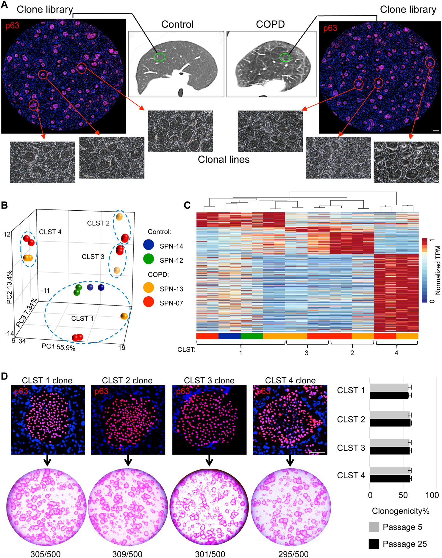
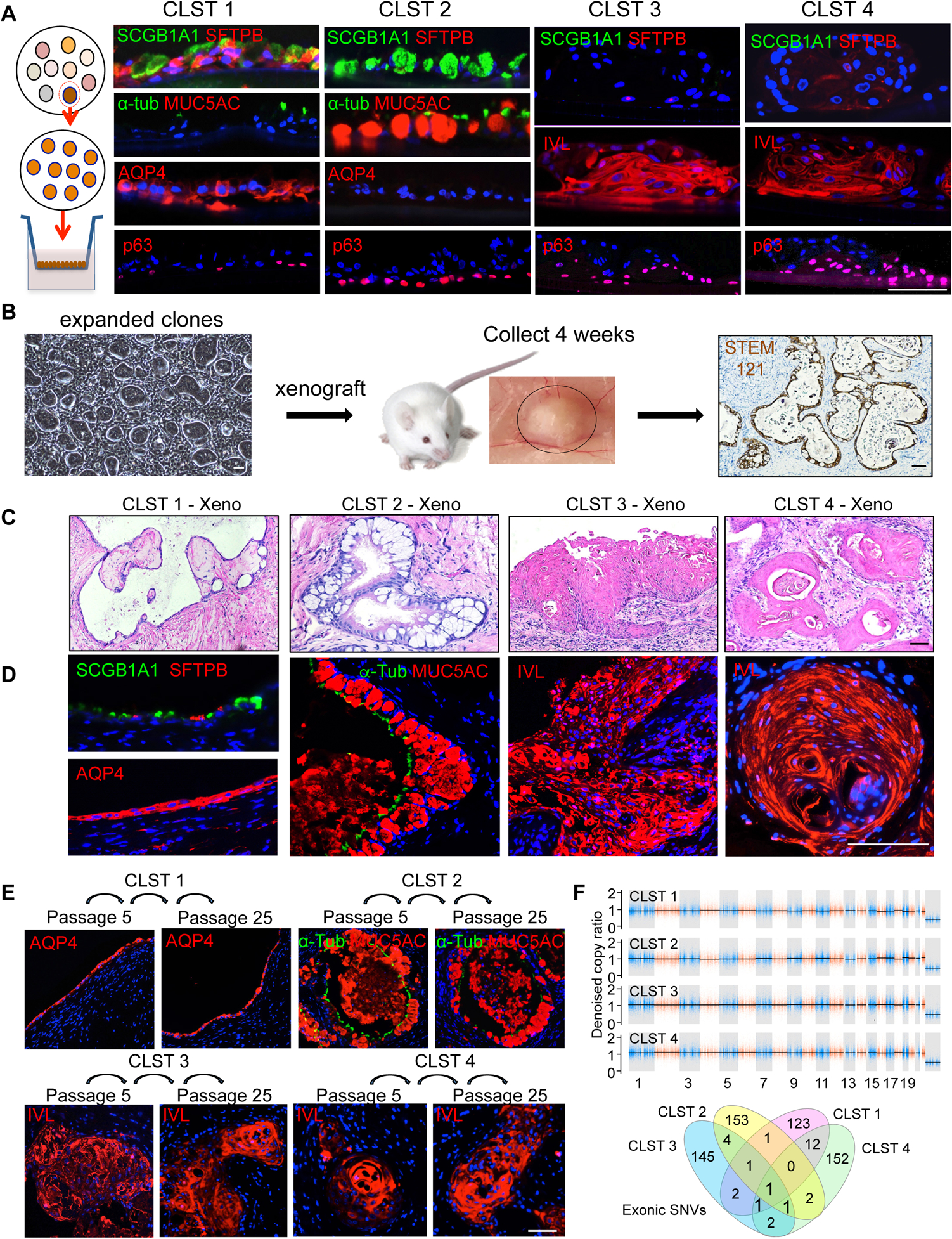
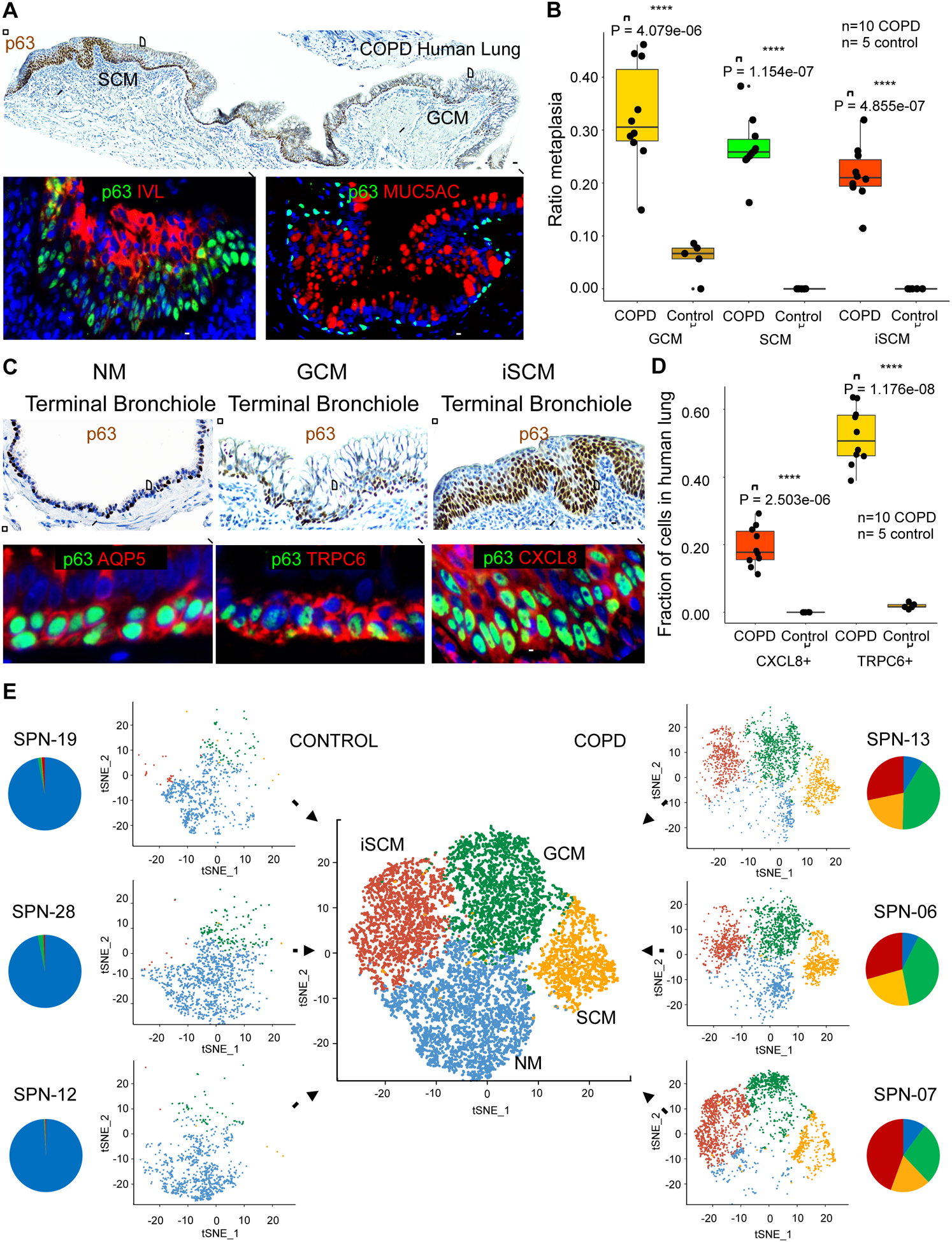
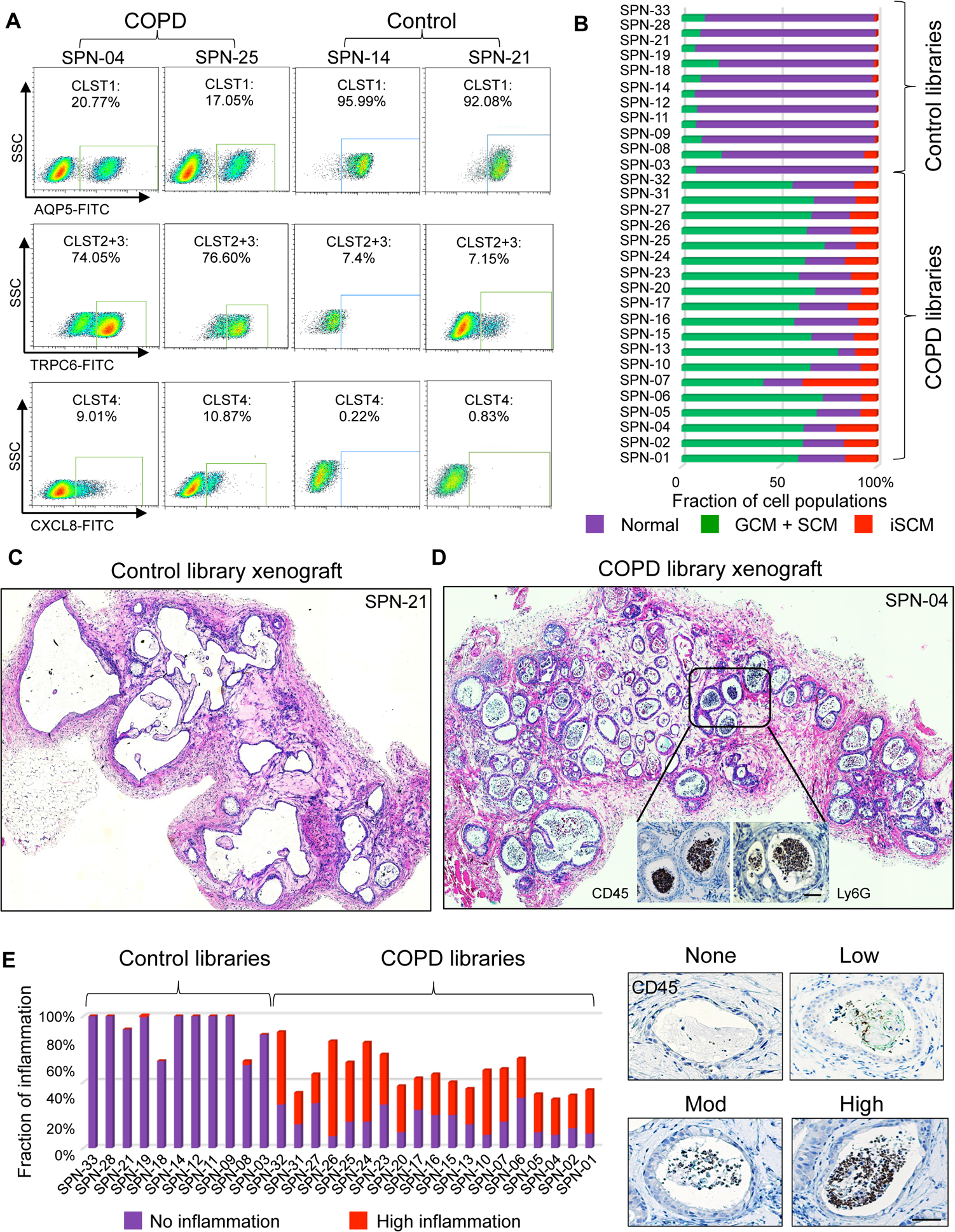
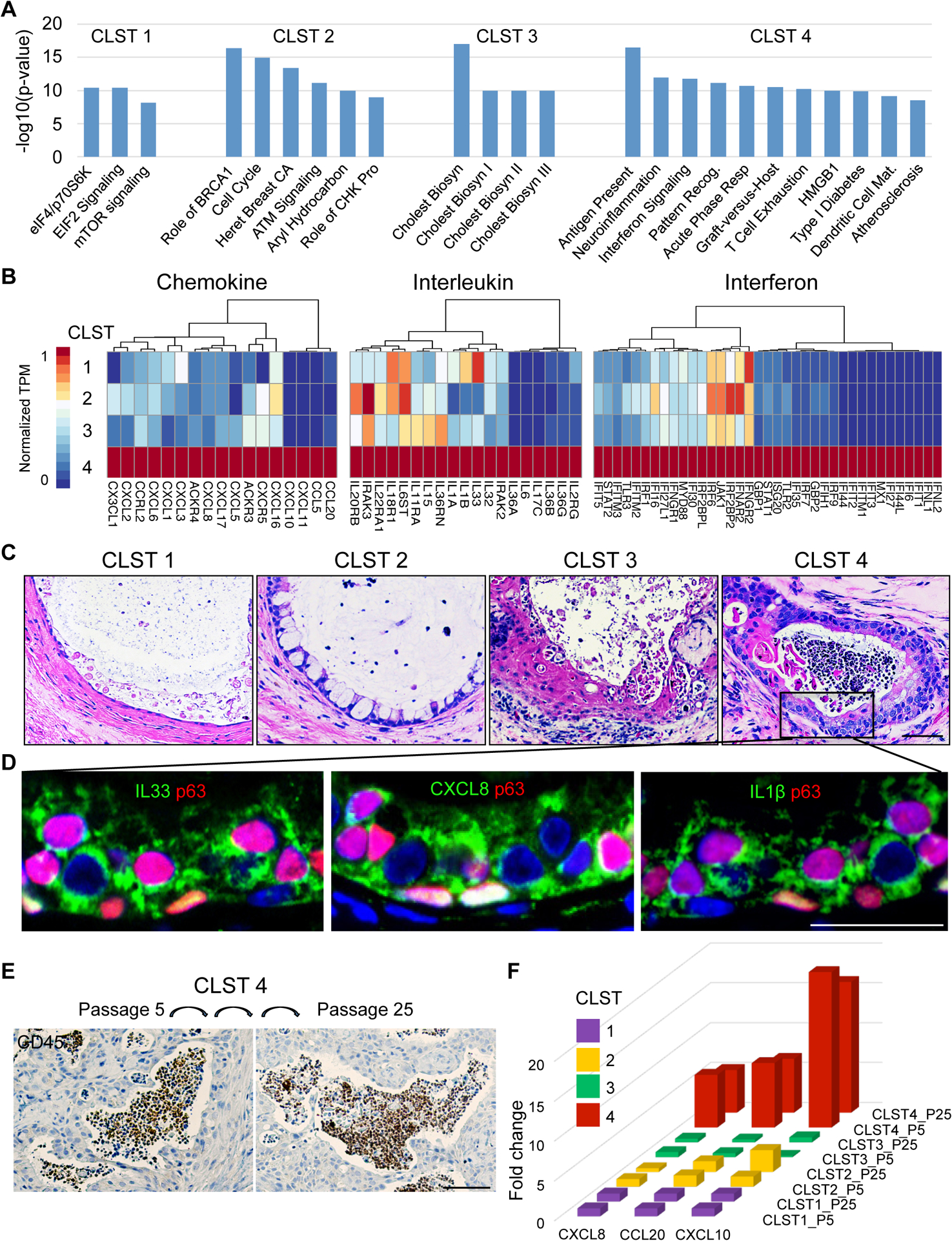
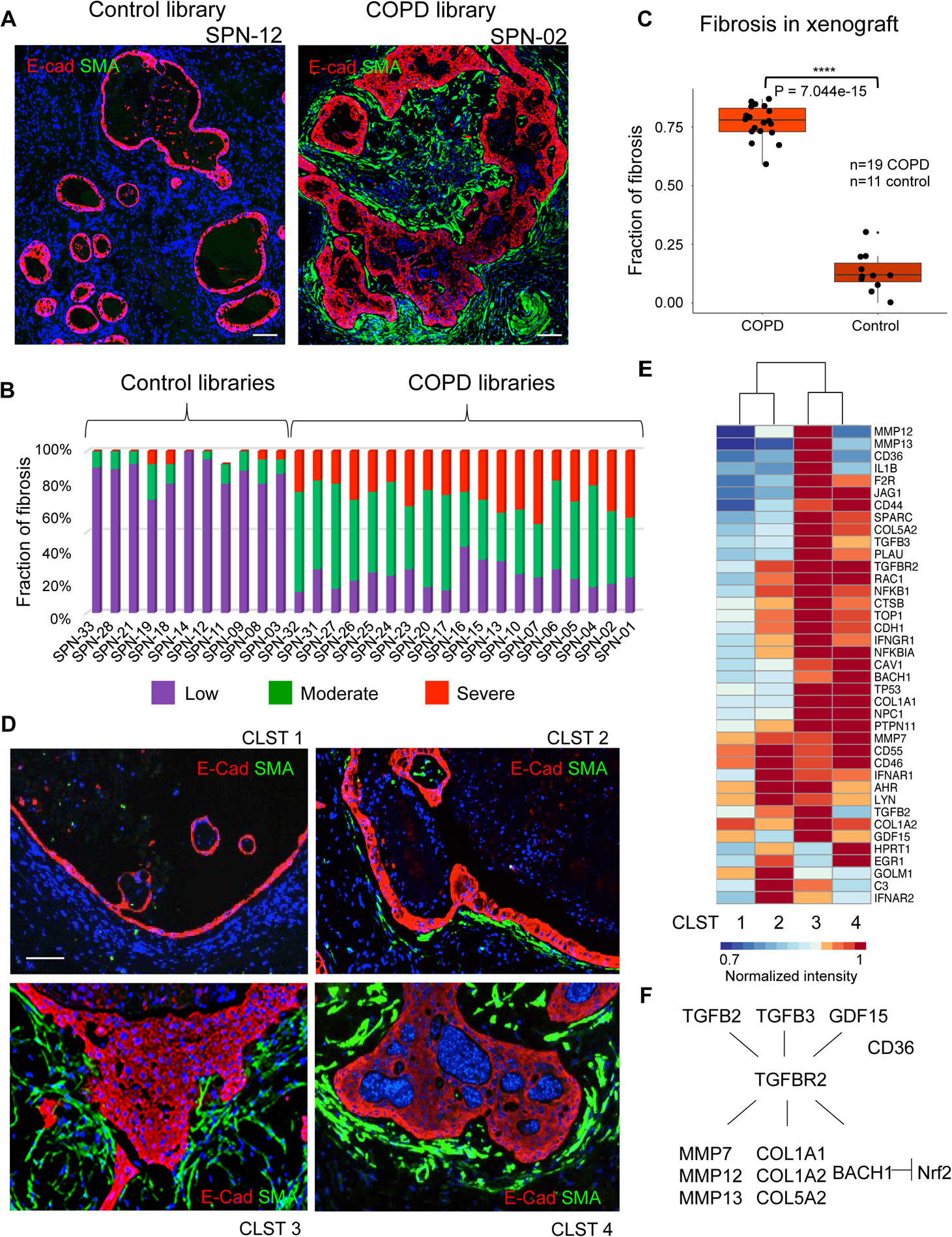
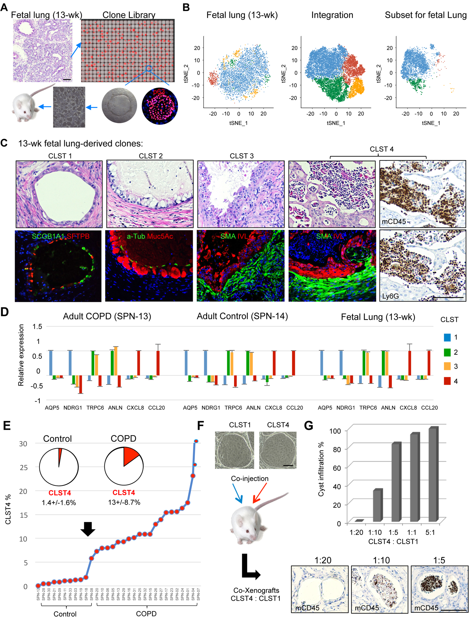
Chronic obstructive pulmonary disease (COPD) is a progressive condition of chronic bronchitis, small airway obstruction, and emphysema that represents a leading cause of death worldwide. While inflammation, fibrosis, mucus hypersecretion, and metaplastic epithelial lesions are hallmarks of this disease, their origins and dependent relationships remain unclear. Here we apply single-cell cloning technologies to lung tissue of patients with and without COPD. Unlike control lungs, which were dominated by normal distal airway progenitor cells, COPD lungs were inundated by three variant progenitors epigenetically committed to distinct metaplastic lesions. When transplanted to immunodeficient mice, these variant clones induced pathology akin to the mucous and squamous metaplasia, neutrophilic inflammation, and fibrosis seen in COPD. Remarkably, similar variants pre-exist as minor constituents of control and fetal lung and conceivably act in normal processes of immune surveillance. However, these same variants likely catalyze the pathologic and progressive features of COPD when expanded to high numbers.

Keywords: COPD; chronic lung disease; fibrosis; inflammation; lung; metaplasia; myofibroblasts; neutrophils; p63; single cell cloning; stem cells.


Conflict of interest statement

Declaration of Interests W.X., F.D.M., W.R., S.W., J.X., M.D., and M.V. have filed patents related to technologies used in the present work. M.V., F.D.M., and W.X. have financial interests in Nüwa Medical Systems, Houston, TX, USA and Tract pharmaceuticals, Houston, TX, USA. Nuwa Medical Systems is a trade name of Tract Pharmaceuticals.

Figures

Figure 1.
Figure 1.. Epithelial clone heterogeneity in COPD
A. Schematic depicting workflow of generating p63+ epithelial colony pools from resected normal and COPD lung tissue. Individual colonies are captured and expanded individually to generate clonal cell lines. Scale bar, 500 μm. B. Principal component analysis of RNAseq differential expression genes (DEGs) (FDR<0.05) derived from multiple clones from control (SPN-12, −14) and COPD (SPN-13, −07) lungs. C. RNAseq expression heatmap of the clones depicted in the PCA plot is shown highlighting Cluster 1 (control) vs Clusters 1–4 in COPD (detailed in Table S4). D. Top, Individual colonies from cloned representatives of Clusters 1–4 stained with antibody to p63 showing uniform nuclear staining. Scale bar, 100 μm. Bottom, Rhodamine red-stained colonies arising on lawns of irradiated 3T3-J2 fibroblasts from 500 cells from each of cloned representatives of Clusters 1–4. Right, Histogram of clonogenicity based on percentage of plated cells that formed colonies using representative clones from Clusters 1–4 at passage 5 (P5) and 25 (P25). Data represented as mean ± SEM; See also Table S1 and Table S4.
Figure 2.
Figure 2.. Commitment of variant clones to metaplastic fates
A. From left, Schematic of clonal expansion and differentiation in air-liquid interface (ALI) cultures assessed by immunofluorescence on histological sections. Cluster 1 clones differentiated to an epithelium characterized by expression of SCGB1A1, SFTPB, α-TUB and AQP4 and the absence of MUC5AC. Cluster 2 clones differentiated into an epithelium characterized by SCGB1A1+, MUC5AC+, AQP4− goblet cells. Cluster 3 and Cluster 4 clones differentiated into squamous epithelia that expressed involucrin (IVL) but not SCGB1A1, AQP4, or SFTPB. Scale bar, 100 μm. B. Schematic of subcutaneous transplants of immature cells from clones or library of clones into immunodeficient mice. Nodules formed at 4 weeks were processed for histology and immunostaining and showed polarized epithelia that reacted with antibodies to the human-specific marker STEM121. Scale bar, 100 μm. C., D. In vivo differentiation following subcutaneous transplantation of cloned representatives of Cluster 1 (SCGB1A1+, SFTPB+, AQP4+), Cluster 2 (MUC5AC+ GCM), and Clusters 3 and 4 (IVL+; SCM). Scale bar, 100 μm. E. Xenografts of Cluster 1–4 clones previously grown in vitro to passage 5 (P5) and passage 25 (P25) showing stability of fate differentiation and expression of Cluster-specific markers. Scale bar, 100um. F. Copy number and single nucleotide variation analysis derived from whole exome sequencing of representative clones of Clusters 1–4 from SPN-13 relative to patient blood. CNV events of larger than 10–20Kb were not detected in any of the clones. Venn diagram of all detected exonic SNVs (synonymous, non-synonymous, indels) in each clone relative to all others. See also Figure S1, Table S2 and Table S5.
Figure 3.
Figure 3.. p63+ cells in COPD metaplasia and clone libraries
A. Top, p63 immunohistochemistry of distal lung of GOLD Stage 4 COPD showing contiguous regions of squamous and goblet cell metaplasia (SCM and GCM) subtended by p63+ basal cells (brown). Scale bar, 200 μm. Bottom, Immunofluorescence micrographs of expanded regions of SCM and GCM stained with antibodies to p63 (green), IVL (red) and MUC5AC (red). Scale bar, 100 μm. B. Box-Whisker plots for the linear occupancy of metaplastic lesions (GCM of P = 4.1e-06, SCM of P = 1.2e-07, and inflammatory SCM of P = 4.9e-07, Student’s t-test) across distal airways ten Stage 4 COPD lungs compared with five normal lungs without disease. C. Top, p63 immunohistochemistry micrographs of regions of COPD distal airways showing, from left, examples of terminal bronchiole, goblet cell metaplasia, and squamous cell metaplasia. Scale bar, 200 μm. Bottom, immunofluorescence micrographs of expanded regions of terminal bronchiole (p63+, Aqp5+), GCM (p63+, TRPC6+), and inflammatory SCM (p63+, CXCL8+). Scale bar, 100 μm. D. Box-Whisker plots of distribution of CXCL8+ (P = 2.5e-06, Student’s t-test) and TRPC6+ basal cells (P = 1.2e-08, Student’s t-test) in 10 cases of Stage 4 COPD distal lung compared with five normal lungs. E. Single and aggregate tSNE profiles of single cell RNAseq data of three COPD and three control clone libraries. Pie charts indicate the fractional contributions of clones of Clusters 1–4 to patient-specific clone libraries (NM, blue; GCM, green, SCM, orange, iSCM, red). See also Figure S2, Figure S3, Table S3, Table S6 and Table S7.
Figure 4.
Figure 4.. Library composition and pro-inflammatory response in xenografts
A. FACS profiles of COPD and control clone libraries using markers established from library scRNAseq and clonal RNAseq including anti-AQP5 (Cluster 1), anti-TRPC6 (Clusters 2+3), and anti-CXCL8 (Cluster 4). B. Histogram compiling FACS quantification data on the relative clone composition of each patient library. C. Histological sections of four-week xenograft of clone libraries from control (SPN-21) showing epithelial cysts devoid of luminal cells. D. Histological sections of four-week xenograft of clone libraries from COPD case (SPN-04) showing epithelial cysts marked by abundant infiltration of CD45+/Ly6G+ leukocytes (insets). E. Histogram depicting the quantification of leukocyte infiltration in xenografts of clone libraries from 11 control and 19 COPD patients based on degree of CD45+ cells in cysts (right). Scale bar, 100 μm. See also Figure S4.
Figure 5.
Figure 5.. Cluster 4 COPD clones are constitutively hyperinflammatory
A. Histogram depicting most significant (P<1.0e-8) pathways determined by Ingenuity Pathway Analysis of RNAseq differentially expressed genes (FDR<0.05) of patient-matched clones representative of Clusters 1–4. B. Differential expression heatmaps of chemokine, interleukin, and interferon genes among RNAseq DEGs (FDR<0.05) of patient-matched clones representative of Clusters 1–4 (SPN-13). C. H&E on sections through four-week xenografts of patient-matched clones of Clusters 1–4 showing that only Cluster 4 clones are accompanied by abundant intraluminal leukocytes. Scale bar, 100 μm. D. Immunofluorescence micrographs of Cluster 4 xenografts revealing high expression in epithelia of inflammatory mediators including IL33, CXCL8, and IL1B. Scale bar, 50 μm. E. CD45 immunohistochemistry of xenografts derived from Cluster 4 clone grown in vitro to passage 5 and to passage 25. Scale bar, 200 μm. F. Histogram of CXCL8, CCL20, and CXCL10 gene expression in clonal representatives of Clusters 1–4 at in vitro passage 5 and passage 25. See also Figure S5.
Figure 6.
Figure 6.. Cluster 3 and 4 clones drive host myofibroblast activation
A. Immunofluorescence micrographs of xenografts derived from control case (SPN-12; left) and COPD case (SPN-02; right) clone libraries stained with antibodies to the myofibroblast marker alpha-smooth muscle actin (SMA). Scale bar, 200 μm. B. Quantification of myofibroblast submucosal accumulation in xenografts based on general scale applied to cysts within 11 control and 19 COPD clone library transplants. C. Box-Whisker plot representation of fibrosis accumulation about cysts in each of 19 COPD and 11 control library xenografts (P = 7.0e-15, Student’s t-test). D. Immunofluorescence micrographs of xenografts derived from patient-matched clones of Clusters 1–4 using antibodies to E-cadherin (ECAD, red) and alpha-smooth muscle actin (SMA, green). Scale bar, 100 μm. E. Differential expression heatmap of fibrosis-related genes (1.5-fold, p<0.05) of xenografts derived from patient-matched clones representative of Clusters 1–4. F. Schematic TGF-β pathway including genes differentially expressed in clones of Clusters 3 and 4. See also Figure S6.
Figure 7.
Figure 7.. Identification of variant clone types in normal and fetal lung
A. Schematic of clone library generation from pseudoglandular fetal lung and analysis by scRNAseq and xenografting. B. Single cell RNA sequencing of clone library from 13-wk fetal lung yielding tSNE profile (left), integration with three adult control and three COPD libraries (middle), and the fetal subset profile based on the integrated profile (right). C. Histology and indicated marker analysis of xenografts of 13-wk fetal clones corresponding to Clusters 1–4. Cluster 4 xenografts are further assessed by immunohistochemistry with antibodies to CD34 and Ly6G. D. Ratio of qPCR-determined marker expression across Cluster 1–4 clones from COPD, Control, and 13-wk fetal lung. Data represented as mean ± SEM. Cluster 1 markers, AQP5 and NDRG1; Clusters 2 and 3, TRPC6 and ANLN; Cluster 4, CXCL8 and CCL20. E. Percentage composition of Cluster 4 clones across 11 Control and 19 COPD clone libraries. F. Schematic for generating xenografts from defined ratios of Cluster 4 and Cluster 1 cells. G. Histogram of quantification of host inflammatory response to co-xenografts of Cluster 4 and Cluster 1 clones based on CD45 and Ly6G monitoring of cystic infiltration by leukocytes. See also Figure S7 and Table S8.

References

    1. Araya J, Cambier S, Markovics JA, Wolters P, Jablons D, Hill A, Finkbeiner W, Jones K, Broaddus VC, Sheppard D, et al. (2007). Squamous metaplasia amplifies pathologic epithelial-mesenchymal interactions in COPD patients. J Clin Invest 117, 3551–3562. - PMC - PubMed
    1. Araya J, and Nishimura SL (2010). Fibrogenic reactions in lung disease. Annu Rev Pathol 5, 77–98. - PubMed
    1. Aschner Y, and Downey GP (2016). Transforming Growth Factor-beta: Master Regulator of the Respiratory System in Health and Disease. Am J Respir Cell Mol Biol 54, 647–655. - PMC - PubMed
    1. Barnes PJ (2016). Inflammatory mechanisms in patients with chronic obstructive pulmonary disease. J Allergy Clin Immunol 138, 16–27. - PubMed
    1. Barnes PJ, Burney PG, Silverman EK, Celli BR, Vestbo J, Wedzicha JA, and Wouters EF (2015). Chronic obstructive pulmonary disease. Nat Rev Dis Primers 1, 15076. - PubMed

Publication types

MeSH terms