Review
. 2021 Jan;19(1):33-44.
doi: 10.5217/ir.2019.09175. Epub 2020 Apr 20.

Endoscopic molecular imaging in inflammatory bowel disease

Affiliations
Review

Endoscopic molecular imaging in inflammatory bowel disease

Nam Seok Ham et al. Intest Res. 2021 Jan.

Abstract

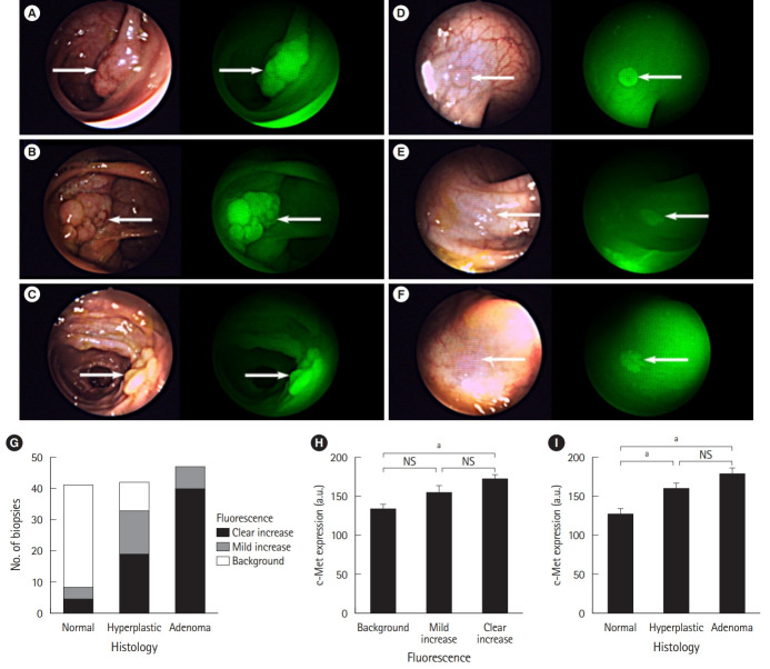
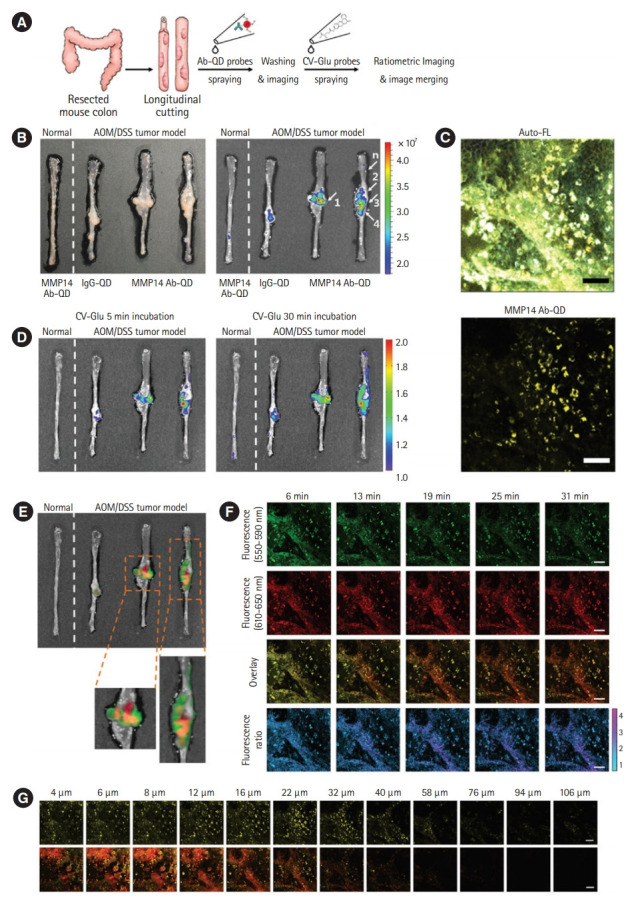
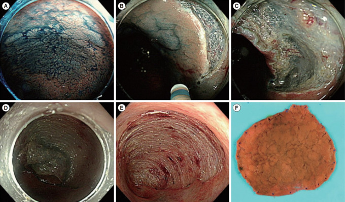
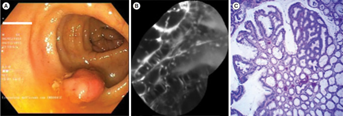
Molecular imaging is a technique for imaging the processes occurring in a living body at a molecular level in real-time, combining molecular cell biology with advanced imaging technologies using molecular probes and fluorescence. Gastrointestinal endoscopic molecular imaging shows great promise for improving the identification of neoplasms, providing characterization for patient stratification and assessing the response to molecular targeted therapy. In inflammatory bowel disease, endoscopic molecular imaging can be used to assess disease severity and predict therapeutic response and prognosis. Endoscopic molecular imaging is also able to visualize dysplasia in the presence of background inflammation. Several preclinical and clinical trials have evaluated endoscopic molecular imaging; however, this area is just beginning to evolve, and many issues have not been solved yet. In the future, it is expected that endoscopic molecular imaging will be of increasing interest among clinicians as a new technology for the identification and evaluation of colorectal neoplasm and colitis-associated cancer.

Keywords: Colitis; Inflammatory bowel disease; Intestinal diseases; Molecular imaging; Neoplasm.


Conflict of interest statement

Conflict of Interest

Myung SJ is an editorial board member of the journal but did not involve in the peer reviewer selection, evaluation, or decision process of this article. He is CEO of EdisBiotech Co. No other potential conflicts of interest relevant to this article were reported.

Figures

Fig. 1.
Fig. 1.
Simultaneous white light (WL) and fluorescent light (FL) images of representative lesions of various morphological and histological subtypes found. (A-C) The lesions shown are clearly visible in WL and show clearly increased fluorescence. (D) A lesion that, although it was visible in WL, had enhanced visibility in FL. (E, F) Images representative of the 9 lesions that were only visible in FL. Polyps are indicated by the white arrows. (A) A 2-cm pedunculated (Paris 0-Ip) tubular adenoma. (B) A 4-cm subpedunculated (Paris 0-Isp) tubulovillous adenoma. (C) A 2-cm sessile (Paris 0-Is) serrated polyp. (D) A 5-mm flat elevated (Paris 0-IIa) tubular adenoma. (E, F) Flat (Paris 0-IIb) tubular adenomas, 5 mm and 4 mm in diameter, respectively. (G) Graph showing the relationship between the degree of fluorescence and the histological diagnosis. (H) Graph showing the relationship between c-Met expression and the degree of fluorescence. (I) Graph showing the relationship between c-Met expression and histological diagnosis. Error bars are means±standard error of the mean. aP<0.001, mixed-model analysis of variance. Adapted from Burggraaf J, et al. Nat Med 2015;21:955-961, with permission from Springer Nature [23].
Fig. 2.
Fig. 2.
(A) Schematic diagram of sequential ex vivo staining with crystal violet (CV)-glutamic acid (Glu) and matrix metalloproteinases 14 (MMP14) antibody-quantum dot (Ab-QD) probes. (B) White light images (left panel) and in vivo imaging system images (right panel) of (from left) a normal colon tissue treated with the MMP14 Ab-QD probe, a tumor tissue treated with IgG-QD conjugates, and two other tumor colons treated with the MMP14 Ab–QD probe. Five regions were histopathologically analyzed (arrows 1, 2, 3, 4, and n). (C) Twophoton microscopy (TPM) images of mouse tumor colon stained by the MMP14 Ab-QD probe. Upper image: autofluorescence (Auto-FL) imaging, lower image: MMP14 Ab-QD probe signals (yellow pseudo-color). (D) Ratiometric signals after spraying all tissues with the CV-Glu probe at 5 and 30 minutes after treatment. (E) Overlay images of the CV-Glu probe radio frequency signal at 30 minutes (green pseudo-color) and MMP14 Ab-QD probe signal (red pseudo-color). (F) Time-dependent TPM images of the CV–Glu probe in the same area shown in panel (C). First row: CV–Glu; second row: CV; third row: overlay images; fourth row: ratiometric images. (G) TPM fluorescence images recorded moving down in the z-direction. First row: MMP14 Ab-QDs (yellow pseudo-color); second row: overlay images of CV-Glu probe. Scale bar: 50 µm. AOM, azoxymethane; DSS, dextran sodium sulfate. Adapted from Park Y, et al. Acs Nano 2014;8:8896-8910, with permission from John Wiley and Sons [24].
Fig. 3.
Fig. 3.
Representative example of endoscopic submucosal dissection for colitis-associated dysplasia. (A) Large, non-ulcerated Paris type O-IIa dysplasia with a distinct border in the rectum. (B) Mucosal incision was performed after submucosal injection. (C) Mild but diffuse submucosal fibrosis and submucosal fat deposition. (D-F) The colonoscope was changed into a gastroscope to expose the submucosal layer more effectively, and en bloc resection was achieved. The final histology revealed low-grade dysplasia (42×40 mm in size, with clear lateral and vertical margins). Adapted from Yang DH, et al. Clin Endosc 2019;52:120-128 [29].
Fig. 4.
Fig. 4.
Dysplastic lesion. (A) White light endoscopic view showing a polypoid lesion (Paris 0-Is) of the transverse colon. (B) After resection and coloration with the 100 μM VRPMPLQ peptide solution, confocal laser endomicroscopy shows active binding of the peptide to dysplastic colonocytes is observed. This along with passive accumulation of the peptide determines an increase in fluorescence. (C) Conventional histology (H&E, ×106) showing low-grade dysplasia. VRPMPLQ is a synthetic peptide conjugated with fluorescein, which shows more selective binding to dysplastic tissue than to normal mucosa. Adapted from De Palma GD, et al. PLoS One 2017;12:e0180509 [67].

References

    1. Drossman DA. Challenges in the physician-patient relationship: feeling “drained”. Gastroenterology. 2001;121:1037–1038. - PubMed
    1. Matloff JL, Abidi W, Richards-Kortum R, Sauk J, Anandasabapathy S. High-resolution and optical molecular imaging for the early detection of colonic neoplasia. Gastrointest Endosc. 2011;73:1263–1273. - PubMed
    1. van Rijn JC, Reitsma JB, Stoker J, Bossuyt PM, van Deventer SJ, Dekker E. Polyp miss rate determined by tandem colonoscopy: a systematic review. Am J Gastroenterol. 2006;101:343–350. - PubMed
    1. Yalamarthi S, Witherspoon P, McCole D, Auld CD. Missed diagnoses in patients with upper gastrointestinal cancers. Endoscopy. 2004;36:874–879. - PubMed
    1. Dacosta RS, Wilson BC, Marcon NE. New optical technologies for earlier endoscopic diagnosis of premalignant gastrointestinal lesions. J Gastroenterol Hepatol. 2002;17 Suppl:S85S104. - PubMed

LinkOut - more resources