Review
doi: 10.3906/sag-2004-160.
Radiological approaches to COVID-19 pneumonia
Affiliations
- PMID: 32299200
- PMCID: PMC7195987
- DOI: 10.3906/sag-2004-160
Item in Clipboard
Review
Radiological approaches to COVID-19 pneumonia
Turk J Med Sci.
.
Abstract
COVID-19 pneumonia has high mortality rates. The symptoms are undiagnostic, the results of viral nucleic acid detection method (PCR) can delay, so that chest computerized tomography is often key diagnostic test in patients with possible COVID-19 pneumonia. In this review, we discussed the main radiological findings of this infection.
Keywords: COVID-19; chest computerized tomography; pneumonia.
This work is licensed under a Creative Commons Attribution 4.0 International License.
Conflict of interest statement
There is no conflict of interest.
Figures
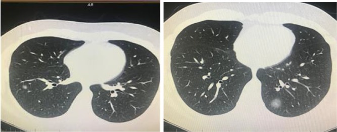
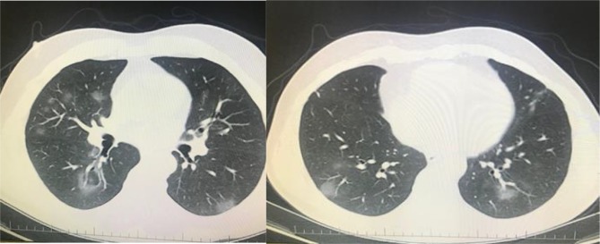
A 32-year-old man was admitted to our clinic with symptoms of fever and cough. PCR was positive for novel coronavirus.
Focal small patchy infiltrate (atypical appearance) was seen in right and left lower lobe (archives of Şule Akçay).
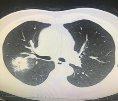
A 66-year-old man’s chest CT imaging, PCR positive,
and CT has right upper lobe consolidation which is atypical
appearance for COVID-19 pneumonia (archives of Şule Akçay).
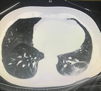
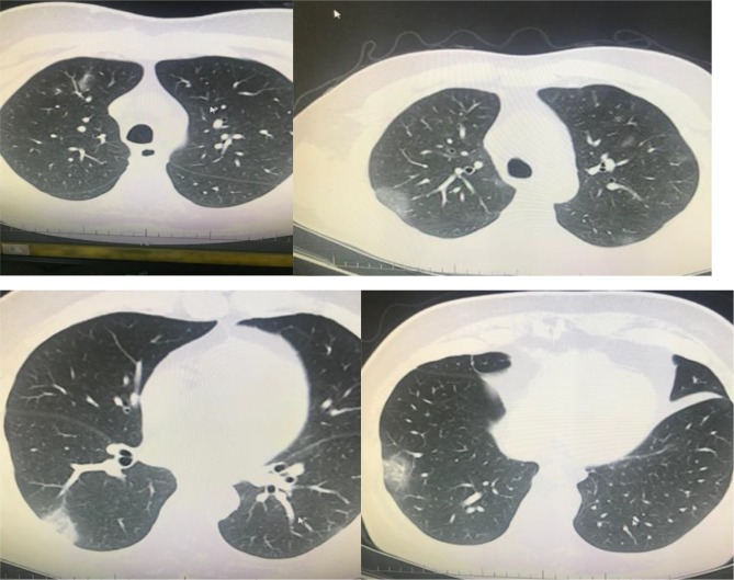
A 66-year-old man with chronic renal failure. PCR was
negative. However, his wife has diagnosed as having COVID-19
pneumonia by PCR positivity. Ground-glass opacity was seen
on left lower lobe, and treatment of COVID-19 pneumonia was
applied (archives of Şule Akçay).
A 47-year-old man with chronic renal failure. PCR was positive. Chest CT revealed bilateral peripheral patchy infiltration
(archives of Şule Akçay).
A 62-year-old man, PCR positive. Bilateral multilobar GGO on his chest CT (archives of Şule Akçay).
A 61-year-old man, PCR positive, unilateral multilobar focal consolidation areas on his chest CT (archives of Şule Akçay).
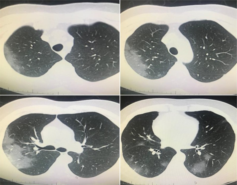
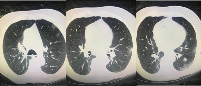
A 72-year-old man, PCR was negative. However, his wife’s swab was PCR positive. His chest CT has GGO in both lungs’
multilobes as typical COVID-19 pneumonia, early pneumonia treatment was essential according this imaging (archives of Şule Akçay).
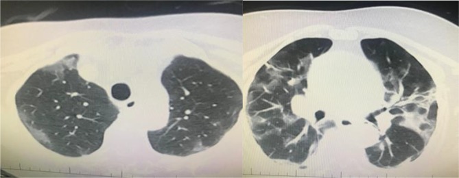
A 55-year-old woman, PCR was positive, bilateral multilobar GGO was seen in her chest CT (archives of Şule Akçay).
Comment in
-
Response to letter to the editor: Radiological approaches to COVID-19 pneumonia.Turk J Med Sci. 2020 Aug 26;50(5):1442-1443. doi: 10.3906/sag-2005-230. Turk J Med Sci. 2020. PMID: 32490639 Free PMC article. No abstract available.
References
-
- Daily COVID-19 statistically case reports. 2020.
-
- Nasir MU J The Role of emergency radiology in COVID-19: from preparedness to diagnosis. Canadian Association of Radiologists Journal. 2020;10 - PubMed
Publication types
MeSH terms
LinkOut - more resources
Full Text Sources
Medical