

Normobaric intermittent hypoxic training regulates microglia phenotype and enhances phagocytic activity
- PMID: 32299228
- PMCID: PMC7221485
- DOI: 10.1177/1535370220919361
Normobaric intermittent hypoxic training regulates microglia phenotype and enhances phagocytic activity
Abstract
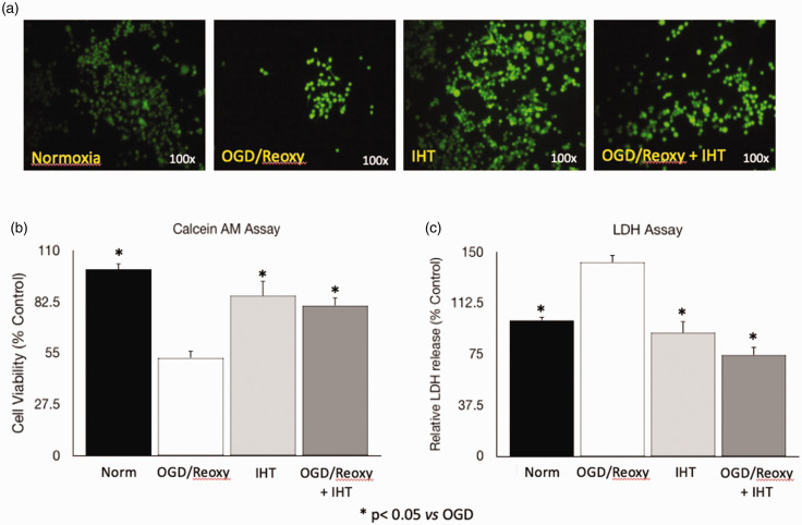
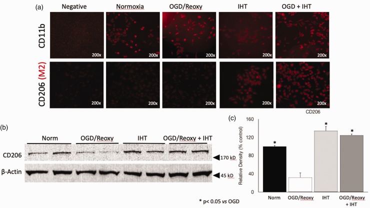
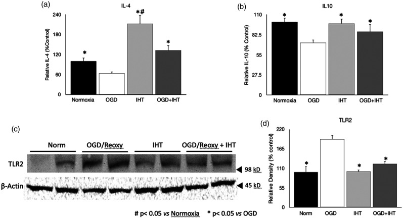
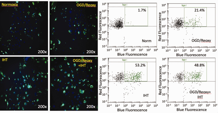
The effects of intermittent hypoxic training or conditioning on many pathological conditions have been widely investigated. One of the pathological conditions dealt with intermittent hypoxic training is ischemic stroke. Well-known mechanisms of intermittent hypoxia-induced protection are related to increased energy metabolism and the enhanced antioxidant effects. In the last decades, the role of microglia in the progress of ischemic stroke-related brain damage has been focused. The dual-edge function of microglia indicates that the microglia-mediated inflammatory response is definitely beneficial in the early stage of ischemic stroke, but long-term activation of microglia is rather detrimental during the recovery process. The effect of IHT on microglia polarization is not investigated. This study focused on whether IHT regulates the polarization of microglia without dampening its classic phagocytic function. This study will provide pivotal information regarding the effects of IHT on the long-term effects on the recovery process from ischemic stroke.
Keywords: Microglia; inflammation; intermittent hypoxia training; ischemic stroke; oxygen–glucose deprivation; phagocytosis.
Figures
References
-
- Kochanek KD, Murphy SL, Xu J, Arias E. Mortality in the United States. NCHS Data Brief 2013; 2014:1–8 - PubMed
-
- Mozaffarian D, Benjamin EJ, Go AS, Arnett DK, Blaha MJ, Cushman M, Das SR, de Ferranti S, Despres JP, Fullerton HJ, Howard VJ, Huffman MD, Isasi CR, Jimenez MC, Judd SE, Kissela BM, Lichtman JH, Lisabeth LD, Liu S, Mackey RH, Magid DJ, McGuire DK, Mohler Cr, 3rd, Moy CS, Muntner P, Mussolino ME, Nasir K, Neumar RW, Nichol G, Palaniappan L, Pandey DK, Reeves MJ, Rodriguez CJ, Rosamond W, Sorlie PD, Stein J, Towfighi A, Turan TN, Virani SS, Woo D, Yeh RW, Turner MB. Heart disease and stroke statistics-2016. Update: a report from the American Heart Association. Circulation 2016; 133:e38–e360 - PubMed
-
- Benjamin EJ, Blaha MJ, Chiuve SE, Cushman M, Das SR, Deo R, de Ferranti SD, Floyd J, Fornage M, Gillespie C, Isasi CR, Jimenez MC, Jordan LC, Judd SE, Lackland D, Lichtman JH, Lisabeth L, Liu S, Longenecker CT, Mackey RH, Matsushita K, Mozaffarian D, Mussolino ME, Nasir K, Neumar RW, Palaniappan L, Pandey DK, Thiagarajan RR, Reeves MJ, Ritchey M, Rodriguez CJ, Roth GA, Rosamond WD, Sasson C, Towfighi A, Tsao CW, Turner MB, Virani SS, Voeks JH, Willey JZ, Wilkins JT, Wu JH, Alger HM, Wong SS, Muntner P. Heart disease stroke statistics-2017. Update: a report from the American Heart Association. Circulation 2017; 135:e146–e603 - PMC - PubMed
-
- Ovbiagele B, Goldstein LB, Higashida RT, Howard VJ, Johnston SC, Khavjou OA, Lackland DT, Lichtman JH, Mohl S, Sacco RL, Saver JL, Trogdon JG. Forecasting the future of stroke in the United States: a policy statement from the American Heart Association and American Stroke Association. Stroke 2013; 44:2361–75 - PubMed
MeSH terms
Substances
LinkOut - more resources
Full Text Sources