. 2020 Jul 17;127(3):379-390.
doi: 10.1161/CIRCRESAHA.119.316369. Epub 2020 Apr 17.

MCUb Induction Protects the Heart From Postischemic Remodeling

Affiliations

MCUb Induction Protects the Heart From Postischemic Remodeling

Jiuzhou Huo et al. Circ Res. .

Abstract

Rationale: Mitochondrial Ca2+ loading augments oxidative metabolism to match functional demands during times of increased work or injury. However, mitochondrial Ca2+ overload also directly causes mitochondrial rupture and cardiomyocyte death during ischemia-reperfusion injury by inducing mitochondrial permeability transition pore opening. The MCU (mitochondrial Ca2+ uniporter) mediates mitochondrial Ca2+ influx, and its activity is modulated by partner proteins in its molecular complex, including the MCUb subunit.

Objective: Here, we sought to examine the function of the MCUb subunit of the MCU-complex in regulating mitochondria Ca2+ influx dynamics, acute cardiac injury, and long-term adaptation after ischemic injury.

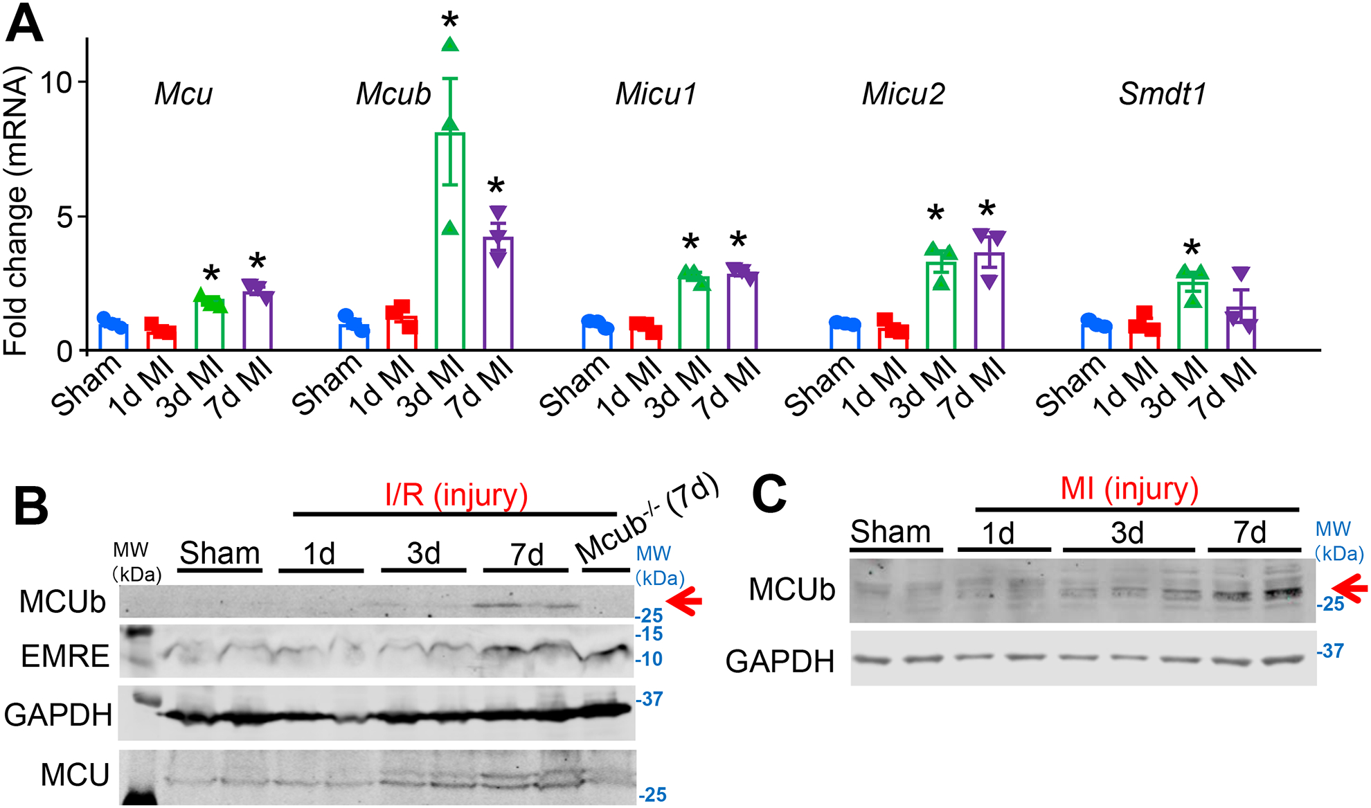
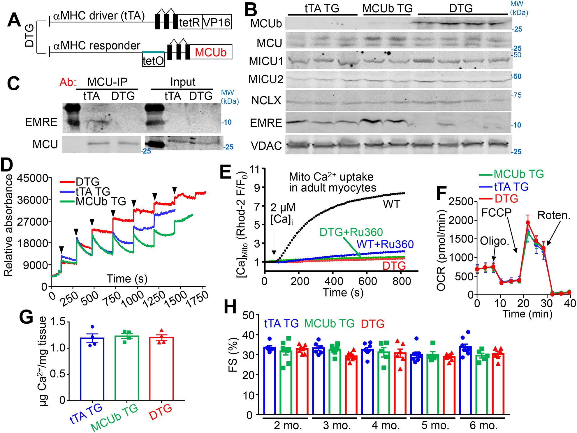
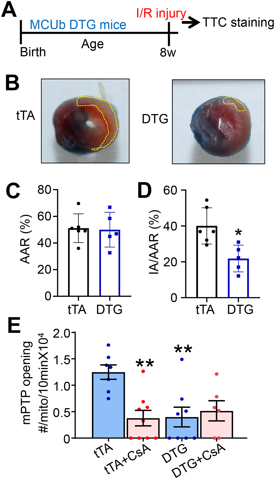
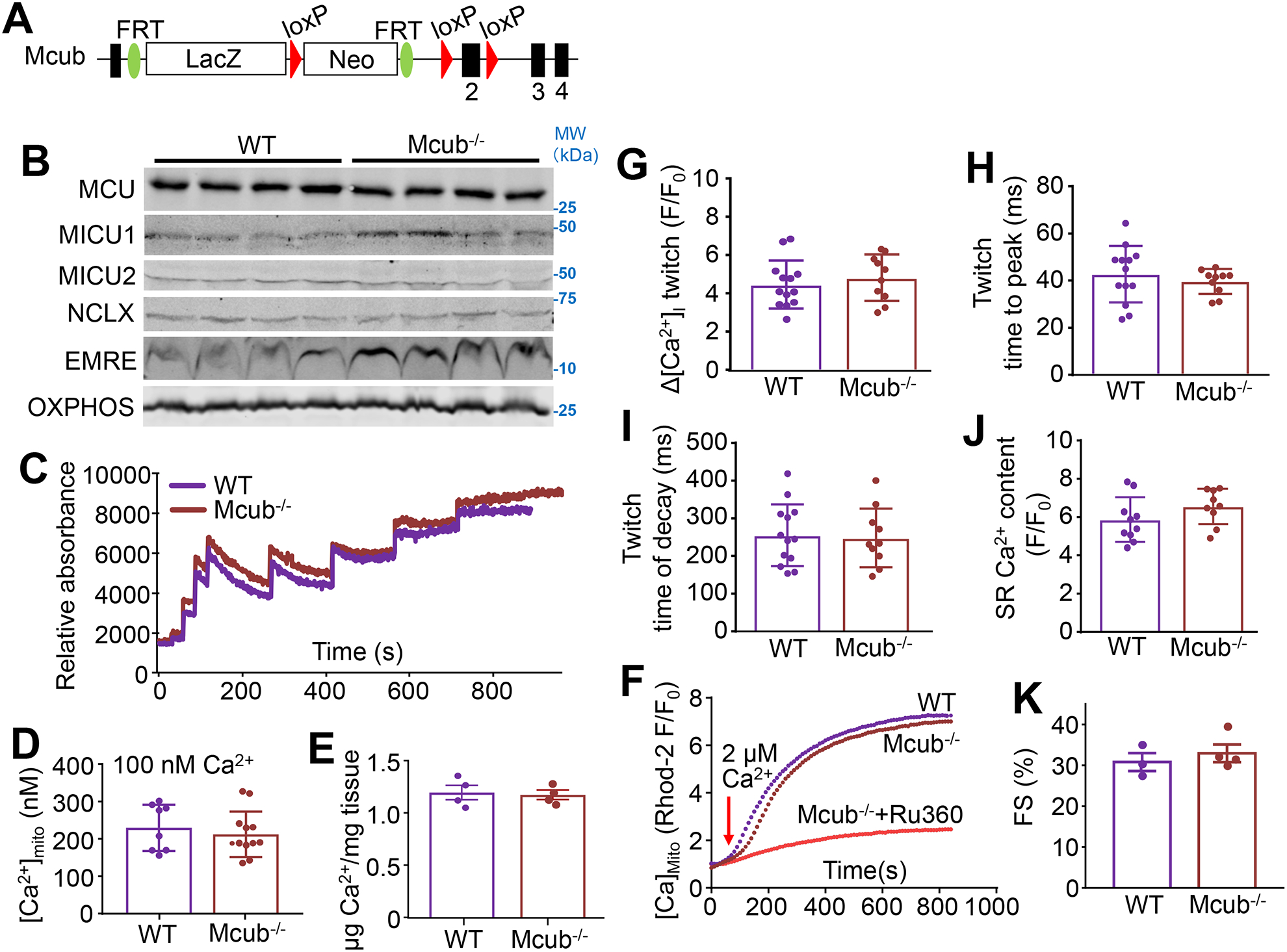
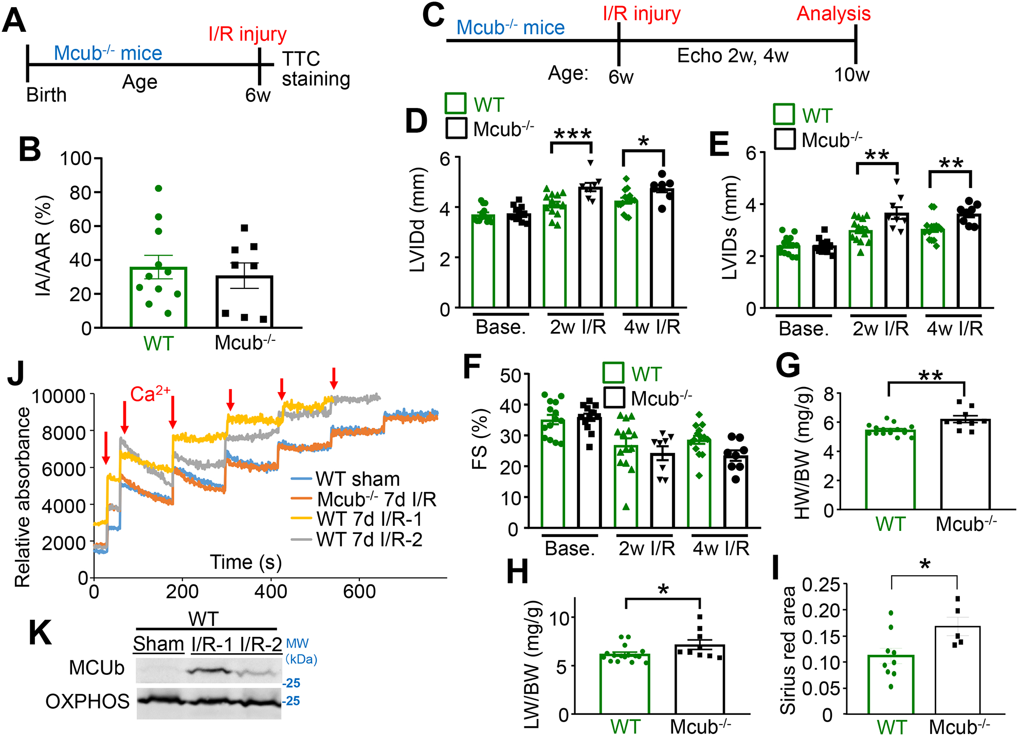
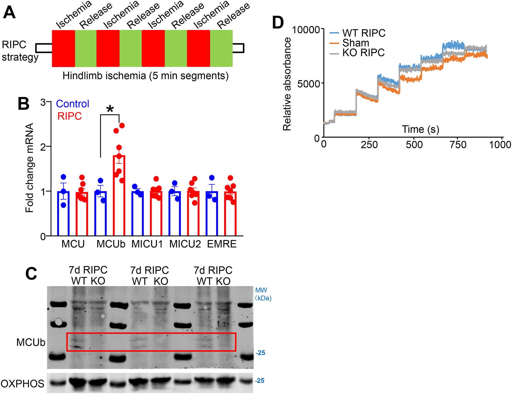
Methods and results: Cardiomyocyte-specific MCUb overexpressing transgenic mice and Mcub gene-deleted (Mcub-/-) mice were generated to dissect the molecular function of this protein in the heart. We observed that MCUb protein is undetectable in the adult mouse heart at baseline, but mRNA and protein are induced after ischemia-reperfusion injury. MCUb overexpressing mice demonstrated inhibited mitochondrial Ca2+ uptake in cardiomyocytes and partial protection from ischemia-reperfusion injury by reducing mitochondrial permeability transition pore opening. Antithetically, deletion of the Mcub gene exacerbated pathological cardiac remodeling and infarct expansion after ischemic injury in association with greater mitochondrial Ca2+ uptake. Furthermore, hindlimb remote ischemic preconditioning induced MCUb expression in the heart, which was associated with decreased mitochondrial Ca2+ uptake, collectively suggesting that induction of MCUb protein in the heart is protective. Similarly, mouse embryonic fibroblasts from Mcub-/- mice were more sensitive to Ca2+ overload.

Conclusions: Our studies suggest that Mcub is a protective cardiac inducible gene that reduces mitochondrial Ca2+ influx and permeability transition pore opening after ischemic injury to reduce ongoing pathological remodeling.

Keywords: calcium; heart; infarction; mitochondria; reperfusion injury.


Conflict of interest statement

DISCLOSURES

All authors confirm no conflict of interest.

Figures

Figure 1.
Figure 1.. MCUb expression is induced following cardiac ischemic injury.
A) MCU-complex components Mcu, Mcub, Micu1, Micu2 and Smdt1 (EMRE) mRNA levels from cardiac left ventricle tissue and isolated myocytes following ischemia reperfusion (I/R) injury for 1-, 3- or 7-days versus sham. n=3 per group. Data presented as mean ± standard deviation of mean (SEM). One-way ANOVA and post hoc Bonferroni test was used for statistical analysis. *p<0.05 versus sham by post hoc Bonferroni’s multiple comparison test. B) Western blot of MCUb, MCU and EMRE from left ventricle of the WT mouse heart following a time-course of I/R injury as shown in days. GAPDH was used as the protein loading control. Mcub−/− heart tissues 7 days after I/R injury is used as an antibody negative control. Arrow shows position of MCUb protein. The molecular weight marker shown is based on the uncropped western blots in which standards can (Online Supplement, and applicable to the remaining western blots in the paper). C) Western blot of MCUb from the heart following a time-course of myocardial infarction (MI) injury as shown. GAPDH was used as the protein loading control.
Figure 2.
Figure 2.. Generation of cardiomyocyte-specific MCUb overexpressing transgenic mice.
A) Schematic of cardiomyocyte-specific MCUb overexpressing mice using a double transgenic (DTG) tet-off system based on the α-myosin heavy chain (α-MHC) promoter. The driver line expresses the tetracycline transactivator cDNA (tTA), and the responder line contains the tet-operator and the MCUb cDNA. B) Western blot of MCUb, MCU, MICU1, MICU2, NCLX and EMRE in cardiac mitochondria from the indicated groups of mice. VDAC was used as a processing and loading control. C) Co-immunoprecipitation of EMRE with an MCU antibody from isolated heart mitochondria from the indicated two groups of mice. Western blotting for MCU from both immunoprecipitated samples as well as input samples is shown as controls. D) Mitochondrial Ca2+ uptake in isolated heart mitochondria from the indicated groups at 3 months of age. Calcium Green-5N was used as the Ca2+ indicator. The arrows indicate addition of 20 μM CaCl2 to the solution. E) Mitochondrial Ca2+ uptake in permeabilized adult cardiomyocytes from hearts of the indicated groups, with or without Ru360 under 2 μmol/L CaCl2 perfusion. F) Oxygen consumption rate (OCR) measurement in cardiac mitochondria from the indicated mouse groups of mice at 3 months of age. n=3 per group. Arrows show the position of the three different drugs given in temporal sequence. G) Quantification of baseline mitochondrial Ca2+ levels in isolated cardiac mitochondria from the indicated groups of mice at 6 months of age. n=4 per group. Data presented as mean ± SEM. One-way ANOVA was used for statistical analysis. H) Echocardiographic measurement of fractional shortening (FS, %) from indicated groups at the indicated ages in months. n=7 in tTA TG group, n=5 in MCUb TG group, and n=6 in DTG group. Data presented as mean ± SEM. Two-way ANOVA was used for statistical analysis, with no significant interactions or effects of variables.
Figure 3.
Figure 3.. MCUb overexpression generates protection from cardiac I/R injury.
A) Temporal strategy of I/R injury in mice for panels “B”, “C” and “D”. Mice at 8 wks of age were challenged with 30 minutes of ischemia followed by 24 hours of reperfusion. B) Hearts were sacrificed for 2,3,5-triphenyltetrazolium chloride (TTC) staining. The yellow dotted area shows ischemic region. C,D) Average area at risk (AAR) and ischemic area (IA)/AAR of hearts from mice subjected to I/R injury from the indicated mouse groups. n=6 in tTA TG group, n=5 in DTG group. Data presented as mean ± SEM. Student’s t-test was used for statistical analysis. *p<0.05 vs tTA. E) Mitochondrial permeability transition pore (mPTP) opening frequency measurements in permeabilized cardiomyocytes. mPTP inhibitor cyclosporine A (CsA) was used as a control. n=7 in tTA TG group, n=9 in tTA group with CsA treatment, n=8 in DTG group, n=6 in DTG group with CsA treatment. Data presented as mean ± SEM. Two-way ANOVA with a post-hoc Bonferroni’s multiple comparison test was performed. A significant interaction was found between treatment and genotype (p<0.05). **p<0.01 vs tTA.
Figure 4.
Figure 4.. Deletion of Mcub does not affect mitochondrial Ca2+ uptake or cardiac function at baseline.
A) Strategy for generating Mcub-null mice with a “knock-out first” allele strategy in which a combined β-galactosidase/neomycin cDNA cassette was inserted between exon 1 and exon 2 of the Mcub gene. B) Western blot of MCU, MICU1, MICU2, NCLX and EMRE in cardiac mitochondria from the indicated groups of mice. OXPHOS antibody was used as a control. C) Mitochondrial Ca2+ uptake assay in isolated heart mitochondria from the indicated groups of mice at 4 months of age. D) Quantification of baseline mitochondrial Ca2+ content ([Ca2+]mito) in permeabilized adult cardiomyocytes from the indicated groups. n=8 in WT group, n=12 in Mcub−/− group. Data presented as mean ± SEM. Student’s t-test was used for statistical analysis. E) Quantification of baseline [Ca2+]mito levels in isolated cardiac mitochondria from the indicated groups of mice at 4 months of age. n=4 per group. Data presented as mean ± SEM. Student’s t-test was used for statistical analysis. F) Measurement of mitochondrial Ca2+ uptake in permeabilized adult cardiomyocytes under 2 μmol/L CaCl2. Ru360 was used as a control. G-I) Measurements Ca2+ transient amplitude, time to peak and twitch decay (tau) from adult cardiomyocytes from hearts of the indicated groups of mice. n=13 in WT group, n=10 in Mcub−/− group. Data presented as mean ± SEM. Student’s t-test was used for statistical analysis. J) Measurements of sarcoplasmic reticulum (SR) Ca2+ content from adult cardiomyocytes from hearts of the indicated groups of mice. n=10 in WT group, n=9 in Mcub−/− group. Data presented as mean ± SEM. Student’s t-test was used for statistical analysis. K) Echocardiographic measurement of fractional shortening (FS, %) from the indicated groups at 4 months of age. n=3 in WT group, n=4 in Mcub−/− group. Data presented as mean ± SEM. Student’s t-test was used for statistical analysis.
Figure 5.
Figure 5.. Cardiac MCUb protein induction protects the heart from damage post I/R injury.
A) Temporal schematic of the I/R injury model in mice for panel “B”. Mice at 6 wks of age were challenged with 60 minutes of ischemia followed by 24 hours of reperfusion. Hearts were sacrificed for TTC staining. B) Both IA and AAR were analyzed and averaged for the indicated groups following I/R injury. n=11 in WT group, n=8 in Mcub−/− group. Data presented as mean ± SEM. Student’s t-test was used for statistical analysis. C) Temporal schematic of the strategy for assessing infarct expansion following I/R injury, for panels “D-H”. Mice at 6 wks of age were challenged with 60 minutes of ischemic injury. Echocardiographic measurements were performed before injury, then 2 and 4 wks of reperfusion. Mice were sacrificed at the 4 wk time point for additional analyses. D-F) Echocardiographic parameters in the indicated groups of mice at the indicated timepoints. LVIDd, end-diastolic left ventricle internal diameter; LVIDs, end-systolic left ventricle internal diameter; FS, fractional shortening. n=14 in WT group, n=12 in Mcub−/− group at baseline; n=14 in WT group, n=8 in Mcub−/− group post injury. Data presented as mean ± SEM. Two-way ANOVA with a post hoc Bonferroni’s multiple comparison test was performed. For “D&E”, there was a significant interaction (p<0.05) between time and genotype variables. For “F”, there was only a significant time effect (p<0.05) as the two-way ANOVA showed no significant interaction. Markings denote the following: *p<0.05, **p<0.01, ***p<0.001. G–H) Heart weight/body weight (HW/BW) ratio and lung weight/body weight (LW/BW) ratio in the indicated groups of mice 4 wks post I/R injury. n=14 in WT group, n=9 in Mcub−/− group. Data presented as mean ± SEM. Student’s t-test was used for statistical analysis. *p<0.05, **p<0.01. I) Quantification of Sirius Red staining of histological sections from hearts of mice 4 wks post I/R injury in the indicated groups. n=14 in WT group, n=9 in Mcub−/− group. Data presented as mean ± SEM. Student’s t-test was used for statistical analysis. *p<0.05. J) Mitochondrial Ca2+ uptake in isolated mitochondria from the left ventricle of hearts in the indicated groups of mice. Mice were challenged with sham or I/R injury, and then collected 7 days post injury. K) Western blot of MCUb in the same isolated hearts used in “J”. OXPHOS antibody was used as the protein loading control.
Figure 6.
Figure 6.. MCUb is induced in the heart by post remote ischemic pre-conditioning from the hindlimb.
A) Temporal strategy of the remote ischemic pre-conditioning (RIPC) in mice for panels “B”, “C”, and “D”. Mice at 9–12 wks of age were challenged with hindlimb femoral artery occlusion for 5 minutes followed by 5 minutes of reperfusion, which was repeated with 4 cycles per day for 7 consecutive days. B) mRNA for MCU-complex components Mcu, Mcub, Micu1, Micu2, and Smdt1 (EMRE) from cardiac left ventricle tissue following the 7-day RIPC protocol, versus sham. n=3 in sham group, n=7 in RIPC group. Data presented as mean ± SEM. Student’s t-test was used for statistical analysis. *p<0.05. C) Western blot of MCUb in the same isolated hearts used in “D”. OXPHOS antibody was used as the protein loading control. The red box shows the area where MCUb protein is induced in the WT heart samples subject to RIPC but not in the Mcub−/− mice (KO). D) Mitochondrial Ca2+ uptake assay in isolated mitochondria from the left ventricle of hearts in the indicated groups of mice. Mice were challenged with sham or the 7-day RIPC protocol, and then hearts were collected and mitochondria were purified for analyses.

Comment in

References

    1. Kalogeris T, Baines CP, Krenz M, Korthuis RJ. Cell biology of ischemia/reperfusion injury. Int Rev Cell Mol Biol. 2012;298:229–317 - PMC - PubMed
    1. Eltzschig HK, Eckle T. Ischemia and reperfusion--from mechanism to translation. Nat Med. 2011;17:1391–1401 - PMC - PubMed
    1. Hausenloy DJ, Yellon DM. Myocardial ischemia-reperfusion injury: A neglected therapeutic target. J Clin Invest. 2013;123:92–100 - PMC - PubMed
    1. Halestrap AP, Clarke SJ, Javadov SA. Mitochondrial permeability transition pore opening during myocardial reperfusion--a target for cardioprotection. Cardiovasc Res. 2004;61:372–385 - PubMed
    1. Halestrap AP. What is the mitochondrial permeability transition pore? J Mol Cell Cardiol. 2009;46:821–831 - PubMed

Publication types

MeSH terms