

Next-generation RNA Sequencing-based Biomarker Characterization of Chromophobe Renal Cell Carcinoma and Related Oncocytic Neoplasms
- PMID: 32299640
- PMCID: PMC8996310
- DOI: 10.1016/j.eururo.2020.03.003
Next-generation RNA Sequencing-based Biomarker Characterization of Chromophobe Renal Cell Carcinoma and Related Oncocytic Neoplasms
Abstract
Background: Renal cell carcinomas (RCCs) are a heterogeneous group of neoplasms. Recent sequencing studies revealed various molecular features associated with histologic RCC subtypes, including chromophobe renal cell carcinoma (ChRCC).
Objective: To characterize the gene expression and biomarker signatures associated with ChRCC.
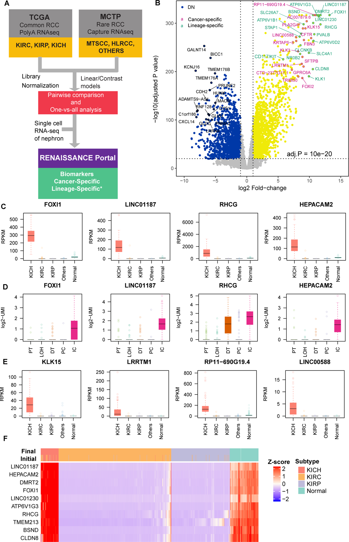
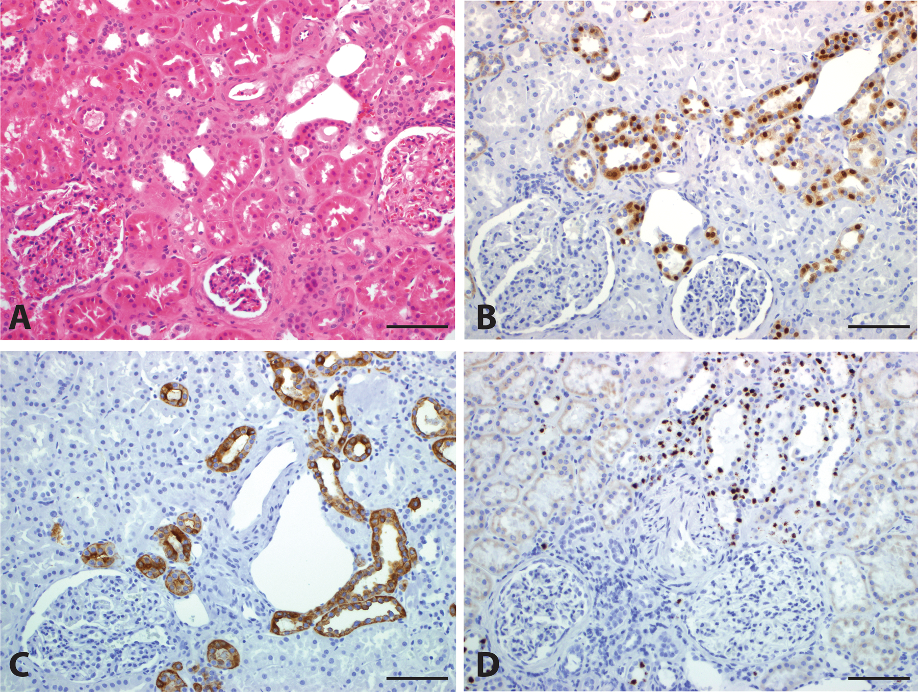
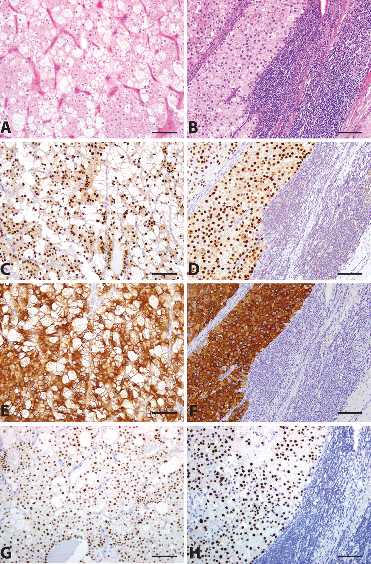
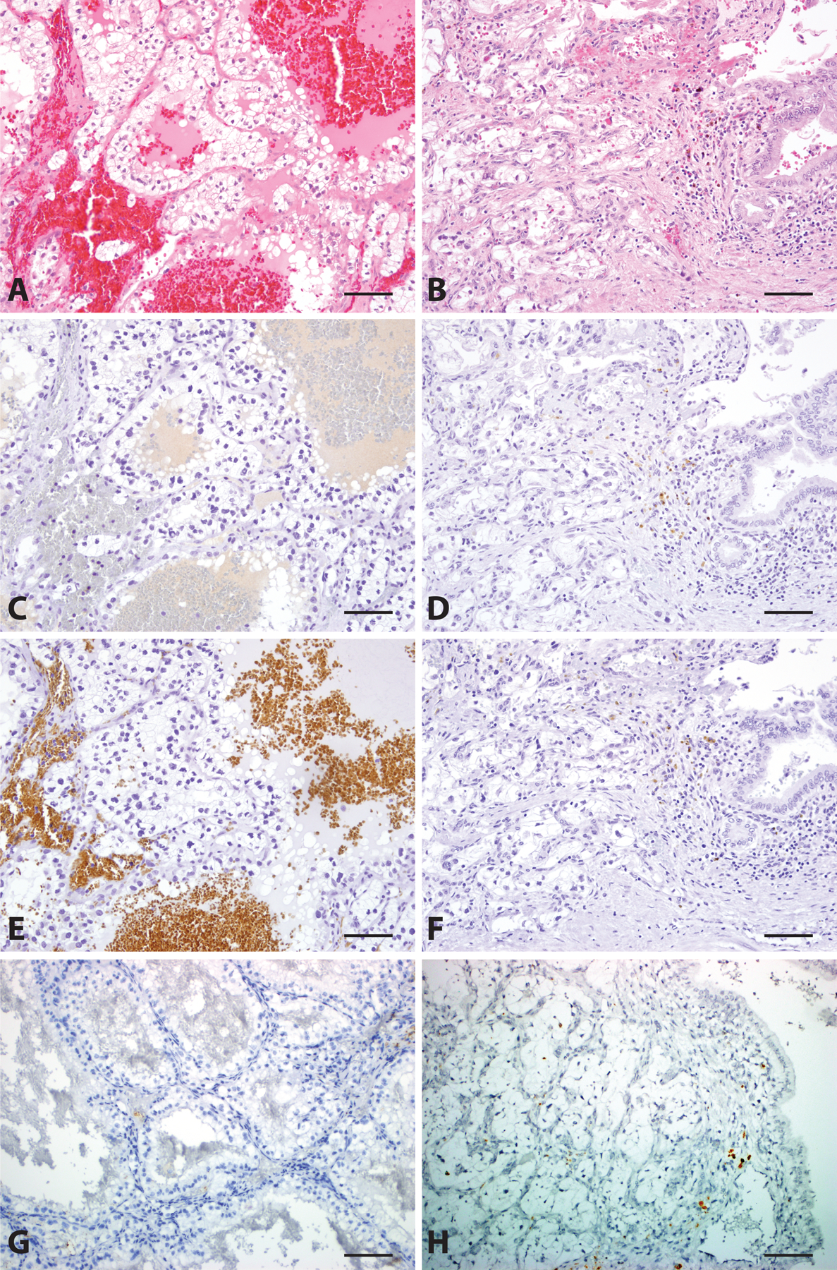
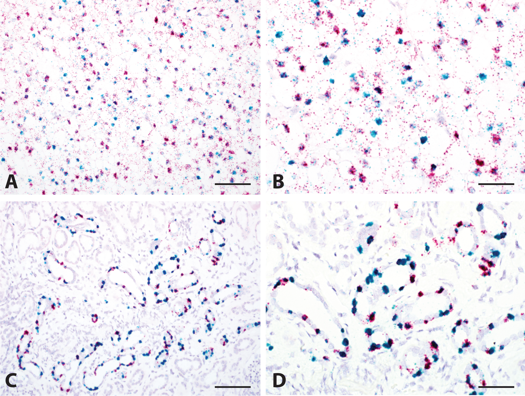
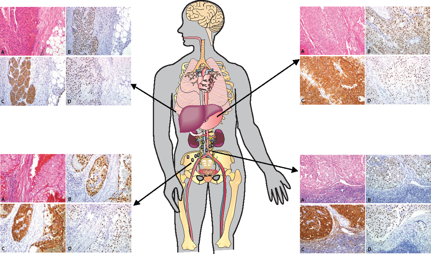
Design, setting, and participants: We performed integrative analysis on RNA sequencing data available from 1049 RCC specimens from The Cancer Genome Atlas and in-house studies. Our workflow identified genes relatively enriched in ChRCC, including Forkhead box I1 (FOXI1), Rh family C glycoprotein (RHCG), and LINC01187. We assessed the expression pattern of FOXI1 and RHCG protein by immunohistochemistry (IHC) and LINC01187 mRNA by RNA in situ hybridization (RNA-ISH) in whole tissue sections representing a cohort of 197 RCC cases, including both primary and metastatic tumors.
Outcome measurements and statistical analysis: The FOXI1 and RHCG IHC staining, as well as the LINC01187 RNA-ISH staining, was evaluated in each case for intensity, pattern, and localization of expression.
Results and limitations: All primary and metastatic classic ChRCCs demonstrated homogeneous positive labeling for FOXI1, RHCG proteins, and LINC01187 transcript. Unclassified RCC with oncocytic features, oncocytoma, and hybrid oncocytic tumor, as well as all but two cases of eosinophilic ChRCC also stained positive. Importantly, metastatic and primary RCC of all other subtypes did not demonstrate any unequivocal staining for FOXI1, RHCG, or LINC01187. In normal kidney, FOXI1, RHCG, and LINC01187 were detected in the distal nephron segment, specifically in intercalated cells. Two cases of eosinophilic ChRCC with focal expression of FOXI1 and LINC01187, and Golgi-like RHCG staining were found to contain MTOR gene mutations upon DNA sequencing.
Conclusions: We demonstrate a pipeline for the identification and validation of RCC subtype-specific biomarkers that can aid in the confirmation of cell of origin and may facilitate accurate classification and diagnosis of renal tumors.
Patient summary: FOXI1, RHCG, and LINC01187 are lineage-specific signature genes for chromophobe renal cell carcinoma.
Keywords: Chromophobe; Classification; Hybrid oncocytic tumor; Immunohistochemistry; Next-generation sequencing; RNA in situ hybridization; Renal cell carcinoma.
Copyright © 2020 European Association of Urology. Published by Elsevier B.V. All rights reserved.
Figures
Comment in
-
Novel Biomarkers in Chromophobe Renal Cell Carcinoma: Distinguishing it from its Mimics.Eur Urol. 2020 Jul;78(1):75-76. doi: 10.1016/j.eururo.2020.03.045. Epub 2020 Apr 21. Eur Urol. 2020. PMID: 32327265 No abstract available.
-
Re: Next-Generation RNA Sequencing-Based Biomarker Characterization of Chromophobe Renal Cell Carcinoma and Related Oncocytic Neoplasms.J Urol. 2020 Dec;204(6):1376. doi: 10.1097/JU.0000000000001281. Epub 2020 Aug 21. J Urol. 2020. PMID: 32955978 No abstract available.
References
-
- Kovacs G, Akhtar M, Beckwith BJ, et al. The Heidelberg classification of renal cell tumours. J Pathol 1997;183:131–3. - PubMed
-
- Udager AM, Mehra R. Morphologic, Molecular, and Taxonomic Evolution of Renal Cell Carcinoma: A Conceptual Perspective With Emphasis on Updates to the 2016 World Health Organization Classification. Arch Pathol Lab Med 2016;140:1026–37. - PubMed
Publication types
MeSH terms
Grants and funding
LinkOut - more resources
Full Text Sources
Medical
Miscellaneous
