. 2020 May 5;117(18):10003-10014.
doi: 10.1073/pnas.1918241117. Epub 2020 Apr 16.

A viral toolkit for recording transcription factor-DNA interactions in live mouse tissues

Affiliations

A viral toolkit for recording transcription factor-DNA interactions in live mouse tissues

Alexander J Cammack et al. Proc Natl Acad Sci U S A. .

Abstract

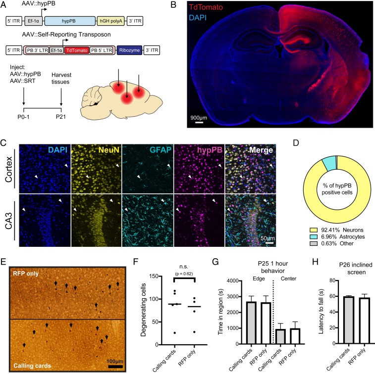
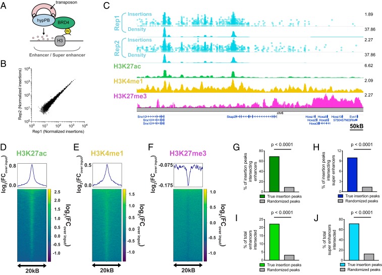
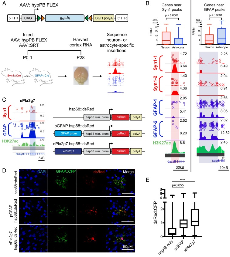
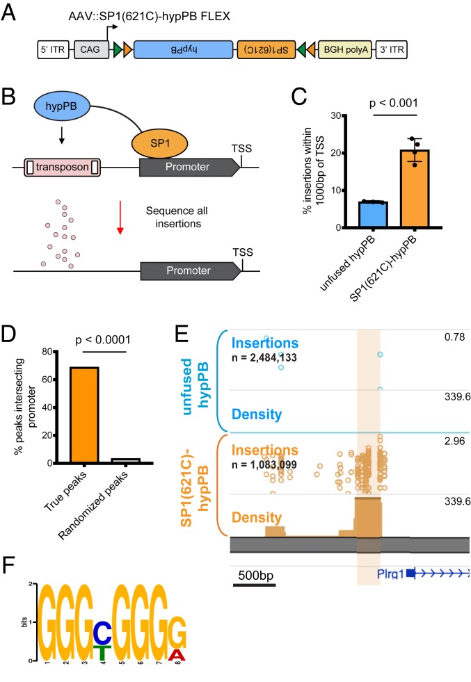
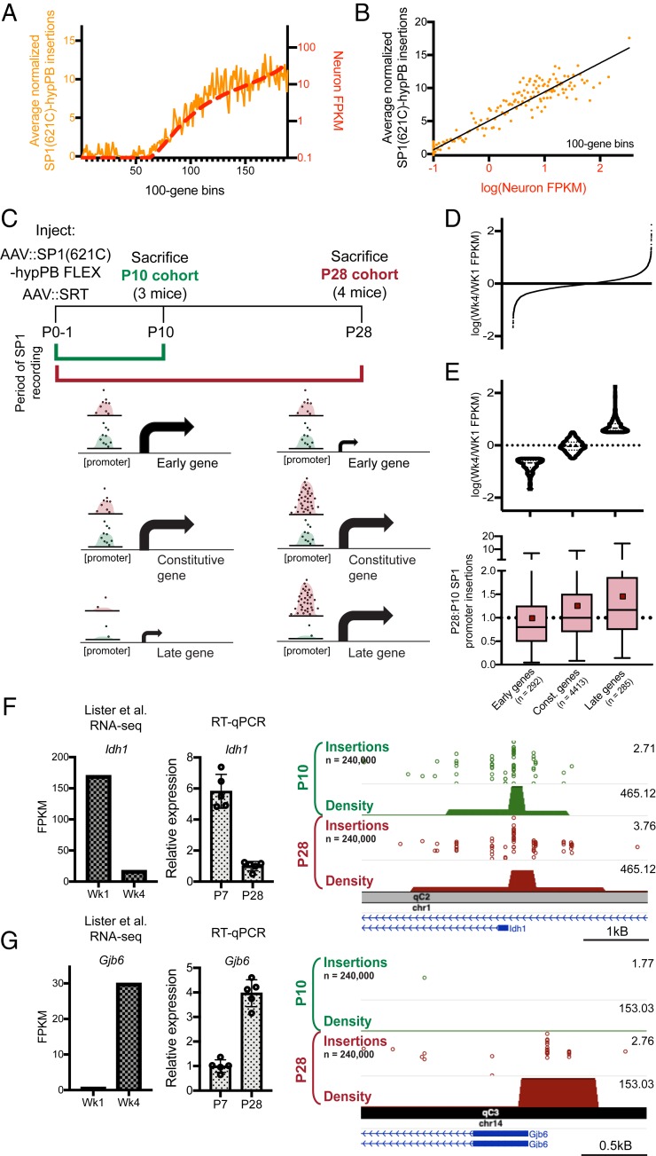
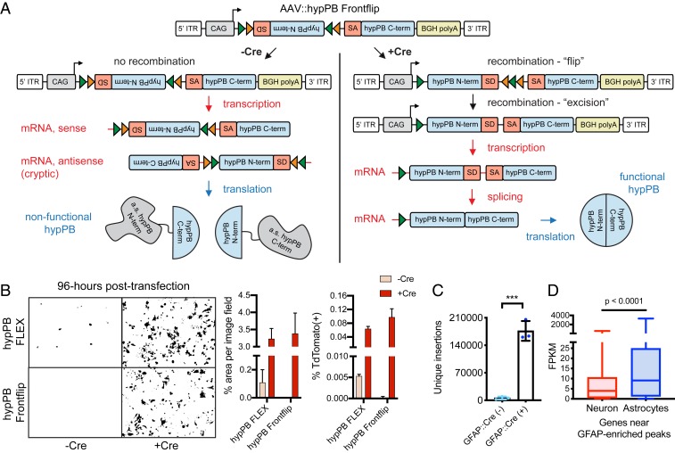
Transcription factors (TFs) enact precise regulation of gene expression through site-specific, genome-wide binding. Common methods for TF-occupancy profiling, such as chromatin immunoprecipitation, are limited by requirement of TF-specific antibodies and provide only end-point snapshots of TF binding. Alternatively, TF-tagging techniques, in which a TF is fused to a DNA-modifying enzyme that marks TF-binding events across the genome as they occur, do not require TF-specific antibodies and offer the potential for unique applications, such as recording of TF occupancy over time and cell type specificity through conditional expression of the TF-enzyme fusion. Here, we create a viral toolkit for one such method, calling cards, and demonstrate that these reagents can be delivered to the live mouse brain and used to report TF occupancy. Further, we establish a Cre-dependent calling cards system and, in proof-of-principle experiments, show utility in defining cell type-specific TF profiles and recording and integrating TF-binding events across time. This versatile approach will enable unique studies of TF-mediated gene regulation in live animal models.

Keywords: brain; enhancer; epigenetics; recording; transcription factor.


Conflict of interest statement

Competing interest statement: R.D.M., A.M., and M.N.W. have filed a patent application on SRT technology. No other authors have disclosures to report.

Figures

Fig. 1.
Fig. 1.
Co-AAV9 intracranial injection efficiently delivers calling cards to the cortex. (A) Experimental paradigm and AAV constructs. Arrows represent approximate AAV injection sites. ITR, inverted-terminal repeat; LTR, long-terminal repeat. (B) Coronal section of a brain injected unilaterally at P0-1 with AAV::hypPB and AAV::SRT, displaying widespread expression of SRT-derived TdTomato fluorescence throughout the brain. Abnormality at right edge is tissue damage that occurred during sectioning and artifact has been removed. DAPI, 4′,6-diamidino-2-phenylindole. (C and D) Coimmunofluorescence showing hypPB expression in neurons and astrocytes. (C) Representative images display colocalization of hypPB with neuronal (NeuN) and astrocyte (GFAP) markers in the cortex and hippocampus. hypPB is myc-tagged, allowing for visualization with myc-specific antibodies. Arrowheads show examples of hypPB-positive astrocytes. (D) The majority of hypPB(+) cells transduced with AAV9 are NeuN(+) neurons and GFAP(+) astrocytes; n = 1,005 myc(+) cells, counted across cortical image fields from 5 mice. (E) Representative images of silver staining in the dorsal cortex to screen for degenerating cells (black arrows) in mice intracranially injected at P0-1 with red fluorescent protein (AAV::RFP only) (Top) or calling cards viruses (AAV::hypPB and AAV::SRT) (Bottom) and killed at P28. (F) Quantification of silver-positive cells in the dorsal cortex revealed injection with either virus produces limited neurotoxicity that did not significantly differ between groups (two-tailed, unpaired Student’s t test, P > 0.05; n.s., not significant). (G and H) Mice injected at P0-1 with AAV calling cards (n = 21) or control, RFP-only (n = 24) viruses displayed no significant differences in anxiety-related behavior (center/edge dwelling) (G) or motor skills (inclined screen test) (H) relative to control. See SI Appendix, Fig. S3 for further behavioral and developmental assessments of these groups. All group comparisons were done with two-tailed, unpaired Student’s t test, with Bonferroni-corrected α = 0.05 as a significance threshold (including all tests in SI Appendix, Fig. S3). kB, kilobase.
Fig. 2.
Fig. 2.
Unfused hypPB-directed calling cards insertions identify active enhancers and super enhancers in the brain. (A) Unfused hypPB endogenously interacts with BRD4 and is redirected toward sites of BRD4 occupancy, i.e., enhancers and super enhancers. (B) Normalized insertion totals in two littermate C57BL/6J mice (Rep1 and Rep2) at 7,031 significantly enriched insertion peaks (P < 10−30) displaying high correlation between replicates (R = 0.994). (CF) Unfused hypPB-directed insertions are highly enriched for the active-enhancer marks H3K27ac and H3K4me1 and depleted for suppressive mark H3K27me3. Representative image (C), heat maps, and enrichment plots (DF) of H3K27ac, H3K4me1, and H3K27me3 density at 7,031 significantly enriched insertion peaks in two littermate mice are shown. In C, the top track of each insertion replicate displays unique insertions, where each circle represents one unique insertion, the y axis represents the number of reads supporting each insertion on a log10 scale, and the bottom track displays normalized local insertion density across the genome (insertions per million per kilobase [kB]). The y axis of ChIP-seq data represents read depth with smoothing filter applied. Heat maps and enrichment plots are centered on insertion peaks and extend 10 kB in either direction. Relative enrichment quantifications displayed in log2(fold change over ChIP-seq input). (G and H) Percentage of 7,031 significantly enriched insertion peaks with at least 1 base pair (bp) intersection with a H3K27ac-marked enhancer or super enhancer. Gray bar represents intersections after randomizing genomic coordinates of insertion peaks. (χ2 test with Yates correction: P < 0.0001.) (I and J) Percentage of H3K27ac-marked enhancers and super enhancers with at least 1-bp intersection with a significantly enriched insertion peak. (χ2 test with Yates correction: P < 0.0001.)
Fig. 3.
Fig. 3.
FLEX calling cards system generates cell type-specific RE profiles. (A) AAV::hypPB FLEX construct and experimental design in Syn1::Cre and GFAP::Cre animals. ITR, inverted terminal repeat. (B, Bottom) Examples of differentially enriched insertion peaks near genes preferentially expressed in neurons (Right) or astrocytes (Left). (B, Top) Quantifications of neuron- and astrocyte-specific expression of genes near GFAP::Cre-enriched [Neuronmedian = 4.16 FPKM, Astrocytemedian = 6.38 FPKM, n = 1,180 genes] (C) or Syn1::Cre-enriched [Neuronmedian = 4.55 FPKM, Astrocytemedian = 0.78 FPKM, n = 540 genes] (D) insertion peaks at a stringent peak-calling significance threshold (P = 10−7) showing significant preferential expression in the expected cell type (two-tailed Mann–Whitney U test: P < 0.0001). (C) A GFAP::Cre-enriched insertion peak proximal to the Pla2g7 gene (ePla2g7; see SI Appendix, Fig. S6E for peak coordinates) was cloned into a plasmid upstream of the hsp68 minimal promoter and a dsRed reporter gene and codelivered along with a GFAP::CFP plasmid to ventricle-proximal glia, including astrocytes, with PALE (46). (D) Expression of dsRed is enhanced by both the canonical GFAP promoter (pGFAP; positive control) and ePla2g7. DAPI, 4′,6-diamidino-2-phenylindole. (E) Quantification of dsRed expression enhancement in CFP(+) astrocytes by pGFAP and ePla2g7; n = 34 to 42 CFP(+) cells from 3 brains per condition (one-way ANOVA with Dunnett’s multiple comparisons test; ****P < 0.0001); pGFAPmean difference = 0.66 (95% CIdifference = 0.01 to 1.33), ePla2g7mean difference = 1.22 (95% CIdifference = 0.58 to 1.86). kB, kilobase.
Fig. 4.
Fig. 4.
Fusion of SP1 to hypPB in AAV calling cards system records SP1 occupancy. (A) Schematic of AAV::SP1(621C)–hypPB FLEX construct. ITR, inverted-terminal repeat. (B) Fusion of the promoter-binding TF SP1 to hypPB directs insertions to promoter-proximal TTAA sites. (C) Percentage of total insertions within 1,000 base pairs (bp) of a TSS, displaying increased promoter-directed insertions upon SP1 fusion as compared with unfused hypPB (n = 3 to 4 mice per group; two-tailed, unpaired Student’s t test: P < 0.001, t = 7.66, degrees of freedom = 5, 95% CIdifference = 9.22 to 18.54; unfused hypPBmean = 6.9%, SP1[621C]–hypPBmean = 20.8%). Error bars represent SDs. Total SP1(621C)–hypPB insertions: 1,083,099; total unfused hypPB insertions: 2,484,133. (D) Percentage of significant SP1 insertion peaks differentially enriched over unfused hypPB (P < 10−15; 1,596 intersecting out of 2,316 total peaks) intersecting promoter-proximal regions (1,000 bp on either side of TSS) compared with randomized peak coordinates (78/2,316). χ2 test with Yates correction: P < 0.0001. (E) Representative insertion peak displaying significantly increased insertion density near the TSS of the Plrg1 gene. (F) Highest information-content motif in the sequences flanking the center of significantly enriched SP1(621C)–hypPB insertion peaks (P < 10−15) is the canonical SP1-binding motif (GGGCGGGG; P < 10−42).
Fig. 5.
Fig. 5.
Longitudinal SP1 profiling reports integrated record of SP1 binding. (A and B) Normalized number of SP1(621C)–hypPB directed insertions at promoter-proximal regions after subtraction of unfused hypPB insertions, versus neuron-specific gene expression for all genes, binned and averaged into 100-gene bins. In A, the left y axis represents the number of promoter insertions normalized to 106 total insertions in the sample, and the right y axis displays neuron-specific RNA expression. Data from ref. . B displays strong correlation of SP1(621C)–hypPB promoter insertions with gene expression after subtraction of unfused hypPB insertions (R = 0.96, P < 0.0001). (C) Experimental paradigm and predicted temporal SP1 occupancy for early-, constitutive-, and late-expressing genes. (D) Distribution of Wk4/Wk1 expression ratios for all expressed and SP1-bound genes. (E, Top) Categorization of genes into “early” [log(Wk4/Wk1 FPKM) < −0.5], “constitutive” [−0.5 < log(Wk4/Wk1 FPKM) < 0.5], and “late” [log(Wk4/Wk1 FPKM) > 0.5] gene sets. Data from ref. . (E, Bottom) SP1-derived promoter insertions for early, constitutive, and late gene sets, demonstrating efficient capture of transient SP1-binding events at early gene promoters and continued integration of constitutive and late gene promoters in the P28 cohort relative to the P10 cohort. One-way ANOVA [F(2, 4,987) = 16.92], P < 0.0001. Early genesmean = 0.98, constitutive genesmean = 1.25, late genesmean = 1.45. Red squares represent means; solid lines represent medians. (F and G) Example of an early-expressed gene (Idh1) displaying equivalent SP1 binding in both cohorts (F) and a late-expressed gene (Gjb6) displaying SP1 binding only in the P28 cohort (G). The left bar graph displays reference RNA-seq read counts from ref. , while the right bar graph displays our own confirmatory RT–quantitative PCR (RT-qPCR) from brains of C57bl6/J mice at P7 and P28. Data from ref. .
Fig. 6.
Fig. 6.
AAV::hypPB Frontflip reduces Cre-independent transposition in vitro and in vivo. (A) Schematic of AAV::hypPB Frontflip. Prior to Cre-mediated recombination, hypPB is split into its N and C termini, with the N terminus in reverse orientation and inside a FLEX cassette. On the 3′ end of the N terminus is the splice donor (SD) and 5′ end of an artificial intron. On the 5′ end of the C terminus is the splice acceptor (SA) and 3′ end of the intron. Upon recombination, the N-terminal fragment is flipped into frame, and the artificial intron is reconstituted. This sequence is then transcribed into mRNA, spliced, and translated into an uninterrupted, functional hypPB protein. ITR, inverted-terminal repeat. (B) hypPB FLEX or hypPB Frontflip plasmids were cotransfected into HEK293T cells along with the BrokenHeart fluorescent transposition reporter. At 96-h posttransfection, Cre(−) background is observed from hypPB FLEX but not hypPB Frontflip. For quantification of fluorescent imaging (left graph): n = 2 wells per condition, 3 images per well. For flow cytometry (right graph): n = 2 wells per condition. (C) AAV::hypPB Frontflip significantly reduced transposition in Cre(−) animals (<10,000 insertions per brain), with a >30-fold increase in insertion total in Cre(+) animals (two-tailed, unpaired Student’s t test: ***P < 0.001). GFAP::Cre(+) vs. (−): P = 0.00031, t = 11.63, degrees of freedom = 4, 95% CIdifference = 129,913 to 211,436. (D) Quantifications of neuron- and astrocyte-specific expression of genes near GFAP::Cre-enriched [Neuronmedian = 3.96 FPKM, Astrocytemedian = 9.10 FPKM, n = 615 genes] insertion peaks, called via AAV::hypPB Frontflip data (P < 10−7), showing significant preferential expression in astrocytes (two-tailed Mann–Whitney U test: P < 0.0001).

References

    1. Spitz F., Furlong E. E. M., Transcription factors: From enhancer binding to developmental control. Nat. Rev. Genet. 13, 613–626 (2012). - PubMed
    1. ENCODE Project Consortium , An integrated encyclopedia of DNA elements in the human genome. Nature 489, 57–74 (2012). - PMC - PubMed
    1. Ernst J., et al. , Mapping and analysis of chromatin state dynamics in nine human cell types. Nature 473, 43–49 (2011). - PMC - PubMed
    1. Heintzman N. D., et al. , Histone modifications at human enhancers reflect global cell-type-specific gene expression. Nature 459, 108–112 (2009). - PMC - PubMed
    1. Mo A., et al. , Epigenomic signatures of neuronal diversity in the mammalian brain. Neuron 86, 1369–1384 (2015). - PMC - PubMed

Publication types