. 2020 Apr 16;10(1):6560.
doi: 10.1038/s41598-020-63482-x.

CDDO-Me Alters the Tumor Microenvironment in Estrogen Receptor Negative Breast Cancer

Affiliations

CDDO-Me Alters the Tumor Microenvironment in Estrogen Receptor Negative Breast Cancer

Michael S Ball et al. Sci Rep. .

Abstract

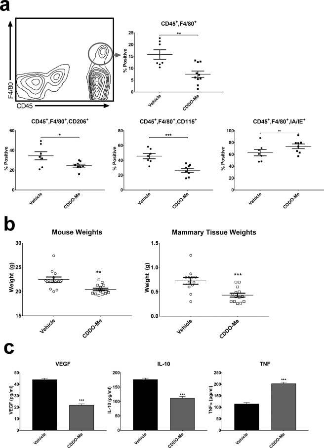
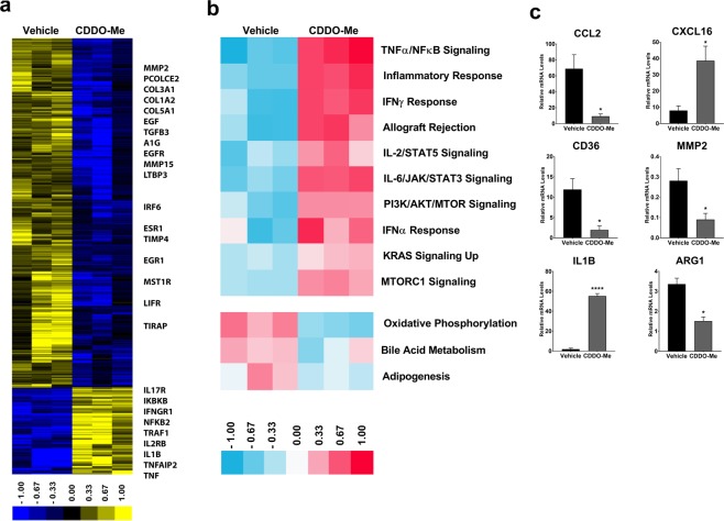
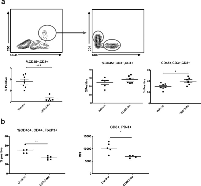
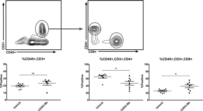
The tumor microenvironment (TME) is an essential contributor to the development and progression of malignancy. Within the TME, tumor associated macrophages (TAMs) mediate angiogenesis, metastasis, and immunosuppression, which inhibits infiltration of tumor-specific cytotoxic CD8+ T cells. In previous work, we demonstrated that the synthetic triterpenoid CDDO-methyl ester (CDDO-Me) converts breast TAMs from a tumor-promoting to a tumor-inhibiting activation state in vitro. We show now that CDDO-Me remodels the breast TME, redirecting TAM activation and T cell tumor infiltration in vivo. We demonstrate that CDDO-Me significantly attenuates IL-10 and VEGF expression but stimulates TNF production, and reduces surface expression of CD206 and CD115, markers of immunosuppressive TAMs. CDDO-Me treatment redirects the TAM transcriptional profile, inducing signaling pathways associated with immune stimulation, and inhibits TAM tumor infiltration, consistent with decreased expression of CCL2. In CDDO-Me-treated mice, both the absolute number and proportion of splenic CD4+ T cells were reduced, while the proportion of CD8+ T cells was significantly increased in both tumors and spleen. Moreover, mice fed CDDO-Me demonstrated significant reductions in numbers of CD4+ Foxp3+ regulatory T cells within tumors. These results demonstrate for the first time that CDDO-Me relieves immunosuppression in the breast TME and unleashes host adaptive anti-tumor immunity.


Conflict of interest statement

Karen Liby is an inventor of patents dealing with chemical synthesis of new triterpenoids and their application in treatment of cancer, as well as in inflammatory diseases, including human kidney disease. No other author has competing interests.

Figures

Figure 1
Figure 1
CDDO-Me redirects cytokine production of F4/80+ TAMs in vivo. (a) Whole mammary/tumor tissue of PyMT mice fed diet with or without 50 mg CDDO-Me/kg diet for 8 weeks was digested to a single cell suspension and analyzed by multicolor flow cytometry for analysis of macrophages. Expression of surface markers is represented as percentage of cells positive for the respective marker vs. vehicle (n = 610 per group). (b) Mouse weights were recorded immediately prior to tissue resection and mammary/tumor tissue weights were recorded immediately after tissue resection (n = 1214 per group). (c) TAMs were isolated using F4/80+ magnetic bead selection and cultured in complete DMEM for 24 hours. Culture supernatants were analyzed by ELISA for murine VEGF, IL-10, and TNF-α (n = 3 per group). *p < 0.05, **p < 0.01, ***p < 0.005 vs. vehicle; ns = nonsignificant. Cytokine data are representative of 3 biologically independent experiments.
Figure 2
Figure 2
CDDO-Me alters transcriptional program of TAMs in vivo. F4/80+ TAMs were isolated from the mammary tissue of 12 week old mice that were fed control or CDDO-Me diet (50 mg/kg diet) for 8 weeks. (a) Total RNA was isolated and analyzed by microarray. (b) Hallmark pathway analysis of differentially expressed genes (DEGs) in microarray was performed. (c) Select mRNA transcripts were validated by Taqman qRT-PCR. *p < 0.05, **p < 0.01, ***p < 0.005 vs. vehicle. Microarray data and qRT-PCR represent the average of three independent experiments.
Figure 3
Figure 3
CDDO-Me increases the proportion of CD8+ T cells and reduces Treg numbers in tumors in vivo. Whole mammary/tumor tissue of mice fed diet with or without 50 mg/kg CDDO-Me for 8 weeks was digested to a single cell suspension and analyzed by multicolor flow cytometry for analysis of CD4 + and CD8+ T cell populations (a). Dual positive CD4/Foxp3 T cell percentages were compared in control and CDDO-Me-treated mice (b). Expression of PD1 on CD8+ T cells was assessed using mean fluorescence intensity (MFI) (b). Where indicated, expression of surface markers is represented as percentage of cells positive for the respective marker (n = 5–10 per group). *p < 0.05, **p < 0.01, ***p < 0.005 vs. vehicle.
Figure 4
Figure 4
CDDO-Me decreases splenic CD4+ and augments CD8+ T cell populations in vivo. Spleens from mice fed control or 50 mg/kg CDDO-Me diet for 8 weeks was digested to a single cell suspension and analyzed by multicolor flow cytometry for analysis of T cell populations. Expression of surface markers is represented as percentage of cells positive for the respective marker (n = 6–8 per group). *p < 0.05 vs. vehicle.

References

    1. Broz ML, Krummel MF. The emerging understanding of myeloid cells as partners and targets in tumor rejection. Cancer Immunol. Res. 2015;3:313–319. doi: 10.1158/2326-6066.CIR-15-0041. - DOI - PMC - PubMed
    1. Schmid P, et al. Atezolizumab and Nab-Paclitaxel in Advanced Triple-Negative Breast Cancer. N. Engl. J. Med. 2018;379:2108–2121. doi: 10.1056/NEJMoa1809615. - DOI - PubMed
    1. Pollard JW. Tumour-educated macrophages promote tumour progression and metastasis. Nat. Rev. Cancer. 2004;4:71–78. doi: 10.1038/nrc1256. - DOI - PubMed
    1. Graves DT, Jiang YL, Williamson MJ, Valente AJ. Identification of monocyte chemotactic activity produced by malignant cells. Science. 1989;245:1490–1493. doi: 10.1126/science.2781291. - DOI - PubMed
    1. Murdoch C, Giannoudis A, Lewis CE. Mechanisms regulating the recruitment of macrophages into hypoxic areas of tumors and other ischemic tissues. Blood. 2004;104:2224–2234. doi: 10.1182/blood-2004-03-1109. - DOI - PubMed

Publication types

MeSH terms