. 2020 Mar 26:2020:8647981.
doi: 10.1155/2020/8647981. eCollection 2020.

Induction of Myogenic Differentiation Improves Chemosensitivity of Chemoresistant Cells in Soft-Tissue Sarcoma Cell Lines

Affiliations

Induction of Myogenic Differentiation Improves Chemosensitivity of Chemoresistant Cells in Soft-Tissue Sarcoma Cell Lines

Lucy E Dawson et al. Sarcoma. .

Abstract

Rhabdomyosarcoma (RMS) and rhabdoid tumors (RT) are rare soft-tissue malignancies with the highest incidence in infants, children, and adolescents. Advanced, recurrent, and/or metastatic RMS and RT exhibit poor response to treatment. One of the main mechanisms behind resistance to treatment is believed to be intratumoral heterogeneity. In this study, we investigated the myogenic determination factor 1 (MYOD1) and Noggin (NOG) markers in an embryonal RMS (ERMS) cell line and an RT cell line and the differential response of the MYOD1 and NOG expressing subpopulations to chemotherapy. Importantly, we found that these markers together identify a subpopulation of cells (MYOD1+ NOG+ cells) with primary resistance to Vincristine and Doxorubicin, two commonly used chemotherapies for ERMS and RT. The chemoresistant MYOD1+ NOG+ cells express markers of undifferentiated cells such as myogenin and ID1. Combination of Vincristine with TPA/GSK126, a drug combination shown to induce differentiation of RMS cell lines, is able to partially overcome MYOD1/NOG cells chemoresistance.


Conflict of interest statement

The authors declare no conflicts of interest.

Figures

Figure 1
Figure 1
MYOD1+ NOG+ cells predominant survival in RD and A-204 cell cultures after treatment with either Vincristine or Doxorubicin. (a) Plots show examples of flow analysis of RD and A-204 cells treated with Vincristine and Doxorubicin. In grey is highlighted the quadrant representing MYOD1+ NOG+ cells. (b) Percentage of live MYOD1+ NOG+ phenotype cells in RD and A-204 cell cultures after 2 days of treatment either with 1 and 10 nM of Vincristine (top panel) or with 0.1 and 1 μM of Doxorubicin (bottom panel). (c) Absolute numbers of total live cells in RD and A-204 cultures treated with 10 nM of Vincristine or 1 μM of Doxorubicin for 2 days. Data are presented as a ratio of absolute numbers of cells in treated cultures to absolute numbers of cells in nontreated cultures (fold change). Data are shown as mean ± standard deviation (N = 11). p < 0.05, ∗∗p < 0.01, ∗∗∗p < 0.001, ANOVA with Tukey's multiple comparison test.
Figure 2
Figure 2
MYOD1+ NOG+ cells express the highest levels of antiapoptotic BCL2 protein among other cells in RD and A-204 cultures. Median fluorescence intensity (MFI) of BCL2 protein in MYOD1/NOG cell subpopulations of RD (left plot) and A-204 (right plot) cell lines. Data are presented as a ratio of BCL2 MFI of MYOD1+ NOG+ cells and MYOD1− NOG+ cells to BCL2 MFI of MYOD1− NOG− cells (fold change), (mean ± standard deviation, N = 9). p < 0.05, ∗∗p < 0.01, ANOVA with Tukey's multiple comparison test).
Figure 3
Figure 3
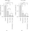
Differentiation therapy increases the effectiveness of chemotherapy. Absolute numbers of total live cells RD (a) and A-204 (b) in cultures treated for 6 days with TPA/GSK126, 10 nM of Vincristine, or combination of Vincristine and TPA/GSK126. Data are presented as a ratio of absolute numbers of cells in treated cultures to absolute numbers of cells in nontreated cultures (fold change) (mean ± standard deviation, N = 4). p < 0.05, ∗∗∗p < 0.001, ANOVA with Tukey's multiple comparison test for the analysis between the groups.

References

    1. Bompas E., Campion L., Italiano A., et al. Outcome of 449 adult patients with rhabdomyosarcoma: an observational ambispective nationwide study. Cancer Medicine. 2018;7(8):4023–4035. doi: 10.1002/cam4.1374. - DOI - PMC - PubMed
    1. Ognjanovic S., Linabery A. M., Charbonneau B., Ross J. A. Trends in childhood rhabdomyosarcoma incidence and survival in the United States, 1975–2005. Cancer. 2009;115(18):4218–4226. doi: 10.1002/cncr.24465. - DOI - PMC - PubMed
    1. Lychou S. E., Gustafsson G. G., Ljungman G. E. Higher rates of metastatic disease may explain the declining trend in Swedish paediatric rhabdomyosarcoma survival rates. Acta Paediatrica. 2016;105(1):74–81. doi: 10.1111/apa.13172. - DOI - PMC - PubMed
    1. Pastore G., Peris-Bonet R., Carli M., Martínez-García C., de Toledo J. S., Steliarova-Foucher E. Childhood soft tissue sarcomas incidence and survival in European children (1978–1997): report from the automated childhood cancer information system project. European Journal of Cancer. 2006;42(13):2136–2149. doi: 10.1016/j.ejca.2006.05.016. - DOI - PubMed
    1. Sultan I., Qaddoumi I., Yaser S., Rodriguez-Galindo C., Ferrari A. Comparing adult and pediatric rhabdomyosarcoma in the surveillance, epidemiology and end results program, 1973 to 2005: an analysis of 2,600 patients. Journal of Clinical Oncology. 2009;27(20):3391–3397. doi: 10.1200/jco.2008.19.7483. - DOI - PubMed

LinkOut - more resources