. 2020 Apr 10;6(15):eaaz2441.
doi: 10.1126/sciadv.aaz2441. eCollection 2020 Apr.

Schizorhodopsins: A family of rhodopsins from Asgard archaea that function as light-driven inward H+ pumps

Affiliations

Schizorhodopsins: A family of rhodopsins from Asgard archaea that function as light-driven inward H+ pumps

Keiichi Inoue et al. Sci Adv. .

Abstract

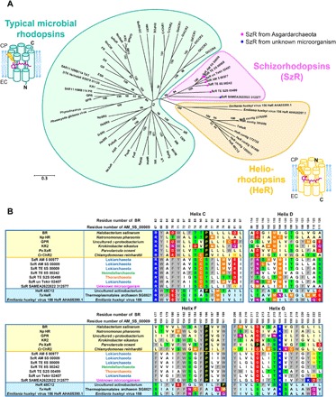
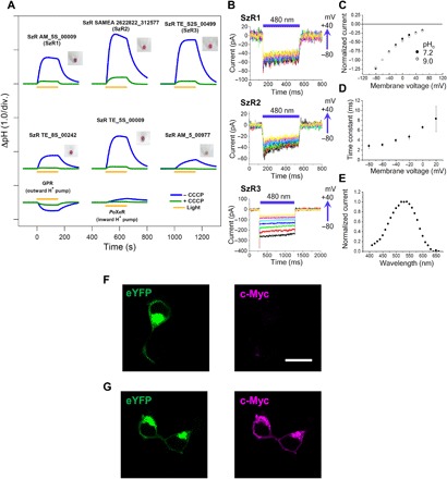
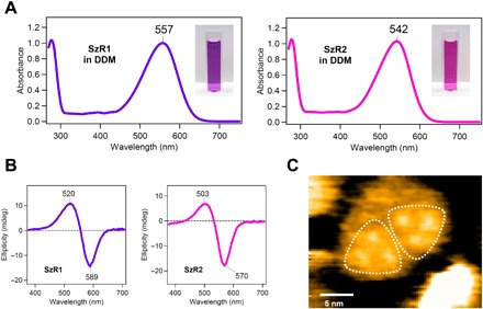
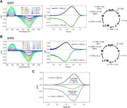
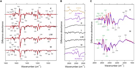
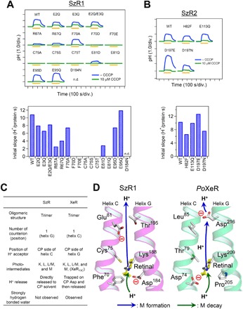
Schizorhodopsins (SzRs), a rhodopsin family first identified in Asgard archaea, the archaeal group closest to eukaryotes, are present at a phylogenetically intermediate position between typical microbial rhodopsins and heliorhodopsins. However, the biological function and molecular properties of SzRs have not been reported. Here, SzRs from Asgardarchaeota and from a yet unknown microorganism are expressed in Escherichia coli and mammalian cells, and ion transport assays and patch clamp analyses are used to demonstrate SzR as a novel type of light-driven inward H+ pump. The mutation of a cytoplasmic glutamate inhibited inward H+ transport, suggesting that it functions as a cytoplasmic H+ acceptor. The function, trimeric structure, and H+ transport mechanism of SzR are similar to that of xenorhodopsin (XeR), a light-driven inward H+ pumping microbial rhodopsins, implying that they evolved convergently. The inward H+ pump function of SzR provides new insight into the photobiological life cycle of the Asgardarchaeota.


Figures

Fig. 1
Fig. 1. Phylogenetic and sequence analyses of SzRs with microbial rhodopsins and HeRs.
(A) Phylogenetic tree of the representative typical microbial rhodopsins (green), HeRs (yellow), and SzRs (pink). The SzRs originated from Asgardarchaeota and from unknown microorganism are indicated with magenta and blue circles, respectively. The molecular orientations of the typical microbial rhodopsins and HeRs are schematically shown. N, N-terminus; C, C-terminus; EC, extracellular side; CP, cytoplasmic side. (B) Multiple amino acid alignment of SzRs with typical microbial rhodopsins and HeRs for the helices C, D, F, and G. Their full sequences are shown in fig. S2.
Fig. 2
Fig. 2. Light-driven active inward H+ transport by SzR.
(A) Ion transport activity assay of SzRs in E. coli cells. The cells were illuminated with light (λ > 500 nm) for 150 s (yellow line). The pictures of the pellets of E. coli cells expressing each SzR are shown next to the corresponding results. (B) Electrophysiological measurements of SzR-driven photocurrent in ND7/23 cells. The cells were illuminated with light (λ = 480 nm, 12.3 mW/mm2) during the time region shown by blue bars. The membrane voltage was clamped from −80 to +100 mV for every 20-mV step. (C to E) I-V plot at pHo 7.2 and 9.0 (C), membrane-voltage dependence of the off-kinetics time constant (D), and action spectrum (E) of the current of SzR3. For action spectrum measurement, the light intensity of each wavelength was adjusted to 0.2 mW/mm2. (F and G) eYFP fluorescence (left, green) and immunofluorescence staining observation of SzR1 with a c-Myc epitope tag at the C terminus in cultured ND7/23 cells (right, magenta) in unpermeabilized (F) and permeabilized conditions with detergent (Triton X-100) (G). Scale bar, 20 μm.
Fig. 3
Fig. 3. Absorption and CD spectra and high-speed AFM imaging of purified SzR proteins.
(A and B) UV-vis absorption (A) and CD (B) spectra of SzR1 (left) and SzR2 (right) in 100 mM NaCl, 20 mM tris-HCl (pH 8.0), and 0.05% DDM. (C) High-speed AFM image of SzR1 trimers in lipid bilayers.
Fig. 4
Fig. 4. Transient absorption changes and the photocycles of SzRs.
(A and B) Transient absorption spectra (left), time evolutions of transient absorption changes at specific wavelengths (middle), and the photocycles determined by analyzing the time evolution with multiexponential functions (right) of SzR1 (A) and SzR2 (B) in POPE/POPG (molar ratio, 3:1) membrane. (C) Transient absorption change of cresol red accompanying the photoreaction of SzR1 in 0.1% DDM. Positive and negative absorption changes at λ = 429 and 573 nm, respectively, indicate the protonation of cresol red.
Fig. 5
Fig. 5. Light-induced low-temperature difference FTIR spectroscopy of SzR1.
(A and B) Light-induced low-temperature K-minus-dark, L-minus-dark, L/M-minus-dark, and M-minus-dark difference FTIR spectra of SzR1 obtained at T = 110, 190, 210, and 230 K, respectively, in the 1800 to 850 (A) and 2630 to 2490 (B) cm−1 regions. In (B), the light-induced low-temperature K-minus-dark FTIR spectra of C1C2 and Rh-PDEtr and low-temperature M-minus-dark FTIR spectrum of Rh-PDEtr were reproduced from (18) and (28). (C) Light-induced low-temperature K-minus-dark and M-minus-dark difference FTIR spectra of SzR1 obtained at T = 110 and 230 K, respectively, in the 2800 to 1760 cm−1 region. The normalization factors multiplied to each spectrum are listed in tables S2 and S3.
Fig. 6
Fig. 6. H+ transport activity of SzR mutants and inward H+ transport model of SzR.
(A and B) Ion transport activity assay in E. coli cells (upper) and initial slopes normalized by the amount of expressed proteins (lower) of the mutants of SzR1 (A) and SzR2 (B). n.d., not determined. (C) Different and similar molecular characters between SzRs and XeRs. (D) H+ transport pathway in SzR1 (left) suggested in this study and in PoXeR (right) (9, 24). The structure of both proteins was drawn on the basis of the x-ray crystallographic structure of NsXeR (PDB code: 6EYU) (10). The proton release and uptake pathways on the M formation and M decay are indicated by blue and green arrows, respectively.

References

    1. Ernst O. P., Lodowski D. T., Elstner M., Hegemann P., Brown L. S., Kandori H., Microbial and animal rhodopsins: Structures, functions, and molecular mechanisms. Chem. Rev. 114, 126–163 (2014). - PMC - PubMed
    1. Philosof A., Béjà O., Bacterial, archaeal and viral-like rhodopsins from the Red Sea. Environ. Microbiol. Rep. 5, 475–482 (2013). - PubMed
    1. Mukherjee S., Hegemann P., Broser M., Enzymerhodopsins: Novel photoregulated catalysts for optogenetics. Curr. Opin. Struct. Biol. 57, 118–126 (2019). - PubMed
    1. Govorunova E. G., Sineshchekov O. A., Hemmati R., Janz R., Morelle O., Melkonian M., Wong G. K.-S., Spudich J. L., Extending the time domain of neuronal silencing with cryptophyte anion channelrhodopsins. eNeuro 5, ENEURO.0174-18.2018 (2018). - PMC - PubMed
    1. Pushkarev A., Inoue K., Larom S., Flores-Uribe J., Singh M., Konno M., Tomida S., Ito S., Nakamura R., Tsunoda S. P., Philosof A., Sharon I., Yutin N., Koonin E. V., Kandori H., Béjà O., A distinct abundant group of microbial rhodopsins discovered using functional metagenomics. Nature 558, 595–599 (2018). - PMC - PubMed

Publication types

LinkOut - more resources